개가시나무 가지로부터 항산화성분의 규명
문 미 연⋅백 종 석⋅김 상 숙⋅장 원 정⋅김 미 선*⋅이 남 호†
제주대학교 자연과학대학 화학과, *제주관광대학 뷰티디자인학과 (2009년 8월 11일 접수, 2009년 8월 26일 수정, 2009년 9월 4일 채택)
Identification of Antioxidative Constituents from The Branches of Quercus gilva Blume
Mi Youn Moon, Jong Seok Baik, Sang Suk Kim, Won Jung Jang, Mi Sun Kim*, and Nam Ho Lee†
Department of Chemistry, Cheju National University, Ara-1, Jeju 690-756, Korea.
*Department of Beauty Design, Cheju Tourism College, Jeju 690-791, Korea (Received August 11, 2009; Revised August 26, 2009; Accepted September 4, 2009)
요 약: 개가시나무 가지의 에탄올 추출물에서 항산화활성을 관찰하였으며, 활성성분을 규명하기 위한 연구를 진행하였
다. 그 결과, 4종류의 화합물을 분리하여 동정하였다. 분리 동정된 성분은 catechin(1),
epi-catechin(2), tyrosol(3) 및 tiliroside(4) 이다. 분리 성분의 항산화활성은 DPPH 라디칼 및 superoxide 음이온 라디칼 소거활성을 이용하여 측정하였 다. 화합물 1, 2, 3, 4는 100 µL/mL 농도에서 각각 94.2 %, 93.4 %, 33.6 %, 11.2 %의 DPPH 라디칼 저해활성을 나타내 었다. 또한, 화합물 1, 2, 3, 4는 200 µL/mL 농도에서 각각 60.2 %, 35.1 %, 20.6%, 4.5 %의 superoxide 음이온 라디칼 저해활성을 나타내었다. 화합물 1 ∼ 4는 개가시나무에서는 처음으로 분리된 물질이다.
Abstract: Investigation of antioxidative constituents from the ethanol extract of
Quercus gilvabranches led to the identification of four compounds; catechin (1),
epi-catechin (2), tyrosol (3) and tiliroside (4). The antioxidative ac- tivity was examined using DPPH radical and superoxide anion radical scavenging method. The isolated compounds 1, 2, 3 and 4 exhibited 94.2 %, 93.4 %, 33.6 % and 11.2 % scavenging activities respectively against DPPH radicals at the concentration of 100 µL/mL. As well, the compounds 1 ∼ 4 showed respectively 60.2 %, 35.1 %, 20.6 %, 4.5 % inhibition activities against superoxide anion radicals at 200 µL/mL. Interestingly, the compounds 1 ∼ 4 were isolated for the first time from
Quercus gilvaBlume.
Keywords: quercus gilva, DPPH radical scavenging activity, superoxide anion radical scavenging activity, catechin, tyrosol
1. Introduction
1)
Quercus gilva, belonging to a family Fagaceae, is an evergreen tall tree. This plant is distributed in the mountain area of Jeju Island in Korea. The lumber of this tree has been utilized as the raw material for household furniture[1]. Previous phytochemical studies have led to the identification of terpenes from the fruit
† 주 저자 (e-mail: [email protected])
of this plant[2,3].
As skin epidermis is exposed to ultraviolet (UV) ra- diation, it accompanies the activation of reactive oxy- gen species (ROS) in the skin cells[4]. The ROS, in- cluding singlet oxygen, superoxide anion radical, hydro- gen peroxide and hydroxyl radical, are highly reactive, and can make a random attack the tissues to cause their oxidative degradation. Oxidative deterioration of lipid in the epidermis[5] as well as protein in the der- mis[6] can lead to generation of sag or wrinkle in the
skin, which is the typical appearance of aged skin. In order to prevent oxidative destruction, antioxidative de- fense system is operating in the body by the action of enzymes such as superoxide dismutase, catalase and glutathione peroxidase. In addition, antioxidative small molecules such as vitamin C and vitamin E are also ef- fective to protect the destruction of oxidative cells. As a supplementary measure to prevent oxidative deterio- ration, use of exterior antioxidant becomes of great im- portance in cosmetic application[7]. Therefore, search for effective and safe antioxidants is necessary.
Natural products developed especially from plant kingdom are recognized as safe and nature-friendly in- gredients in cosmetic industries. Thus, development of antioxidative extracts as well as isolated constituents from plants has practical values. We are continuously searching plants available in Jeju to find bioactive nat- ural ingredients[8-10]. Jeju Island has diversity of a plant community with more than 1,800 plant spe- cies[11]. As we found that the ethanol extract of Q.
gilva branches exhibited significant antioxidative activ- ities, in this study, we isolated and identified the active constituents present in this plant.
2. Materials and Methods
2.1. Reagents and Equipments
All solvents were of analytical grade. 1H (400.00 MHz) and 13C (100.60 MHz) nuclear magnetic reso- nance (NMR) spectra were recorded on a JEOL, JNM-LA 400 instrument, with chemical shift data re- ported in ppm relative to the solvent used. 2D NMR spectra were recorded on the same instrument using field gradient FG2 (inverse) probe. Normal and re- versed phase (RP) column chromatography (CC) were performed using Merck silica gel (0.063 ∼ 0.200 mm) and RP-18 silica gel (230 ~ 400 mesh) respectively.
Silica gel 60 F254 coated on aluminium plates for thin layer chromatography (TLC) was supplied by Merck.
Gel filtration chromatography (GFC) was performed using Sephadex LH-20 (0.100 ~ 0.025 mm) obtained from Fluka. 1,1-Diphenyl-2-picrylhydrazyl (DPPH) was purchased from Aldrich Chem. Co. Ascorbic acid
were purchased from Sigma Chemical Co.
2.2. Plant Material
The branches of Q. gilva were collected in February 2006 from Halla botanical garden in Jeju, Korea. A voucher specimen (J-064) was prepared and deposited at the Laboratory of Natural Product Chemistry, De- partment of Chemistry, Cheju National University.
2.3. Extraction and Isolation
The dried branches of Q. gilva (705 g) was cut into small pieces and extracted with 70 % ethanol (14 L) at room temperature for 7 days. The extract was fil- tered and evaporated to a gummy mass in a rotary evaporator under vacuum at a maximum temperature of 40 ℃. The ethanol extract (22 g) was partitioned between water and n-hexane and the aqueous part was further portioned between ethyl acetate and water and then n-butanol and water.
A part of ethyl acetate-soluble extract (3.3 g) was subjected to column chromatography (CC) over re- versed phase (RP) silica gel (SiO2) eluting with gra- dient H2O-MeOH to yield nine fractions (fr. I ∼ IX).
The first fraction (fr. I, 143 mg), eluted with 20 % methanol, was applied to GFC over Sephadex LH-20 with CHCl3/MeOH (4/1) to give the compound 3 (8.3 mg). The second fraction (fr. II) and third fraction (fr.
III) were identified as the compounds 1 (725 mg) and 2 (359 mg) respectively. The fraction V (250 mg) was applied GFC with CHCl3/MeOH (4/1) to produce sev- en fractions, among which the fifth fraction (fr. V-5) was further SiO2 column chromatographed with the same solvent system to yield the compound 4 (11.8 mg).
Compound 1 1H-NMR (400 MHz, CD3OD) δ 2.50 (1H, dd, J = 16.1, 8.1 Hz, H-4a), 2.84(1H, dd, J = 16.1, 5.4 Hz, H-4b), 3.98(1H, ddd, J = 7.8, 7.8 and 5.4 Hz, H-3), 4.56(1H, d, J = 7.6 Hz, H-2), 5.85(1H, d, J
= 2.4 Hz, H-8), 5.92(1H, d, J = 2.4 Hz, H-6), 6.71 (1H, dd, J = 8.0, 2.0 Hz, H-6'), 6.76(1H, d, J = 8.0 Hz, H-5'), 6.83(1H, d, J= 2.0 Hz, H-2'); 13C-NMR(100 MHz, CD3OD) δ 28.5(C-4), 68.8(C-3), 82.8(C-2),
95.5(C-8), 96.3(H-6), 100.8(C-10), 115.2(C-2'), 116.1 (C-5'), 120(C-6'), 132.2(C-1'), 146.1(C-3'), 146.2 (C-4'), 156.9(C-9), 157.5(C-7), 157.5(C-5).
Compound 2 1H-NMR (400 MHz, CD3OD) δ 2.73 (1H, dd, J = 16.8, 3.0 Hz, H-4a), 2.85(1H, dd, J = 16.8, 4.7 Hz, H-4b), 4.18(1H, ddd, J = 1.5, 3.7 and 3.8 Hz, H-3), 4.81(1H, d, J = 1.5 Hz, H-2), 5.91(1H, d, J
= 2.2 Hz, H-6), 5.93(1H, d, J = 2.2 Hz, H-8), 6.75 (1H, d, J = 8.3 Hz, H-5'), 6.79(1H, dd, J = 8.3, 2.0 Hz, H-6'), 6.96(1H, d, J = 2.0 Hz, H-2'); 13C-NMR (100 MHz, CD3OD) δ 29.2(C-4), 67.5(C-3), 79.9(C-2), 95.9 (C-8), 96.4(C-6), 100.0(C-10), 115.3(C-2'), 115.9(C- 5'), 119.4(C-6'), 132.3(C-1'), 145.7(C-3'), 145.9(C-4'), 157.3(C-9), 157.6(C-7), 158.0(C-5).
Compound 3 1H-NMR (400 MHz, CD3OD) δ 2.70 (1H, t, J = 7.1 Hz, H-7), 3.67(1H, t, J = 7.1 Hz, H-8), 6.68(2H, d, J = 8.0 Hz, H-2 & H-6), 7.00(2H, d, H-3
& H-5) 13C-NMR (100 MHz, CD3OD) δ 39.4(C-7), 64.6 (C-8), 116.4(C-3 & C-5), 130.4(C-1), 130.8(C-2 & C- 6), 157.6(C-4).
Compound 4 1H-NMR (400 MHz, CD3OD) δ 3.45 ∼ 3.48(4H, m, H-2'', H-3'', H-4'', H-5'') 4.18(1H, dd, J = 11.6, 2.2 Hz, H-6''a), 4.29(1H, dd, J = 11.6, 6.6 Hz, H-6''b), 5.21(1H, d, J = 7.6 Hz, H-1''), 6.07(1H, d, J
= 16.1, H-8'''), 6.11(1H, d, J = 2.0 Hz, H-8), 6.26(1H, d, J = 2.0 Hz, H-6), 6.78(2H, d, J = 8.3 Hz, H-3''' &
5'''), 6.80(2H, d, J = 8.3 Hz, H-3' & 5'), 7.30(2H, d, J
= 8.3 Hz, H-2''' & 6'''), 7.40(1H, d, J = 16.1 Hz, H-7''') 7.97(2H, d, J = 8.3 Hz, H-2' & 6'); 13C-NMR (100 MHz, CD3OD) δ 64.3(C-6''), 71.7(C-4''), 75.7 (C-2''), 75.8(C-5''), 78.0(C-3''), 95.0(C-8), 100.3(C-6), 104.1(C-1''), 105.4(C-10), 114.7(C-8'''), 116.0(C-3' &
5'), 116.8(C-3''' & 5'''), 122.7(C-1'), 127.0(C-1'''), 131.2 (C-2''' & 6'''), 132.2(C-2' & 6'), 135.2(C-3), 146.6 (C-7'''), 158.5(C-2), 159.2(C-5), 161.2(C-4'''), 161.5(C- 4'), 161.9(C-7), 162.9(C-9), 168.8(C-9'''), 179.3 (C-4).
2.4. DPPH Free Radical Scavenging Activity Test The free radical scavenging activity was assayed us- ing deep blue 1,1-diphenyl-2-picrylhydrazyl (DPPH)
radicals[12]. Sample stock solutions (1 mg/mL) were diluted to final concentrations of 100, 50, 10 and 5 g/mL in 70 % ethanol or DMSO. 0.5 mL of 0.2 mM DPPH ethanol solution was added to 1 mL of sample solutions of different concentrations, shaken well by vortex, and allowed to react at room temperature. The absorbance values were measured after 10 min at 525 nm by UV/Vis spectrophotometer. The free radical scavenging activity of samples was calculated accord- ing to the formula:
DPPH radical scavenging activity (%) =
[1 - (Abssample - Absblank)/Abscontrol] × 100
where Abssample is the absorbance of the experimental sample, Absblank is the absorbance of the blank, Abscontrol is the absorbance of the control.
2.5. Superoxide Anion Radical Scavenging Activity The superoxide anion radical was generated via a xanthine-xanthine oxidase system and assayed by the reduction of nitroblue tetrazolium (NBT)[13]. The re- action mixture was prepared with 108 uL of buffer (200 mM sodium phosphate, pH 7.5), 2 uL of NBT (50 mM), 30uL of EDTA (5 mM), 20 uL of xanthine (5 mM) in 1 N NaOH, 20 uL of xanthine oxidase in buffer (0.5 U/mL) and 20 uL of isolated compounds 1 ∼ 4.
The absorbance values were measured at 540 nm by UV/Vis spectrophotometer. The superoxide anion radi- cal scavenging activity of samples was calculated ac- cording to the formula:
Superoxide anion radical scavenging activity (%) = [1 - (Abssample - Absblank)/Abscontrol] × 100
where Abssample is the absorbance of the experimental sample, Absblank is the absorbance of the blank, Absontrol
is the absorbance of the control.
3. Results and Discussion
3.1. Isolation and Structure Identification
The plant extract (22 g) was prepared using 70 %
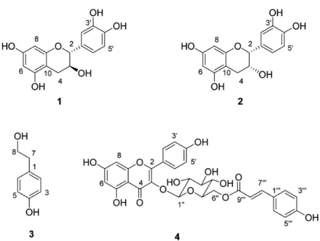
Figure 1. Structures of compounds isolated from
Q. gilva.
ethanol from the branches of Q. gilva (705 g) at room temperature. The ethanol extract was tested for its an- tioxidative activities using DPPH radical scavenging method. Ascorbic acid was used as a positive control in this experiment. The extract showed strong radical scavenging activities. Compared to ascorbic acid (SC50, 50 % scavenging concentration, 4.2 µg/mL), ethanol extract exhibited an activity with SC50 value of 13.5 µg/
mL. As strong radical scavenging activities were ob- served in this extract, the fractionation was conducted to identify the active constituents. The ethanol extract was partitioned into n-hexane, ethyl acetate (EtOAc) and n-butanol. The EtOAc fraction was subjected to repeated column chromatography over silica gel as well as Sephadex LH-20 to result in the isolation of four compounds (1 ∼ 4) (Figure 1).
The compound 1 was isolated as the major compo- nent in the EtOAc-soluble fraction. Inspection of 13C and DEPT NMR spectra indicated the presence of 15 carbons including 12 aromatic and three aliphatic car- bons in the compound 1. On the 1H NMR spectrum, signals at δ 6.71 (dd, J = 8.0, 2.0, C-6'), 6.76 (d, J = 8.0, C-5') and 6.83 (d, J = 2.0, C-2') were observed in the lower field, which suggested the presence of 1,3,5- trisubstituted benzene ring. In addition, meta-coupled
aromatic proton peaks (δ 5.85 and 5.92, J = 2.4 Hz) showed the structural feature of another benzene ring.
In higher field, proton signals for one methylene (δ 2.50 and 2.84) and two oxygen-bearing methines (δ 3.98, 4.56) are detected to be coupled each other, which suggested they are in the same spin system. Based on this spectroscopic information, the compound 1 was identified as catechin. The large coupling constant (J
= 7.6 Hz) of H-2 (δ 4.56), indicating the trans-con- formation between H-2 and H-3, also verified the ster- eochemistry of catechin (1). The structure of this known compound was further identified by comparing the obtained data with those of literature values[14].
The compound 2 was also isolated as the major con- stituents in this ethyl acetate fraction. Inspection of 1H and 13C NMR spectra revealed that signals of com- pound 2 were almost identical to those of compound 1.
This suggested that compounds 1 and 2 share the same molecular skeleton. The distinguishable difference was observed at δ 4.81 (H-2) with small coupling constant (J = 1.5 Hz), which indicated the cis-conformation be- tween H-2 and H-3. Therefore, the compound 2 was identified as C-2 steroisomer of 1, epi-catechin[14].
The compound 3 showed six peaks for four aromatic and two aliphatic methylene carbons in 13C and DEPT
Figure 2. DPPH radical scavenging activities of the isolated compounds 1-4 (100 µg/mL) from the
Q. gilvabranch.
NMR spectra. Examination of 1H NMR spectrum pro- posed the existence of protons for 1,4-disubstituted symmetric benzene (δ 6.68 and 7.00, J = 8.0 Hz) and for a molecular subunit of -CH2CH2OH. The attachment of OH at C-4 was inferred by down field shifted carbon signal at δ 157.6. From this data, the compound 3 was identified as 2-(4-hydroxyphenyl)-1-ethanol, tyrosol[15].
The compound 4 showed 26 signals accounting for two carbonyl carbons, 18 sp2 carbons, and six oxy- gen-bearing aliphatic carbons in 13C NMR spectrum.
Inspection of aliphatic carbons in 13C spectrum coupled with corresponding protons in 1H NMR spectrum re- vealed that a glucose unit is present in compound 4.
The large coupling constant (J = 7.6 Hz) at δ 5.21 (H-1'') showing HMQC relation with δ 104.1 (C-1) indicted that the glucose is in β-configuration. 1H NMR signals corresponding to meta-coupled protons at δ 6.11 (1H, J = 2.0) and 6.26 (1H, J = 2.0) as well as ortho-coupled symmetric protons at δ 6.80 (2H, J
= 8.3) and 7.97 (2H, J = 8.3) is indicative of kaemp- ferol moiety in 4. This assumption was supported by the observation of 13C NMR signals for a carbonyl car- bon at δ 179.3 (C-4), for oxygen-bearing aromatic carbons at δ 159.2 (C-5), 161.9 (C-7), 162.9 (C-9) and 161.5 (C-4'), as well as for the characteristic olefin carbons at δ 158.5 (C-2) and 135.2 (C-3). Examina- tion of 1H signals at 6.07 (d, J = 16.1 Hz, C-8''') and 7.40 (d, J = 16.1 Hz, C-7''') proposed the existence of trans-olefin. Coupled with 13C signals for ester carbon
Figure 3. Superoxide anion radical scavenging activities of the isolated compounds 1-4 (200 µg/mL) from the
Q. gilvabranch.
at δ 168.8 (C-9''') and p-hydroxyphenyl carbons at δ 116.8, 127.0, 131.2 and 161.2, the presence of p-cou- maroyl ester moiety is strongly suggested. Combined all of these informations, the compound 4 was identified as kaempferol-3-O-(6''-coumaroyl)glucopyranoside, tiliro- side[16].
3.2. DPPH Radical and Superoxide Anion Radical Sc- avenging Acitivities
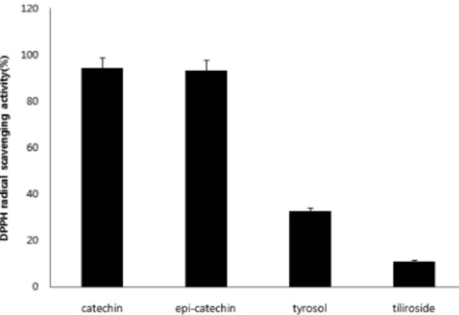
The antioxidative activities were examined DPPH free radical and superoxide anion radical scavenging method. The DPPH radical scavenging activity can be followed by a loss of DPPH absorbance at 525 nm. The isolated compounds 1, 2, 3 and 4 exhibited 94.2 %, 93.4
%, 33.6 % and 11.2 % scavenging activities respec- tively against DPPH radicals at the concentration of 100 µg/mL. The superoxide anion radical was gen- erated via a xanthine-xanthine oxidase system. The scavenging activities were assayed by the reduction of nitroblue tetrazolium (NBT) as determined by absorb- ance values measured at 540 nm. The isolated com- pounds 1 ∼ 4 showed respectively 60.2 %, 35.1 %, 20.6
%, 4.5 % inhibition activities against superoxide anion radicals at 200 µg/mL.
4. Conclusion
In the present study, we have isolated four com-
pounds (1 ∼ 4) from the ethanol extract prepared from the branches of Q. gilva. It is interesting to note that the compounds 1 ∼ 4 were isolated for the first time from this plant. From isolation of the constituents as well as evaluation of their activities, it was con- cluded that catechin (1) and epi-catechin (2) are pres- ent as the major antioxidative components in the extract. Also, tyrosol (3) present as a minor component showed a moderate antioxidative activity. This in- formation will be potentially useful when utilizing the extract of this plant as an industrial ingredient in the future.
Acknowledgements
This research work was supported by grant from the Ministry of Knowledge and Economy, Korea.
References
1. Y. N. Lee, Flora of Korea, Kyohak Publishing Co.
Seoul, Korea, 68 (2004).
2. T. Yasuhide, K. Yashikai, S. Jiro, T. Ichiro, and I.
Hidei, Studies on the constituents of Quercus spp.
VII. triterpenes of Quercus gilva blume, Yakugaku Zassi, 96, 1213 (1976).
3. I. Hidei, T. Yasuhide, K. Yashiaki, and I. Yoichi, Structure of gilvanol, a new triterpene isolated from Quercus gilva blume, Chem. Pharm. Bull., 26, 331 (1978).
4. P. M. Tyrell and P. Mireille, Single oxygen involve- ment in the inactivation of cultured human fibro- blasts by UVA (334 nm, 365 nm) and near-visible (405 nm) radiations, Photochem. Photobiol., 49, 407 (1989).
5. K. Chiba, T. Stone, K. Kawakami, and M. Onue, Skin roughness and wrinkle formation induced by repeated application of squalene monohydroperoxide to the hairless mouse, Exp. Dermatol., 8, 471 (1999).
6. H. Tanaka, T. Okada, H. Konishi, and T. Tsuji, The effect of reactive oxygen species on the biosyn-
thesis of collagen and glucosaminoglycans in cul- tured human dermal fibroblasts, Arch. Dermatol.
Res., 285, 352 (1993).
7. N. Noguchi, Y. Iwaki, M. Takahashi, E. Komuro, Y.
Kato, K. Tamura, O. Cynshi, T. Kodama, and E.
Niki, 2,3-Dihydro-5-hydroxy-2,2-dipentyl-4,6-tert- butylbenzofuran: design and evaluation as a novel radical-scavenging antioxidant against lipid perox- idation, Arch. Biochem. Biophys., 342, 236 (1997).
8. J. M. Kim, R. K. Ko, J. W. Hyun, and N. H. Lee, Identification of new dibenzofurans from Distylium racemosum, Bull. Korean Chem. Soc., 30, 261 (2009).
9. R. K. Ko, S. Lee, C. G. Hyun, and N. H. Lee, New dibenzofurans from the branches of Distylium race- mosum sieb. et Zucc, Bull. Korean Chem. Soc., 30, 1376 (2009).
10. N. Sultana and N. H. Lee, New phenylpropanoids from Sasa quelpaertensis nakai with tyrosinase in- hibition activities, Bull. Korean Chem. Soc., 30, 1729 (2009).
11. Y. N. Lee, K. S. Lee, and Y. H. Shin, Wild Plants of Jeju Island, Yeomiji Botanical Garden, Jeju, Korea, 1 (2001).
12. N. Sultana and N. H. Lee, Antielastase and free radical scavenging activities of compounds from stems of Cornus kousa, Phytotherapy Res., 21, 1171 (2007).
13. M. E. Hildago, E. Fernandez, W. Quilhot, and E.
Lissi. Antioxidant capacity of depsodes and de- psidones. Phytochemistry, 37, 1585 (1994).
14. I. H. Kang, J. H. Cha, S. W. Lee, H. J. Kim, S. H.
Kwon, I. H. Ham, B. S. Hwang, and W. K. Whang, Isolation of anti-oxidant from domestic Cratagus pinnatifida bunge leaves, Kor. J. Pharmacogn., 36, 121 (2005).
15. N. F. Komissarenko, E. V. Krivoruchko, V. S.
Kislichenko, and V. N. Kovalev, Tyrosol from Ribes nigrum, Chem. Natural Compound, 33, 97 (1997).
16. K. Y. Jung, S. R. Oh, S. H. Park, I. S. Lee, K. S.
Ahn, J. J. Lee, and H. Lee, Anti-complement ac- tivity of tiliroside from the flower bud of Magnolia fargesii, Biol. Pharm. Bull., 21, 10 (1998).