†Corresponding Author:So-Yeon Yoon (Department of Design & Environmental Analysis, Cornell University) E-mail:[email protected]
TEL:+1-607-255-8467 FAX:+1-607-255-0305
Neuroaesthetics: A Concise Review of the Evidence Aimed at Aesthetically Sensible Design
Yun Jung Choi*․So-Yeon Yoon*†
*Department of Design & Environmental Analysis, Cornell University
Abstract
In recent years, advancing technology and growing interest in neuromarketing and neurobranding have led to foundational research that facilitates a better understanding of consumers’ affective responses and unconscious information processing. However, the areas of aesthetics and design have remained largely unaffected by such advances and implications. The purpose of this study is to present a systematic review of the neuroscientific evidence aimed at sensible design for design and marketing researchers interested in exploring neuroaesthetics, an interdisciplinary area by nature. Sciencedirect, EBSCO, and the Google Scholar database were searched in February 2014 to select and review previous studies of aesthetics involving neuroscience. Twenty-eight studies were reviewed and divided into two categories: reward system and emotion. In addition to discussions on previous approaches, future research directions focusing on the process of aesthetic judgments (e.g., design elements, marketing stimuli) are proposed.
Key words: Neuroaesthetics, Review, Brain science, Neuromarketing, Neuropackaging, Emotion
1. Introduction
Despite the numerous new products introduced every day, only a few are adopted by the masses then even fewer become part of a trend. To develop sensible designs that will be accepted by the masses, companies invest substantial resources toward consumer research.
Consumer preferences or judgments of product design, especially aesthetic judgments, have posed constant challenges as significant research issues. To better understand consumers’ positive responses to and adoption of design, researchers have begun to pay more attention to neuroscientific methods that can shed light on the hidden processes of the brain.
The recent development of neuroscience allows researchers to effectively and more accurately measure brain activities related to visual information processing.
Although there has been a growing interest in neuromarketing and neurobranding, which has established a foundation for better understanding of user emotions and unconscious information processing, scholars have yet to explore the area of aesthetics.
Neuroaesthetics, as an interdisciplinary field, refers to the neural approaches to aesthetics. This new field utilizes methods of neuroscience to identify main issues in aesthetics. Zeki (2003), who first coined the term neuroaesthetics, highlighted a parallel between elements of art and the basic neural mechanism. They have a common aim to understand the true nature of objects or spaces in the world. The human brain organizes the sensual elements of information such as color, sound, and taste to understand the character of objects.
Similarly, designers experiment with different information elements to produce the best results (Chatterjee, 2011).
Then, is there any universal good design? Can neuroscience reveal aesthetically sensible design?
The purpose of this study is to present a systematic review of the neuroscientific evidence aimed at sensible design. Given the increased interest, along with technological timeliness, it is important to overview and
to understand the current status of research and implications to review major experimental studies and address the future directions in neuroaesthetics.
The present review includes basic facts about the human brain structures relevant to neuroaesthetics, as well as a discussion of major research issues closely relevant to neuroaesthetics, particularly with regard to visual aesthetics. A review of previous literature across multiple disciplines was conducted by searching databases (i.e., Sciencedirect, EBSCO, and Google Scholar) using the keywords neuroaesthetics, neural / neuroscience/brain, and beauty / design for full-text, peer-reviewed articles in English. After screening the abstracts to determine each study’s relevance, 28 articles were further reviewed. The literature was categorized into two main subjects related to neuroaesthetics: reward system and emotion. The purpose of this review is to synthesize the findings regarding the process of aesthetic judgment (i.e., design elements and marketing stimuli) and to suggest future directions for researchers.
The key contributions of this review include a better understanding of the emerging field of neuroaesthetics for future studies.
2. Basic Facts - the Human Brain
Before exploring the basic principles of neuroscience for design, it is necessary to have an understanding of the human brain. The brain is largely composed of neurons and glial cells. Neurons are specialized to pass or block signals to other target cells across synapses, which are structures that permit an electrical or chemical signal to pass between nerve cells.
Specifically, chemical signal transduction between
synapses involves various neurotransmitters. The state of
the human mind varies according to the types of
neurotransmitters that are activated. For example, it is
commonly recognized that the release of dopamine is
closely related to pleasure and consumer satisfaction
(Fugate, 2007).
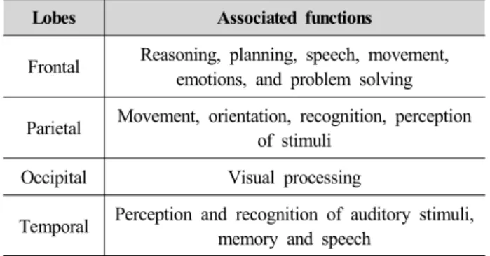
The cerebral cortex, the dominant feature of the human brain and the region of interest in neuroaesthetics, includes four lobes: frontal, parietal, occipital, and temporal. These lobes are involved with the perception and interpretation of sensations.
Specifically, the occipital lobe is the visual center of the brain, whereas the temporal lobe is involved with hearing and memory. The parietal lobe is associated with attention, spatial cognition, and muscular movement. The frontal lobe controls the information stored in the occipital and temporal lobes (Plassmann, O’Doherty, Shiv, & Rangel, 2008). Another important brain structure, the limbic system, includes the amygdala, corpus striatum, and basal ganglia. According to studies in affective neuroscience, responses related to brands such as brand familiarity or preference are associated with the limbic system (Walla, Mavratzakis,
& Bosshard, 2013). The limbic system is also referred to as the “emotional brain”. From evolution of brain, structures that are directly linked to critical skills and senses for survival such as the cerebellum associated with regulation and coordination of movement and balance are known to be much older than other structures, thus referred to as “old brain” (Ornstein, 1991). According to Ornstein (1991), our old brain is concerned exclusively with survival. The limbic system, the emotional brain is the evolutionarily old brain.
Damasio (2005) states that emotion, feeling and biological regulation all play a role in human reasons;
the lowly orders of our organism are in the loop of higher reason. In other words, the emotional brain related to survival plays an important role in decision making as illustrated in Figure 1.
LeDoux (1998) points out that the amygdala-located in the old brain has a greater influence on the cortex than the cortex has on the amydala, allowing emotional arousal to dominate and control thinking. Neuroscientists have established we make decisions in an emotional manner and then rationally justify the decisions. Often
the final decision is triggered by the old brain that doesn’t even understand words (Renvoise, 2007).
Noninvasive neuroscientific methods include the use of electroencephalography (EEG), magneto-encephalography (MEG), positron emission topography (PET), and functional magnetic resonance imaging (fMRI). Of these methods, fMRI is the most commonly used in consumer neuroscience. The fMRI provides us with visual data about the change of blood flow in the brain (Morin, 2011). The main advantage of fMRI is spatial resolution better than that of EEG; whereas, the disadvantages include cost and a relatively slow temporal resolution.
Figure 1. Human brain system
Table 1. Associated functions of cerebral cortex sections
Lobes Associated functions
Frontal Reasoning, planning, speech, movement, emotions, and problem solving
Parietal Movement, orientation, recognition, perception of stimuli
Occipital Visual processing
Temporal Perception and recognition of auditory stimuli, memory and speech
Previous studies in neuroaesthetics reported that visual information processing (the judgment of the attractiveness of faces, places, and objects) involves several parts of the prefrontal cortex (Plassmann, Kenning, Deppe, Kugel, & Schwindt, 2008).
Specifically, orbitofrontal, insular, medial prefrontal, and
posterior cingulate cortices and the ventral striatum have
been discussed in terms of their relevance to the reward system (Deppe et al., 2005; Walla et al., 2013).
Emotional response to an object has been reported to involve the activation of the anterior medial temporal lobe, medial and orbitofrontal cortices, and subcortical structures (Chatterjee, 2011). Dorsolateral prefrontal and medial frontal cortices and parietal circuits are involved in decision-making processes such as consumer preferences or judgments about an object. Given these research findings, it is apparent that basic knowledge of the brain would facilitate more effective design research into human aesthetic experiences.
3. Where we’ve been:
review of selective studies
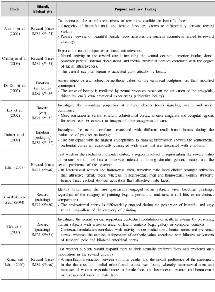
In this section, we review previous neuroaesthetic studies. Table 2 presents a brief overview of major studies that met our selection criteria.
3.1. Reward system
Reward system refers to the complex network of the various brain structures involved in processing rewards (Hubert & Kenning, 2008). The perception of rewards as visual stimuli positively or negatively reinforces human behavior as pursuing rewards or avoiding punishments, respectively. Understanding the reward system in the human brain is closely linked to studying consumer evaluation of objects that drives particular human behavior such as product consumption.
Most of the neuroaesthetic studies have discussed the close relationship between reward systems and aesthetic experiences (Cinzia & Vittorio, 2009). That is, the more attractive the visual stimuli are perceived to be, the higher reward value (i.e., the desire to acquire an aesthetic object) in areceiver’s brain. The beauty of the object—a central concept in aesthetics—activates the neural circuitry known as a reward system, including
the ventral striatum (the nucleus accumbens), the obifrontal cortex, and amygdala (Deppe et al., 2005;
Walla et al., 2013). Visual stimuli that has been known to activate the reward system in previous studies include beautiful faces (Aharon et al., 2001; Bray &
O’Doherty, 2007; Cloutier, Heatherton, Whalen, &
Kelley, 2008; Ishai, 2007; Kranz & Ishai, 2006; J.
O’Doherty et al., 2003; J. P. O’Doherty, 2004; Senior, 2003; Tsukiura & Cabeza, 2011; Winston, O’Doherty, Kilner, Perrett, & Dolan, 2007; Zaidel & Cohen, 2005), attractive paintings (Deppe et al., 2007; Vartanian &
Goel, 2004), packaging (Reimann, Zaichkowsky, Neuhaus, Bender, & Weber, 2010; Stoll, Baecke, &
Kenning, 2008), and conspicuous products such as sports cars (Erk, Spitzer, Wunderlich, Galley, & Walter, 2002; Plassmann, Kenning, & Ahlert, 2007).
The encoding of visual stimuli as rewards seems to be primarily involved in the orbitofrontal cortex (Plassmann, O’Doherty, & Rangel, 2007). The orbitofrontal cortex is closely linked to numerous other brain structures and enables individuals to memorize the reward aspects of sensory stimuli (Hubert & Kenning, 2008). Several studies (Deppe, Schwindt, Kugel, Plassmann, & Kenning, 2005; Honea & Horsky, 2012;
Plassmann, Kenning, et al., 2008) showed that attractive faces activated areas within the orbitofrontal cortex. A study conducted by Kawabata and Zeki (2007) reported different brain activations for beautiful, neutral, and ugly paintings in medial orbitofrontal cortex. Also, Ambler et al. (2010) and Erk et al. (2002) demonstrated the important role of the orbitofrontal cortex in the aesthetic judgments of car images or advertising stimuli.
Chatterjee et al. (2009) suggested that part of the
ventral striatum might be associated with not only
perceptual processing of the beauty of faces, but also
automatic responses to the beauty of objects. This result
implies the extended role of the ventral striatum in
aesthetic experiences. Furthermore, some studies pointed
to the relevance of the nucleus accumbens, which is
part of the ventral striatum, to the anticipation and
Study Stimuli,
Method (N) Purpose and Key Finding
Aharon et al.
(2001)
Reward (face) fMRI (N=23)
To understand the neural mechanisms of rewarding qualities in beautiful faces:
- Categories of beautiful male and female faces are shown to differentially activate reward system.
- Passive viewing of beautiful female faces activates the nucleus accumbens related to reward circuitry.
Chatterjee et al.
(2009)
Reward (face) fMRI (N=13)
Explore the neural responses to facial attractiveness
- Neural activity in the reward circuit including the ventral occipital, anterior insular, dorsal posterior parietal, inferior dorsolateral, and medial prefrontal cortices correlated with the degree of facial attractiveness.
- The ventral occipital region is activated automatically by beauty.
Di Dio et al.
(2007)
Emotion (sculpture) fMRI (N=14)
Assess objective and subjective aesthetic values of the canonical sculptures vs. their modified counterparts
- The sense of beauty is mediated by neural processes based on the activation of the amygdala, driven by one’s own emotional experiences (subjective beauty).
Erk et al.
(2002)
Reward (car) fMRI (N=12)
Investigate the rewarding properties of cultural objects (cars) signaling wealth and social dominance
- More activation in ventral striatum, orbitofrontal cortex, anterior cingulate and occipital regions for sports cars in contrast to images of other categories of cars.
Hubert et al.
(2009)
Emotion (packaging) fMRI (N=11)
Investigate the neural correlates associated with different retail brand frames during the evaluation of product packaging
- The participant with the highest susceptibility to framing information showed the ventromedial prefrontal cortex is reciprocally connected with areas that are associated with emotions.
Ishai (2007) Reward (face) fMRI (N=40)
Test whether the medial orbitofrontal cortex, a region involved in representing the reward value of various stimuli, exhibits a three-way interaction among stimulus gender, beauty, and the sexual preference of the observer
- In heterosexual women and homosexual men, attractive male faces elicited stronger activation than attractive female faces; whereas, in heterosexual men and homosexual women, attractive female faces evoked stronger activation than attractive male faces.
Kawabaki and Zeki (2004)
Reward (painting) fMRI (N=10)
Identify brain areas that are specifically engaged when subjects view beautiful paintings, regardless of the category of painting (e.g., a portrait, a landscape, a still life, or an abstract composition)
- The orbito-frontal cortex is differentially engaged during the perception of beautiful and ugly stimuli, regardless of the category of painting.
Kirk et al.
(2009)
Reward (painting) fMRI (N=14)
Investigate the neural system supporting contextual modulation of aesthetic ratings by presenting human subjects with artworks under different contexts (e.g., gallery or computer context) - Contextual modulation correlated with activity in the medial orbitofrontal cortex and prefrontal
cortex; whereas, the context, independent of aesthetic value, correlated with bilateral activations of temporal pole and bilateral entorhinal cortex.
Kranz and Ishai (2006)
Reward (face) fMRI (N=40)
Test whether subjects would respond more to their sexually preferred faces and predicted such modulation in the reward circuitry
- A significant interaction between stimulus gender and the sexual preference of the participant in the thalamus and medial orbitofrontal cortex was found, whereby heterosexual men and homosexual women responded more to female faces and heterosexual women and homosexual men responded more to male faces.
Table 2. Overview of selected studies
Study Stimuli,
Method (N) Purpose and Key Finding
Morris et al.
(1998)
Emotion (face) PET (N=5)
Test a neural role for the human amygdala in processing emotional facial expressions
- The result showed the involvement of the amygdala in the differential neural response to fearful and happy expressions.
O’Doherty et al. (2003)
Reward (face) fMRI (N=25)
Investigate brain regions that respond to attractive faces that manifested either a neutral or mildly happy facial expression
- Responses in the medial orbitofrontal cortex (OFC) were enhanced by a smiling facial expression, suggesting that the reward value of an attractive face as indexed by medial OFC activity is modulated by a perceiver-directed smile.
Reimann et al.
(2010)
Reward (packaging) fMRI (N=17)
Explore aesthetic experiences by investigating behavioral, neural, and psychological properties of package design
- Aesthetic packaging designs result in increased activation in the nucleus accumbens and the ventromedial prefrontal cortex associated with the reward value.
Stoll et al.
(2008)
Reward (packaging) fMRI (N=11)
Investigate the different neural processing of negative visual stimuli vs. positive ones - The study found that attractive and unattractive packages are able to trigger different cortical
activity changes.
- Significant activity changes within regions of reward processing
Vartanian and Goel (2004)
Reward (painting) fMRI (N=12)
Determine the neuroanatomical correlates of aesthetic preference for abstract paintings
- Activation in right caudate nucleus decreased in response to decreasing preference, and activation in bilateral occipital gyri, left cingulate sulcus, and bilateral fusiform gyri increased in response to increasing preference.
Winston et al.
(2007)
Emotion (face) fMRI (N=28)
Address whether the amygdala is sensitive to reward value in faces, indexed as facial attractiveness
- The amygdala, the region associated with social and emotional perception, showed a significant nonlinear response profile, with greatest responses to attractive and unattractive faces, relative to those of medium attractiveness.
prediction of rewards (Hubert & Kenning, 2008).
Aharon et al. (2001) reported that the nucleus accumbens of male participants was activated during the perception of attractive female faces. Next, the amygdala is a subcortical structure in the medial temporal lobe of the brain (Walla et al., 2013). The amygdala seems to be involved in the emotional valence attached to the visual stimuli. McClure et al.
(2011) pointed out that the activation of the amygdala may be linked to the perceived strength of consumer affective response to stimuli.
3.2. Emotion
Another important research issue in neural visual-information processing includes the emotions evoked by stimuli. Emotion-related responses through
self-reports can be easily biased due to cognitive influences. Thus, research about emotion needs to combine traditional research methods with neuroscience.
Several brain structures including the orbitofrontal cortex, the anterior and posterior cingulate, the ventral striatum, and the amygdala have been discussed as mediating emotional responses to visual stimuli (Chatterjee, 2011). In particular, the insula and the amygdala have been known to mediate aesthetic preferences (Cinzia & Vittorio, 2009).
The amygdala plays an important role in visual
emotional discrimination (Walla et al., 2013). Di Dio et
al. (2007) found that subjective aesthetic preference
about sculptures influenced the activation of the
amygdala. This result shows a possible link between an
aesthetic object and emotion. Other neuroimaging
studies have demonstrated the direct and indirect
involvement of the amygdala in emotional behavior by identifying various pathways of visual emotion perception (Morris et al., 1998; Winston et al., 2007). It is clear the amygdala plays a mediating role in the emotional valence of visual stimuli; however, it remains unclear which visual elements are involved in this process and how they function. Another noticeable brain structure related to emotion is the prefrontal cortex.
Many studies in neuroscience have suggested that the prefrontal cortex is associated with various features of affective processing such as the approach or withdrawal component of emotion (Walla et al., 2013).
4. Where we’re going: future directions
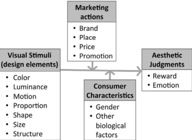
Previous studies have focused on the identification of neural activation in response to attractive objects.
Despite the contribution of these neuroscience studies to aesthetic judgments, it remains open how neuroscientific outcomes relate to previous aesthetic studies in visual-information processing. Future studies should explore what exact visual information is processed in the brain and whether activity in the brain structure related to the reward system and emotion is based on the difference in value of the predicted experience or the difference in other extrinsic information such as brand or price. Thus, it is important for future research to explore affective information processing resulting from cues from more varied stimuli. It might be a good starting point for future study to investigate the process by which the brain encodes “pleasure” or “pleasantness”
from design cues such as shape or color, marketing cues such as price or brand, and consumer cues such as gender or age. The research framework for neuroaesthetics is conceptualized in Figure 2.
Figure 2. A framework for neuroaesthetics research