Yeungnam Univ J Med 2016;33(1):13-20
High levels of carcinoembryonic antigen and smoking might be markers of colorectal adenoma in Korean males aged 40-49 years
In Cheol Yoon, Jeong Hyeon Cho, Heejin Choi, Young Hoon Choi, kyu Min Lim, Sung Hwa Choi, Jae Ho Han, Hyeon Ju Jeong, Hong Sub Lee
Department of Internal Medicine, Myongji Hospital, Seonam University College of Medicine, Goyang, Korea Background: Prevalence of adenoma in males aged 40-49 years in Korea was higher than expected. The aim of this study was to investigate the prevalence and risk factors of colorectal adenoma in males aged 40-49 years.
Methods: Total 1,902 asymptomatic subjects with a mean age of 47.9±6.7 years, who underwent a screening colonoscopy in a health promotion center of Myongji Hospital from 2010 to 2013 were enrolled in this study.
We conducted a case-control study to determine the risk factors for adenoma. The subjects were classified into two groups (adenoma vs. controls). To validate the diagnostic value of carcinoembryonic antigen (CEA) for adenoma, area under the receiver operating characteristic curve (AUROC) was calculated.
Results: At least one colorectal adenoma was identified in 385 subjects (20.2%). Among these 385 subjects, 372 subjects were found to have a non-advanced adenoma, 13 subjects had an invasive adenoma. One subject had cancer. Male sex, age, smoking, metabolic syndrome, and elevated CEA level were significantly associated with a colorectal adenoma in univariate analysis. However, metabolic syndrome was not significant in multivariate analysis. In the male group, the AUROC of CEA for colorectal adenoma was 0.600 (0.543 to 0.656) in non-smokers under 50 years of age, and 0.615 (0.540 to 0.690) in smokers under 50 years of age.
Conclusion: Male sex, smoking, and high levels of CEA seem to be associated with colorectal adenoma.
High levels of CEA and smoking may be diagnostic markers for any colorectal adenoma in Korean males aged 40-49 years.
Keywords: Carcinoembryonic antigen; Colorectal adenoma; Colonoscopy; Risk factors; Smoking
Received: November 20, 2015, Revised: March 15, 2016 Accepted: March 16, 2015
Corresponding Author: Hong Sub Lee, Department of In- ternal Medicine, Myongji Hospital, Seonam University College of Medicine, 55 Hwasu-ro 14beon-gil, Deogyang-gu, Goyang 10475, Korea
Tel: +82-31-810-5335, Fax: +82-31-969-0500 E-mail: epoch0123@naver.com
INTRODUCTION
Colorectal cancer (CRC) incidence is rapidly increasing in many Asian countries, with rates approaching those of Western countries [1]. In South Korea, CRC was the 3rd most com- monly diagnosed cancer in 2011, and the incidence of CRC has continued to increase in Korea, with a higher increase in men compared with women [2]. CRC screening is recom- mended for average-risk persons beginning at age 50 accor-
ding to the guidelines of many professional societies [3-5].
The most reliable method for early detection of precancerous lesions among the CRC screening methods is colonoscopy which is associated with reduced CRC mortality [6].
Recently, it was reported that the prevalence of overall
colorectal neoplasia in men aged 30-39 years exhibiting all
risk factors was not lower than that in average-risk women
aged >50 years [7]. In another study, the authors recom-
mended earlier colorectal screening before 50 years in males
with metabolic syndrome and history of smoking [8]. How-
ever, it was reported that the prevalence of adenomas was
similar between individuals aged 40-49 years and those aged
50-59 years, although the prevalence of advanced neoplasia
in the 50-59 years age group may be higher than that in
the 40-49 years age group [9]. Also, colonoscopic detection
of CRC is uncommon in asymptomatic persons 40-49 years of
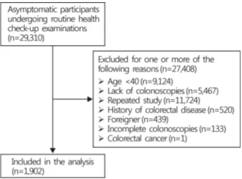
Fig. 1. Flow diagram for selection of study participants. Asympto- matic participants undergoing routine health check-up examina- tions from January 2010 until December 2013 at Myongji Hospital, Goyang, Korea (n=29,310).
age [10]. In Korea, although the prevalence of non-advanced adenoma in males aged 40-49 years is higher than that in Western countries, the prevalence of advanced adenoma and adenocarcinoma is lower [11,12]. Although gender, smoking, and metabolic syndrome are important risk factors of colo- rectal neoplasm, the benefits of earlier screening colonoscopy in subjects with these risk factors remain unclear.
Therefore, the aim of the present study was to investigate the prevalence and risk factors of colorectal adenoma in males aged 40-49 years.
MATERIALS AND METHODS
1. Study population
The study population consisted of subjects who had under- gone general check-ups at the health promotion center of Myongji Hospital, Goyang, Korea from January 2010 until December 2013 (n=29,310). We excluded subjects with the following characteristics: age under 40 years (n=9,124), sub- jects who did not receive a colonoscopy (n=5,467), subjects who received a colonoscopy more than 2 times (n=11,724), subjects who were not Korean (n=439), and subjects who had an incomplete colonoscopy result (n=133). Total 1,902 sub- jects who underwent first colonoscopy were included in the final analysis (Fig. 1).
The general check-ups included physical examination (in- cluding anthropometric measurement, body composition ana-
lysis, blood pressure), blood tests including metabolic profiles and tumor markers, such as carcinoembryonic antigen (CEA), esophagogastroduodenoscopy, abdominal sonography, plain chest radiography, and electrocardiography. An examinee could select additional screening tests, such as colonoscopy, compu- ted tomography, and so on.
This study was approved by the institutional review board of the Myongji Hospital.
2. Data collection and definitions
All examinees were instructed to complete a self-adminis- tered health questionnaire before the health check-up. The questionnaire included past history of colorectal disease, drug history, family history of CRC, the amount of alcohol con- sumption, smoking status, and whether regular exercise was performed or not, and so on. If subjects had undergone two or more colonoscopies during the study period, result of the first colonoscopy was considered in the analysis. Heavy alco- hol consumption was defined as drinking >80 g of alcohol per day. Smoking status was divided into three categories:
(non-smoker, <20 pack years, ≥20 pack years)
The percentage of total body mass was calculated by body composition analysis (Inbody 720, BiospaceCo.Ltd., Seoul, South Korea). Arterial stiffness checked in the carotid artery was stratified into two stages (normal vs. abnormal) by pulse wave velocity (PWV) determined with an automatic device (Nippon Colin, Komaki, Japan). Body mass index was calcu- lated as weight (kg) divided by height squared (m
2).
Metabolic syndrome was diagnosed if three or more of the following five factors were satisfied [13]: (1) high blood pres- sure or use of antihypertensive drugs, (2) elevated triglycerides
≥150 mg/dL or use of drugs for elevated triglycerides, (3) reduced high-density lipoprotein cholesterol level (<40 mg/dL in males, <50 mg/dL in females), (4) elevated fasting glucose or use of anti-diabetic medication, and (5) central obesity de- fined for the Korean population as a waist circumference of
≥90 cm for males and ≥85 cm for females [14].
Colonoscopies were performed by experienced gastroente-
rologists after bowel preparation with 4 L of polyethylene gly-
col solution (Colyte, Taejun, Seoul, South Korea). Advanced
adenoma was defined as adenomas with a diameter ≥10 mm,
and/or with villous component ≥25%, and/or with high grade
dysplasia.
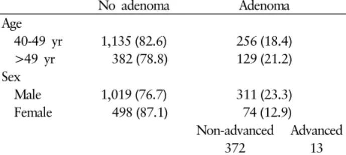
Table 1. Prevalence of colorectal adenoma in the study population
No adenoma Adenoma
Age
40-49 yr 1,135 (82.6) 256 (18.4) >49 yr 382 (78.8) 129 (21.2)
Sex
Male 1,019 (76.7) 311 (23.3)
Female 498 (87.1) 74 (12.9)
Non-advanced Advanced
372 13
Values are presented as number (%).
3. Statistical analysis
Data was divided into two groups (no adenoma vs. any ad- enoma). Continuous variables were compared using Student’s t-test. All continuous variables are expressed as means±S.E.M for each group. Categorical variables between the two groups were compared using the Pearson’s chi-squared test. For each variable, the odds ratio and 95% confidence interval were reported. The receiver operating characteristic curve (ROC curve) through logistic regression was calculated to obtain the cut-off value according to smoking status. p -values <0.05 were considered statistically significant. SPSS for Windows version 11 (SPSS Inc., Chicago, IL, USA) was used for all analyses.
RESULTS
1. Prevalence of colorectal adenoma in the study population
The study included 1,902 asymptomatic subjects who un- derwent health check-ups in a single endoscopy unit via screening colonoscopy (Fig. 1). Also, 1,391 (males:females=
967:424) subjects were 40-49 years of age, and 511 (males:
females= 363:148) subjects were older than 49 years. Table 1 show the prevalence of colorectal adenoma in the study population. At least one colorectal adenoma was identified in 385 patients (20.2%). The prevalence of colorectal adenoma was 23.3% in the male group (311 out of 1,330), and 12.9%
in the female group (74 out of 572). The prevalence of colo- rectal adenoma in subjects 40-49 years of age and in those
≥50 years was 18.4% (256 out of 1,391) and 21.2% (129 out of 511), respectively. Among the subjects with one or more colorectal neoplasms, 372 subjects were found to have a non- advanced adenoma, 13 subjects had an invasive adenoma.
One patient had a cancer.
Advanced adenoma (n=13) and colorectal adenocarcinoma (n=1) were found only in male subjects. Seven out of 13 subjects with advanced adenoma belong to male group 40-49 years of age (0.7%, 7 out of 967).
2. Risk factors of colorectal adenoma in the study population
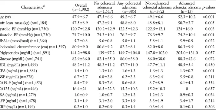
Table 2 and 3 show the differences in demographics and clinical characteristics between the subjects with adenomatous polyps and controls. Male sex, smoking status, existence of metabolic syndrome, and arterial stiffness were significantly associated with colorectal adenoma in univariate analysis. The prevalence of overall colorectal adenoma increase as age, soft lean mass, systolic blood pressure, diastolic blood pressure, glycosylated hemoglobin (HbA1c), abdominal circumference, CEA level, and prostate specific antigen (PSA) level increased.
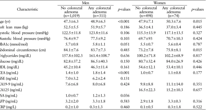
3. Risk analysis for colorectal adenoma by gender
The risk factors for colorectal adenoma in both genders are showed in Table 4 and 5. Age was significantly associated with colorectal adenoma regardless of gender. Arterial stiff- ness and CEA were significantly associated with colorectal adenoma in male subjects, but a similar effect was not seen in female subjects. PSA was significantly associated with colo- rectal adenoma in the male group. Smoking (≥20 pack years) and HbA1c tended to be associated with colorectal adenoma in the male group. In the female group, abdominal circum- ference was significantly associated with colorectal adenoma, but not in the male group.
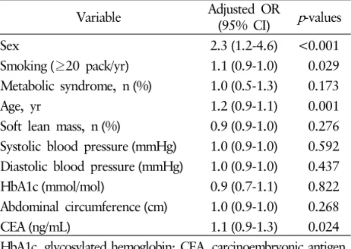
According to multiple logistic regression, adenoma was sig- nificantly associated with male sex, age, smoking, and CEA level (Table 6).
To validate the values of CEA for adenoma, area under the ROC curve (AUROC) was calculated (Table 7). AUROC of CEA for colorectal adenoma in non-smoking male subjects (n=585) under 50 years of age was 0.600 (95% CI, 0.543- 0.656). AUROC of CEA for colorectal adenoma in smoking male subjects (n=376) under 50 years of age was 0.615 (95%
CI, 0.540-0.690). The cut off value of high CEA level was 2.5 ng/mL in smokers and 1.0 ng/mL in non-smokers. The sen- sitivity and specificity of CEA level was 70.9% and 57.9%
in smokers, and 76.8% and 64.8% in non-smokers.
Table 2. Characteristics of study participants by colorectal adenoma status (categorical variables)
Characteristic
a)Overall (n=1,902)
No colorectal adenoma (n=1,517)
Any colorectal adenoma (n=385)
Non-advanced colorectal
adenoma (n=372)
Advanced colorectal adenoma
(n=13)
Adjusted OR (95% CI) p -values Male 1,330 (69.9) 1,019 (67.2) 311 (80.8) 298 (80.1) 13 (100) 2.0 (1.5-2.7) <0.001 Alcohol consumer (n=1,897) 919 (48.4) 739 (48.8) 180 (46.9) 172 (46.4) 8 (61.5) 0.9 (0.7-1.1) 0.491 Smoking (≥20 pack-years)
(vs never) (n=1,902) 461 (24.2) 351 (23.1) 110 (28.5) 107 (35.0) 3 (23.0) 1.3 (1.0-1.7) 0.028 History of hypertension
(n=1,901) 511 (26.9) 403 (26.6) 108 (28.1) 101 (27.2) 7 (53.8) 1.0 (0.8-1.3) 0.562 History of diabetes (n=1,901) 167 (8.8) 133 (8.8) 34 (8.8) 33 (8.9) 1 (7.7) 1.0 (0.6-1.4) 1.000 History of dyslipidemia
(n=1,901) 159 (8.4) 130 (8.6) 29 (7.5) 28 (7.5) 1 (7.7) 0.8 (0.5-1.3) 0.606 History of CVA (n=1,901) 21 (1.1) 16 (1.1) 5 (1.3) 5 (1.3) 0 (0) 1.2 (0.4-3.3) 0.596 History of angina or myocardial
infarction (n=1,901) 78 (4.1) 63 (4.2) 15 (3.9) 15 (4.0) 0 (0) 0.9 (0.5-1.6) 0.887 Regular exercise (n=1,897) 1,582 (83.4) 1,264 (83.5) 318 (82.8) 308 (83.0) 10 (76.9) 0.9 (0.7-1.2) 0.759 Metabolic syndrome (n=1,902) 407 (21.4) 307 (20.2) 100 (26.0) 95 (25.5) 5 (38.5) 1.3 (1.0-1.7) 0.018 Stool OB (n=638) 161 (25.2) 133 (25.3) 28 (24.8) 27 (24.5) 1 (33.3) 0.9 (0.6-1.5) 1.000 Arterial stiffness (n=723) 303 (41.9) 220 (39.3) 83 (50.9) 81 (50.6) 2 (66.6) 1.6 (1.1-2.2) 0.009 Values are presented as number (%).
a)
All participants had data on age, sex. The number in parentheses in this column indicate the number of participants with data available for each of the variables.
OR, odds ratio; CI, confidence interval; CVA, cerebral vascular accident; Stool OB, stool occult blood.
Table 3. Characteristics of study participants by colorectal adenoma status (continuous variables) Characteristic
a)Overall
(n=1,902)
No colorectal adenoma (n=1,517)
Any colorectal adenoma (n=385)
Non-advanced colorectal adenoma
(n=372)
Advanced colorectal adenoma
(n=13) p -values
Age (yr) 47.9±6.7 47.5±6.6 49.2±6.7 49.1±6.6 52.3±10.2 <0.001
Soft lean mass (kg) (n=1,184) 47.5±8.9 47.2±9.1 48.8±8.0 48.8±8.1 50.7±3.7 0.005 Systolic BP (mmHg) (n=1,750) 120.7±12.8 120.2±12.9 122.5±12.3 122.5±12.1 124±16.0 0.003 Diastolic BP (mmHg) (n=1,750) 74.7±10.0 74.3±10.1 76.2±9.7 76.3±9.7 74.2±10.4 <0.001 HbA1c (mmol/mol) (n=1,364) 5.7±0.8 5.6±0.8 5.8±1.1 5.8±1.1 5.9±0.9 0.022 Abdominal circumference (cm) (n=1,597) 80.9±9.0 80.6±9.2 82.2±8.1 82.0±8.0 86.3±9.9 0.002 Triglycerides (mg/dL) (n=1,891) 141.2±98.8 139±97.2 149.7±104.0 147.8±102.0 205.0±131.0 0.057 Glucose (mg/dL) (n=1,741) 82.9±36.0 82.1±35.0 86.0±38.0 86.0±38.0 88.3±42.6 0.072 HDL (mg/dL) (n=1,499) 48.2±11.2 48.3±11.2 47.7±11.0 47.7±11.1 48.5±5.4 0.450 CEA (ng/mL) (n=1,881) 1.4±1.0 1.3±1.0 1.6±1.3 1.6±1.3 1.3±0.7 <0.001
NSE (ng/mL) (n=278) 6.7±2.7 6.8±2.8 6.2±2.3 6.3±2.4 5.5±0.8 0.211
CA19-9 (ng/mL) (n=1,453) 8.4±7.9 8.3±7.6 8.6±8.8 8.7±8.9 6.1±4.3 0.554
CA125 (ng/mL) (n=446) 16.4±21 16.5±22.3 15.2±10.3 15.2±10.3 0 0.657
PSA (ng/mL) (n=1,279) 1.0±0.9 1.0±0.7 1.2±1.3 1.2±1.3 0.9±0.3 0.034
AFP (ng/mL) (n=1,478) 3.1±1.9 3.1±2.0 3.3±1.9 3.3±1.9 3.4±1.7 0.214
CRP (mg/L) (n=1,194) 0.2±1.0 0.2±0.9 0.3±1.4 0.3±1.4 0.1±0.1 0.306
Values are presented as mean±standard deviation.
a)