Risk factors for adjacent segment degeneration: more than 5 years after fusion
Asian Spine Journal
Asian Spine Journal
273Copyright Ⓒ 2013 by Korean Society of Spine Surgery
This is an Open Access article distributed under the terms of the Creative Commons Attribution Non-Commercial License (http://creativecommons.org/licenses/by-nc/3.0/) which permits unrestricted non-commercial use, distribution, and reproduction in any medium, provided the original work is properly cited.
Asian Spine Journal • pISSN 1976-1902 eISSN 1976-7846 • www.asianspinejournal.org
Received Jun 20, 2013; Revised Aug 16, 2013; Accepted Aug 20, 2013 Corresponding author: Byung Joon Shin
Department of Orthopaedic Surgery, Soonchunhyang University College of Medicine, 59 Daesagwan-ro, Yongsan-gu, Seoul 140-743, Korea
Tel: +82-2-709-9250, Fax: +82-2-794-9414, E-mail: [email protected]
Analysis of Risk Factors for Adjacent Segment Degeneration Occurring More than 5 Years after Fusion with Pedicle Screw Fixation for
Degenerative Lumbar Spine
Jaewan Soh
1, Jae Chul Lee
2, Byung Joon Shin
21Department of Orthropaedic Surgery, Soonchunhyang University Hospital, Soonchunhyang University College of Medicine, Cheonan, Korea
2Department of Orthopaedic Surgery and Spine Center, Soonchunhyang University College of Medicine, Seoul, Korea
Study Design: A retrospective study.
Purpose: We investigated the risk factors in adjacent segment degeneration (ASD) after more than 5 years of follow-up of lumbar spinal fusion.
Overview of Literature: There are many concerns regarding ASD followed by lumbar spinal fusion. However, there is a great deal of dispute about the risk factors.
Methods: A total of 55 patients who were followed up for more than 5 years after lumbar fusion were observed. Gender, age, resi- dence, fusion method, number of fusion segments and radiological measurements were analyzed. In the radiological measurement, disc height, lumbar lordotic angle (LLA), fusion segment lordotic angle and fusion segment lordotic angle per level (FSLA per level) were estimated. In preoperative MRI, Pfirrmann’s classification was used. The clinical result was evaluated by the criteria of Kim and Kim. Statistical univariate analysis was performed with the chi-square test by using SPSS ver. 12.0. Multivariate logistic regression analysis was conducted with SAS ver. 9.
Results: There were 21 patients with adjacent segment degeneration. Further, there was little relationship between ASD and gender, age, residence, fusion method, number of fusion segments, degree of preoperative adjacent disc degeneration in MRI, or preoperative and postoperative LLA. However, the frequency of ASD was significantly low in cases where FSLA per level was >15° (p=0.009). There was no significant relationship between ASD and the clinical result.
Conclusions: In patients followed up for more than 5 years after lumbar spinal fusion, the most important factor in the prevention of ASD was the restoration of FSLA per level to >15°.
Keywords: Lumbar; Adjacent segment degeneration; Fusion; Pedicle screw fixation
Clinical Study Asian Spine J 2013;7(4):273-281 • http://dx.doi.org/10.4184/asj.2013.7.4.273
ASJ A SJ
Introduction
When a structural deformation or nerve compression
is so severe that simple decompression alone does not produce satisfactory outcomes for degenerative lumbar disease, extensive decompression and lumbar vertebral
treatments.
Although some fusion methods and fixation systems have been developed to achieve successful fusion in spinal surgery, a long-term follow-up after a strong fu- sion has revealed degenerative changes at the adjacent segments due to the loss of mobility of the fusion site and the mechanical load caused thereby [1,2]. Degenera- tive changes at the adjacent segments include segmental instability, spinal stenosis, intervertebral disc lesion, retro-spondylolisthesis and fracture [3-5]. These poor outcomes are known to follow the accelerated degenera- tive changes at the adjacent segments after fusion [2,6,7].
As these complications are observed during a long-term follow-up, a cautious application of the fusion itself as well as new alternatives have been suggested. Therefore, measures to reduce and treat degenerative changes after fusion are discussed, along with the increased interest in causative factors related to the prevention and treatment reported by many studies. Nevertheless, these are still controversial.
On the basis of previous studies, in order to determine its causative factors, we statistically analyzed the correla- tion between possible factors and the degenerative change in adjacent segments among patients with radiographic changes during middle- or long-term follow-up of over 5 years after fusion with pedicle screw. In addition, we investigated the correlation between a radiological de- generative change at the adjacent segments and the actual clinical symptoms in order to show whether the radio- logical change was an index of the actual abnormality. We define that the degenerative change in the adjacent seg- ment with a radiographic change is ‘adjacent segment de- generation’, and the adjacent segment degeneration with clinical symptom is ‘adjacent segment disease’.
Materials and Methods
1. Materials
The subjects of this study were 55 patients who had un- dergone pedicle screw fixation and spine fusion of three or fewer segments due to degenerative lumbar disease.
The patients had been followed up for over 5 years. Their mean age at operation was 50.2 years (range, 34–67 years) and they consisted of 18 males and 37 females. Their mean follow-up period was 8 years and 6 months (range,
orthopedic surgeon. The fusion methods were posterolat- eral fusion and posterior lumbar interbody fusion in 24 and 31 cases, respectively (Table 1).
2. Methods
The 55 subjects were retrospectively investigated with their medical records and radiological findings.
Criteria of degenerative change at adjacent segments:
radiological degenerative change at the adjacent seg- ments was considered to exist when anterior or posterior displacement of >3 mm was found on the X-ray of the sagittal plane of the closest upper segment and the closest lower segment at the last follow-up, when the height of the intervertebral disc relative to that of the upper inter- body had declined by 20% and when a segmental motion instability of more than 15° was observed on the X-ray of the sagittal plane with flexion and extension.
1) Patient-related factors
Gender, age and lifestyle by residential area were analyzed as patient-related factors, which could have some influ- ence. Age was examined by dividing the patients into two groups: ≥50 years and <50 years of age (mean, 50 years).
The effect of differences in lifestyle was examined by clas- sifying residential areas into urban and rural categories.
2) Preoperative lumbar factors
Magnetic resonance imaging (MRI) was used to investi- gate whether there had been a degree of preoperative ad- jacent disc degeneration. Patients recording grade ≥III in the five-grade classification of Pfirrmann et al. [8] based on MRI were considered to have a degenerative change.
In addition, the preoperative lumbar lordotic angle was measured by Cobb’s angle made by the upper endplate of the first lumbar vertebra and the upper endplate of the sacrum. It was classified, with its mean value of 32° as a standard, into ≥32° and <32°; moreover, its correlation with a degenerative change at the adjacent segments was assessed.
3) Surgery-related factors
As factors related to surgical treatment, fusion method (posterolateral fusion or posterior lumbar interbody fu- sion) and the number of fusion segments (one, two and three segments) were analyzed.
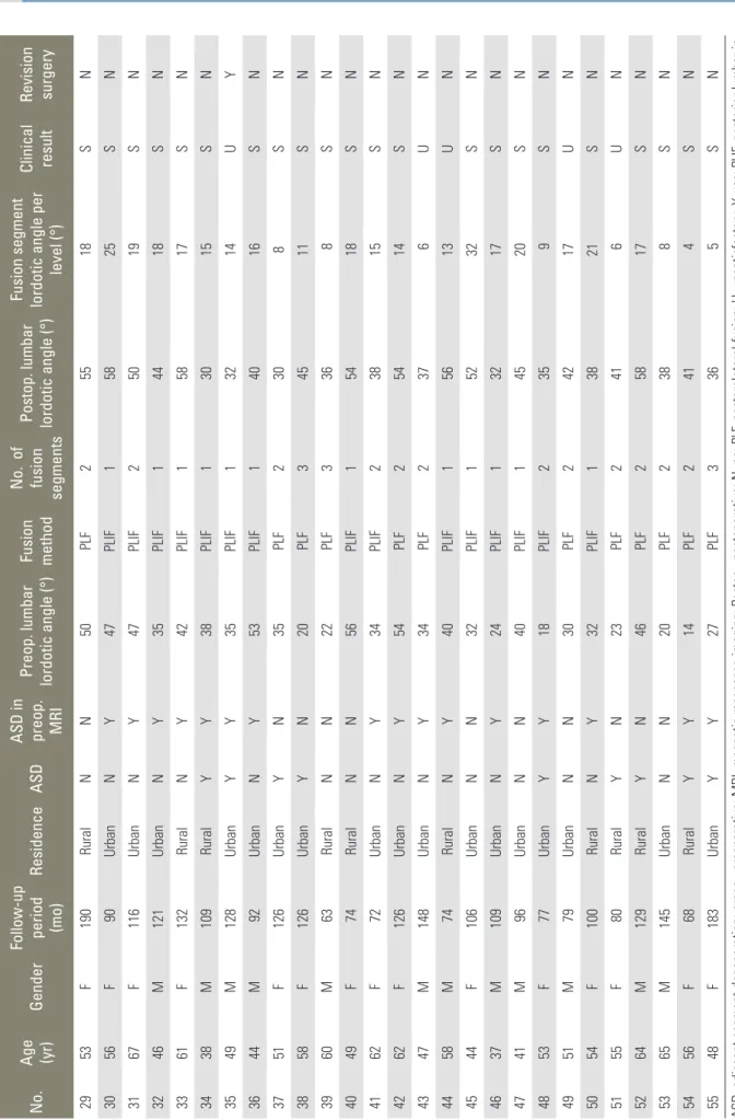
Table 1. Summerized data of 55 patients followed up for over 5 years after spinal fusion No.Age (yr)GenderFollow-up period (mo)
ResidenceASD
ASD in preop. MRI Preop. lumbar lordotic angle (°) Fusion method
No. of fusion segments
Postop. lumbar lordotic angle (°) Fusion segment lordotic angle per
level (°)
Clinical result Revision surgery
163F153UrbanNN30PLF13222UY 241F58UrbanNY38PLIF13320SN 340F113RuralYN24PLF21815SN 456M134RuralYY42PLF25215SN 559F60RuralNN46PLIF14825SN 665F114RuralYY19PLIF23011UY 759F60RuralYY42PLIF15019SN 855F72UrbanNY32PLIF15432SN 947F121UrbanNN41PLF23824SN 1057F68RuralNN32PLF23812SN 1144F101UrbanNY26PLIF14817SN 1265F98UrbanNY45PLIF35412SN 1361F74UrbanNN64PLIF15018UN 1453M129UrbanYY21PLF233 4SN 1542F87UrbanYN30PLIF14010SN 1642F132UrbanYN32PLF1325SN 1738F120UrbanNN33PLIF24012SN 1851F126UrbanYY34PLIF14230SN 1937F91UrbanNY55PLIF14316SN 2047F126RuralYN52PLIF15312SN 2159F89RuralNN14PLF13925UN 2256F57RuralNY25PLF237 6UN 2334M105UrbanNN10PLF33012SN 2456M60UrbanNN22PLF22812SN 2559M125UrbanYN18PLF23010SN 2636F84RuralNY5PLIF22310UN 2756M75UrbanYN30PLIF14510SN 2862F60UrbanYY17PLIF224 9SN (Continued to the next page)
No.Age (yr)GenderFollow-up period (mo)
ResidenceASD
ASD in preop. MRI Preop. lumbar lordotic angle (°) Fusion method
No. of fusion segments
Postop. lumbar lordotic angle (°) Fusion segment lordotic angle per
level (°)
Clinical result Revision surgery
2953F190RuralNN50PLF25518SN 3056F90UrbanNY47PLIF15825SN 3167F116UrbanNY47PLIF25019SN 3246M121UrbanNY35PLIF14418SN 3361F132RuralNY42PLIF15817SN 3438M109RuralYY38PLIF13015SN 3549M128UrbanYY35PLIF13214UY 3644M92UrbanNY53PLIF14016SN 3751F126UrbanYN35PLF2308SN 3858F126UrbanYN20PLF34511SN 3960M63RuralNN22PLF336 8SN 4049F74RuralNN56PLIF15418SN 4162F72UrbanNY34PLIF23815SN 4262F126UrbanNY54PLF25414SN 4347M148UrbanNY34PLF237 6UN 4458M74RuralNY40PLIF15613UN 4544F106UrbanNN32PLIF15232SN 4637M109UrbanNY24PLIF13217SN 4741M96UrbanNN40PLIF14520SN 4853F77UrbanYY18PLIF235 9SN 4951M79UrbanNN30PLF24217UN 5054F100RuralNY32PLIF13821SN 5155F80RuralYN23PLF241 6UN 5264M129RuralYN46PLF25817SN 5365M145UrbanNN20PLF238 8SN 5456F68RuralYY14PLF241 4SN 5548F183UrbanYY27PLF336 5SN ASD, adjacent segment degeneration; preop., preoperative; MRI, magnetic resonance imaging; Postop., postoperative; N, no; PLF, posterolateral fusion; U, unsatisfactory; Y, yes; PLIF, posterior lumbar terbody fusion; S, satisfactory.
Table 1. Continued
4) Postoperative radiological change-related factors Measures from the radiological images taken just after surgery were evaluated as surgical outcomes. First, the postoperative lumbar lordotic angle was classified, with its mean value of 40° as a standard, into ≥40° and <40°, and the meaning of each group was analyzed. Next, the fusion segment lordotic angle per level was calculated by dividing the lordotic angle of the fusion site or a Cobb’s angle between the upper endplate of the fusion segment and the lower endplate of the fusion segment by the num- ber of fused segments. It was also divided, with its mean of 15°, into ≥15° and <15°, and its correlation with the degenerative change of adjacent segments was assessed.
5) Correlation between radiological degenerative change in adjacent segment and clinical symptoms
The relationship between degenerative change in the ad- jacent segments and clinical outcome was assessed. The clinical outcomes were divided into satisfactory (excellent, good) and unsatisfactory (fair, poor) on the basis of the assessment base of the criteria of Kim and Kim [9].
6) Statistical analysis
To verify the significance of each factor, a univariate analysis was performed with the chi-square test by using SPSS ver. 12.0 (SPSS Inc., Chicago, IL, USA). Multivari- ate logistic regression analysis including all factors was also performed with SAS ver. 9 (SAS Institute, Cary, NC, USA), and the odds ratio of the significant factors was calculated. The significance level was p<0.05.
Results
1. Radiological degenerative change at adjacent segments Degenerative change at the adjacent segments was ob- served in a total of 21 cases. The change occurred at the upper segments (14 of retrolisthesis, seven of decreased height of the intervertebral disc, seven of segmental mo- tion instability, and one of spondylolisthesis) in 18 cases, at the lower segments (one of retrolisthesis, two of de- creased height of the intervertebral disc and one of spon- dylolisthesis) in four cases, and there was one case that showed the change at both the upper and lower segments.
2. Analysis on causative factors 1) Patient-related factors
According to the analysis of patient-related factors, the subjects included 18 males and 37 females, and the post- operative degenerative change was found in seven males and 14 females; hence, there was no significant difference by gender (p=0.940). The age of the subjects was ≥50 years and <50 years in 34 and 21 cases, respectively. The degenerative change did not show any significant differ- ence by age as it was found in 14 out of the 34 cases with the age of ≥50 years and in seven out of the 21 aged <50 years (p=0.561). The relationship between degenerative change and differences in residential area as lifestyle was examined. The change was observed in 12 of 35 residents of urban areas and nine of 20 resident in rural areas; there was no statistically significant correlation (p=0.431) (Table 2).
2) Preoperative lumbar factors
When the influence of the degree of preoperative adjacent disc degeneration was investigated by MRI, the degen- erative change at the adjacent segments was found at the last follow-up in 11 out of 29 cases with and ten out of 26 without preoperative degenerative change; the difference was not statistically significant (p=0.968). In addition, the degenerative change was observed in eight and 13 among 27 and 28 cases with the preoperative lumbar lordotic angle of ≥32° and <32°, respectively; hence, there was no significant difference (p=0.200) (Table 2).
3) Surgery-related factors
When the difference by the fusion method was investigat- ed, the degenerative change was found in 11 and 10 out of 24 and 31 cases treated with posterolateral fusion and posterior lumbar interbody fusion, respectively, and the difference was not statistically significant (p=0.304). As for the number of fusion segments, one-segment fusion and two- and three-segment fusions were performed in 26 and 29 cases, respectively, and the degenerative change was observed in eight and 13, respectively. There was also no significant difference (p=0.284) (Table 2).
4) Postoperative radiological change-related factors When the influence of the postoperative lumbar lordotic angle on the degenerative change at the adjacent seg- ments was investigated, the change was found in ten and
and the difference was not significant (p=0.551). In addi- tion, the fusion segment lordotic angle per level recorded
≥15° and <15° in 28 and 27 cases, respectively, and the degenerative change was shown in six and 15 cases, re- spectively. Thus, their difference was statistically signifi- cant (p=0.009) (Table 2).
5) Correlation between radiological degenerative change of adjacent segments and clinical symptoms
For the correlation between the degenerative change of adjacent segments and clinical outcomes, the change was shown in 18 out of 44 cases with satisfactory clinical out- comes and three out of 11 with unsatisfactory outcomes.
As there was no significant difference, the radiological change did not imply unsatisfactory clinical outcomes (p=0.405) (Table 2).
Among the 21 cases with change at the adjacent seg- ments, three needed a revision surgery (14.3%, 5.5% out of the total subjects); two of these were surgically treated for spinal stenosis at the upper adjacent segments, and the other one was treated for segmental instability.
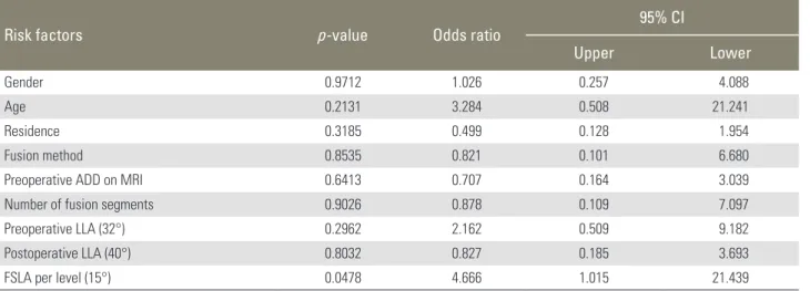
When the multivariate logistic regression analysis on risk factors was conducted–with independent variables as gender, age, residential area, fusion method, degree of preoperative adjacent disc degeneration on MRI, the number of fusion segments, and the lumbar lordotic an- gle and the fusion segment lordotic angle per level at the fusion site after the surgery–the fusion segment lordotic angle per level of <15° increased the risk of degenera- tive change at the adjacent segments 4.666 times (range, 1.015–21.439 times) (Table 3).
Discussion
Decompression and lumbar vertebral fusion have been widely conducted as surgical treatment for lumbar degen- erative change. Spinal fusion provoked a conflict between benefits secured just after the surgery and future prob- lems. The complications of lumbar vertebral fusion, such as intervertebral disc degeneration at adjacent segments, instability, fatigue and fracture, were observed during middle- and long-term follow-up [3-5]. Although many researchers have pointed out the degenerative lesions at the adjacent segments occurring frequently after fusion as major causes of these late complications [2,6,7], their causes, frequencies and risk factors are still controversial.
geration
Risk factors No. of
patients ASD p-value
Patient-related factors
Gender 0.940
Male 18 7
Female 37 14
Age (yr) 0.561
<50 21 7
≥50 34 14
Residence 0.431
Urban 35 12
Rural 20 9
Preoperative lumbar factors
Preoperative ADD on MRI 0.986
Yes 29 11
No 26 10
Preoperative LLA 0.200
<32° 28 13
≥32° 27 8
Surgery-related factors
Fusion method 0.304
PLF 24 11
PLIF 31 10
Nor of fusion segments 0.284
Single 26 8
2 or 3 29 13
Postoperative radiological-related factors
Postoperative LLA 0.551
<40° 26 11
≥40° 29 10
FSLA per level 0.009
<15° 27 15
≥15° 28 6
Correlation between ASD and clinical symptom
Clinical result 0.405
Satisfactory 44 18
Unsatisfactory 11 3
ASD, adjacent segment degeneration; ADD, adjacent disc degenera- tion; MRI, magnetic resonance imaging; LLA, lumbar lordotic angle;
PLF, posterolateral fusion; PLIF, posterior lumbar interbody fusion;
FSLA, fusion segment lordotic angle.
It is the predominant view that the degenerative lesion at the adjacent segments can be part of normal aging, and that the reduced mobility and the mechanical load following lumbar fusion accelerates the degeneration [1- 3,10,11]. This biomechanical change at the adjacent seg- ments is affected by the range of fused segments and the sagittal angle, and stronger fixation of fused segments is known to be associated with a larger effect as more stress is put on the adjacent segments [12,13]. Cunningham et al. [14] reported that the pressure of the adjacent inter- vertebral disc became larger by 45% in their cadaveric study, and Lee and Langrana [15] found that the load at the adjacent segments was raised in their biomechanical study on lumbosacral fusion. For the influence of gender, Kumar et al. [16] and Ha et al. [17] showed no significant difference in the rate of the degenerative change by gen- der, while Etebar and Cahill [18] insisted that the rate of the change at the adjacent segments was higher in females after menopause. This study did not reveal any significant difference by gender.
Many researchers believe that older age leads to more change at the adjacent segments [10,18-21]. As reasons for that, Aota et al. [20] pointed out that it was more dif- ficult for the spine to adapt to postoperative biomechani- cal change in the elderly, whereas Etebar and Cahill [18]
reported that osteoporosis negatively affected the existing degenerative procedure. However, we found no signifi- cant correlation between age and the degenerative change at the adjacent segments. As for the effect of lifestyle, Gil- let [22] and Cho et al. [23] reported that differences in lifestyle could influence the adjacent segments, and Ahn
et al. [24] reported that manual workers and residents in rural areas had around 47 times higher risk compared with those in urban areas. However, no significant corre- lation with residential area was observed in this study.
When the cases with preoperative instability or inter- vertebral disc degeneration at the adjacent segments were reviewed, Aota et al. [20] reported that instability dete- riorated after the surgery in all cases with preoperative anterior displacement of ≥3 mm. Ha et al. [21] showed that preoperatively, more severe degenerative change in the adjacent joints was associated with more radiologi- cal change in the joints, and no degenerative change was observed for more than 5 years of follow-up in cases without preoperative degenerative change in the joints.
However, this study did not show any direct correlation between preoperative adjacent disc degeneration on MRI and postoperative degenerative change at the adjacent segments.
Schlegel et al. [13] revealed that the change at the adja- cent segments occurred early in the fusion when a device was used for fixation, and Rahm and Hall [2] insisted that posterior intervertebral body fusion increased the load over adjacent segments because the remaining mobility after bone fusion was excluded along with stronger initial fixation. However, Kim et al. [25] and Ha et al. [21] found no significant difference in the frequency of the degen- erative change by the fixation system or fusion method.
This study also showed that the correlation between the fusion method and degenerative change was not signifi- cant. Moreover, there was no significant difference in the degenerative change by the number of fusion segments.
Table 3. Multivariate logistic regression analysis including all risk factors
Risk factors p-value Odds ratio 95% CI
Upper Lower
Gender 0.9712 1.026 0.257 4.088
Age 0.2131 3.284 0.508 21.241
Residence 0.3185 0.499 0.128 1.954
Fusion method 0.8535 0.821 0.101 6.680
Preoperative ADD on MRI 0.6413 0.707 0.164 3.039
Number of fusion segments 0.9026 0.878 0.109 7.097
Preoperative LLA (32°) 0.2962 2.162 0.509 9.182
Postoperative LLA (40°) 0.8032 0.827 0.185 3.693
FSLA per level (15°) 0.0478 4.666 1.015 21.439
CI, confidence interval; ADD, adjacent disc degeneration; MRI, magnetic resonance imaging; LLA, lumbar lordotic angle; FSLA, fusion segment lor- dotic angle.
sisted that the changes at the adjacent segments became larger due to more stress on the segments for multi-level fusion, Kettler et al. [26] and Ha et al. [17] reported that there was no correlation between the number of fusion segments and the change. This study also indicated no significant correlation between them.
For the change at the adjacent segments by the sagit- tal angle, reduced lordotic angle has been reported to promote the degenerative change early by leading to a concentrated load of segmental motion at the adjacent segments [27,28]. Herkowitz and Kurz [29], Cho et al. [23]
and Ahn et al. [24] stated that it was critical to maintain the lumbar lordotic angle during follow-up after fusion, and decreased lordotic angle eventually stimulated the degenerative change at the adjacent segments. For the segmental sagittal angle, Ahn et al. [19] found that the decrease in the fusion segment lordotic angle by 10° was associated with 3.2 times the increase in the degenerative change. In this study, the degenerative change was ob- served more frequently in the cases where the fusion seg- ment lordotic angle per level was <15° after the surgery.
The incidence rate of the degenerative change at the adjacent segments after lumbar vertebral fusion has been reported variously as between 19.4% and 40%
[3,20,21,25]. When it was investigated whether the de- generative change provoked clinical symptoms, Booth et al. [6] reported that a radiological degenerative change was found in many cases in >5 years after lumbar verte- bral fusion; however, there were almost no cases with this symptom; other previous studies also revealed no correla- tion between radiological change and clinical symptoms [2,18,23,30]. This study found that unsatisfactory out- comes were observed in only 11 among 21 cases with the degenerative change at the adjacent segments, and that the correlation between radiological change and surgical outcomes was not statistically significant. However, it was reported that if clinical symptoms were observed again along with the degenerative change at the adjacent seg- ments, surgical treatments were necessary in 8% to 16.8%
of cases [14,20,25]. This study also revealed that three (14.3%) out of the 21 cases needed surgical treatment.
Conclusions
During the long-term follow-up after pedicle screw fixation and fusion, gender, age, residential area, fusion
gree of preoperative adjacent disc degeneration on MRI showed no significant relationship with the postoperative degenerative change at the adjacent segments; however, the correlation between the fusion segment lordotic angle per level and the postoperative degenerative change was significant. Therefore, efforts to restore the fusion seg- ment lordotic angle per level to >15° are most important and are considered to be able to reduce the degenerative change at the adjacent segments.
Conflict of Interest
No potential conflict of interest relevant to this article was reported.
Acknowledgments
This work was supported by the Soonchunhyang Univer- sity Research Fund.
References
1. Boden SD, McCowin PR, Davis DO, Dina TS, Mark AS, Wiesel S. Abnormal magnetic-resonance scans of the cervical spine in asymptomatic subjects. A prospective investigation. J Bone Joint Surg Am 1990;72:1178-84.
2. Rahm MD, Hall BB. Adjacent-segment degeneration after lumbar fusion with instrumentation: a retro- spective study. J Spinal Disord 1996;9:392-400.
3. Brunet JA, Wiley JJ. Acquired spondylolysis after spi- nal fusion. J Bone Joint Surg Br 1984;66:720-4.
4. Leong JC, Chun SY, Grange WJ, Fang D. Long-term results of lumbar intervertebral disc prolapse. Spine (Phila Pa 1976) 1983;8:793-9.
5. Whitecloud TS 3rd, Davis JM, Olive PM. Operative treatment of the degenerated segment adjacent to a lumbar fusion. Spine (Phila Pa 1976) 1994;19:531-6.
6. Booth KC, Bridwell KH, Eisenberg BA, Baldus CR, Lenke LG. Minimum 5-year results of degenerative spondylolisthesis treated with decompression and instrumented posterior fusion. Spine (Phila Pa 1976) 1999;24:1721-7.
7. Frymoyer JW, Matteri RE, Hanley EN, Kuhlmann D, Howe J. Failed lumbar disc surgery requiring second operation. A long-term follow-up study. Spine (Phila
Pa 1976) 1978;3:7-11.
8. Pfirrmann CW, Metzdorf A, Zanetti M, Hodler J, Boos N. Magnetic resonance classification of lumbar intervertebral disc degeneration. Spine (Phila Pa 1976) 2001;26:1873-8.
9. Kim NH, Kim DJ. Anterior interbody fusion for spondylolisthesis. Orthopedics 1991;14:1069-76.
10. Quinnell RC, Stockdale HR. Some experimental ob- servations of the influence of a single lumbar floating fusion on the remaining lumbar spine. Spine (Phila Pa 1976) 1981;6:263-7.
11. Yang SW, Langrana NA, Lee CK. Biomechanics of lumbosacral spinal fusion in combined compression- torsion loads. Spine (Phila Pa 1976) 1986;11:937-41.
12. Okuda S, Iwasaki M, Miyauchi A, Aono H, Morita M, Yamamoto T. Risk factors for adjacent segment degeneration after PLIF. Spine (Phila Pa 1976) 2004;29:1535-40.
13. Schlegel JD, Smith JA, Schleusener RL. Lumbar mo- tion segment pathology adjacent to thoracolumbar, lumbar, and lumbosacral fusions. Spine (Phila Pa 1976) 1996;21:970-81.
14. Cunningham BW, Kotani Y, McNulty PS, Cappucci- no A, McAfee PC. The effect of spinal destabilization and instrumentation on lumbar intradiscal pressure:
an in vitro biomechanical analysis. Spine (Phila Pa 1976) 1997;22:2655-63.
15. Lee CK, Langrana NA. Lumbosacral spinal fu- sion. A biomechanical study. Spine (Phila Pa 1976) 1984;9:574-81.
16. Kumar MN, Baklanov A, Chopin D. Correlation be- tween sagittal plane changes and adjacent segment degeneration following lumbar spine fusion. Eur Spine J 2001;10:314-9.
17. Ha Ky, Kim YH, Kang KS. Surgery for adjacent seg- ment changes after lumbosacral fusion. J Korean Soc Spine Surg 2002;9:332-40.
18. Etebar S, Cahill DW. Risk factors for adjacent- segment failure following lumbar fixation with rigid instrumentation for degenerative instability. J Neuro- surg 1999;90:163-9.
19. Ahn DK, Lee S, Jeong KW, Choi DJ, Cha SK, Cho KH. Degenerative change of adjacent segments ac- cording to the fusion method after L4-5 segmental fusion: comparative study of posterolateral fusion
and posterior lumbar interbody fusion. J Korean Or- thop Assoc 2006;41:281-7.
20. Aota Y, Kumano K, Hirabayashi S. Postfusion in- stability at the adjacent segments after rigid pedicle screw fixation for degenerative lumbar spinal disor- ders. J Spinal Disord 1995;8:464-73.
21. Ha KY, Kim KW, Park SJ, Lee YH. Changes of the adjacent-unfused mobile segment after instrumental lumbar fusion: more than 5-years follow-up. J Korean Soc Spine Surg 1998;5:205-14.
22. Gillet P. The fate of the adjacent motion segments af- ter lumbar fusion. J Spinal Disord Tech 2003;16:338- 45.
23. Cho JL, Park YS, Han JH, Lee CH, Roh WI. The changes of adjacent segments after spinal fusion:
follow-up more than three years after spinal fusion. J Korean Soc Spine Surg 1998;5:239-46.
24. Ahn DK, Lee S, Jeong KW, Park JS, Cha SK, Park HS.
adjacent segment failure after lumbar spine fusion:
controlled study for risk factors. J Korean Orthop As- soc 2005;40:203-8.
25. Kim HT, Kang DW, Yoo CH, Jeoung JH, Chang SA.
Late changes at the adjacent segments to lumbar fu- sions. J Korean Soc Spine Surg 1996;3:1-10.
26. Kettler A, Wilke HJ, Haid C, Claes L. Effects of specimen length on the monosegmental motion behavior of the lumbar spine. Spine (Phila Pa 1976) 2000;25:543-50.
27. Dekutoski MB, Schendel MJ, Ogilvie JW, Olsewski JM, Wallace LJ, Lewis JL. Comparison of in vivo and in vitro adjacent segment motion after lumbar fu- sion. Spine (Phila Pa 1976) 1994;19:1745-51.
28. Grouw AV, Nadel CI, Weierman RJ, Lowell HA. Long term follow-up of patients with idiopathic scoliosis treated surgically: a preliminary subjective study.
Clin Orthop Relat Res 1976;(117):197-201.
29. Herkowitz HN, Kurz LT. Degenerative lumbar spon- dylolisthesis with spinal stenosis. A prospective study comparing decompression with decompression and intertransverse process arthrodesis. J Bone Joint Surg Am 1991;73:802-8.
30. Ghiselli G, Wang JC, Bhatia NN, Hsu WK, Dawson EG. Adjacent segment degeneration in the lumbar spine. J Bone Joint Surg Am 2004;86:1497-503.