8
Immune Network
Introduction
CD30 belongs to tumor necrosis factor (TNF)/
nerve growth factor (NGF) receptor superfamily (1,2) and was originally discovered on Hodgkin and Reed-Sternberg cells in Hodgkin's lymphoma (3). It is now known as an activation antigen that is expressed on normal T an B lymphocytes following antigenic or mitogenic stimulation (2-5).
Studies with human T cell clones revealed CD30 to be preferentially expressed by Th2 clones (4,5) and CD30 signaling induces Th2 differentiation (6).
Consequently, despite several controversial reports (7,8), CD30 has long been proposed as a marker of type 2 helper T (Th2) cells. After commitment, only Th2 cells express CD30 but not Th1 (9) and CD30
expression is regulated by cytokines in a very similar fashion to Th2 differentiation. For instances, CD30 expression is upregulated by IL 4 and downregulated by IFNγ(9), which are critical cytokines for Th2 and Th1 differentiation, respectively. Furthermore, CD28 stimulation is responsible for CD30 expression (10) and also differentiation of Th2 rather than Th1 cells (11,12) through acceleration of IL 4 receptor sen- sitivity (13). These data strongly implicate that CD30 may play an important role in Th2 differentiation.
At a clonal level, CD30 signaling have shown pleiotropic and controversial effects depending on cell types. Binding of CD30 ligand (CD30 L) to its receptor induces apoptosis (14-16) and also enhances cell proliferation (10,14,17). Antibody production of murine B cell can be enhanced by CD30 ligation (18), however, similar effects on human B cells could not be observed (19). CD30 functions also include NF-B activation (20), cytokine production (21), and inhibition of cytotoxicity of NK cell line through downregulation of CD28 expression (22). CD30-deficient mice showed partial defect in negative selection of thymus cells
Expression on Murine T Cells
Sang-Yun Nam
Department of Microbiology, School of Natural Science, Jeonju University, Jeonju, Korea
ABSTRACT
Background: CD30 is a member of TNF receptor family and expressed on lymphocytes and other hematopoietic cells following activation as well as Hodgkin and Reed- Sternberg cells in Hodgkin's lymphoma. In this study, CD30-mediated regulation of cell adhesion molecule expression on normal activated mouse T cells was investigated.
Methods: Mouse T cells were activated with anti-CD3 antibody for induction of CD30, which was cross-linked by immobilized anti-CD30 antibody. Results: High level of CD30 expression on T cells was observed on day 5, but only little on day 3 even under culture condition resulting in an identical T cell proliferation, indicating that CD30 expression requires a prolonged stimulation up to 5 days. Cross-linking of CD30 alone altered neither proliferation nor apoptosis of normal activated T cells. Instead, CD30 appeared to promote cell adherence to culture substrate, and considerably upregulated ICAM-1 and, to a lesser extent, ICAM-2 expression on activated T cells, whereas CD2 and CD18 (LFA-1) expression was not affected. None of cytokines known as main regulators of ICAM-1 expression on tissue cells (IL 4, IFNγ and TNFα) enhanced ICAM-1 expression in the absence of CD30 signals. On the other hand, addition of NF-κB inhibitor, PDTC (0.1 mM) completely abrogated the CD30-mediated up- regulation of ICAM-1 expression, but not CD2 and ICAM-2 expression. Conclusion:
This results support that CD30 upregulates ICAM-1 expression of T cell and such regulation is not mediated by higher cytokine production but NF-κB activation.
Therefore, CD30 may play important roles in T-T or T-B cell interaction through regulation of ICAM-1, and -2 expression. (Immune Network 2003;3(1):8-15)
Key Words: CD30, cell adhesion molecules, ICAM-1 (CD54), T cells
Correspondence to: Sang-Yun Nam, Department of Microbiology, School of Natural Science, Jeonju University, Jeonju 560-759, Korea. (Tel) 063-220-2518, (Fax) 063-220-2054, (E-mail) sangyun@
www.jeonju.ac.kr
This work was supported by Korean Research Foundation Grant (KRF-2000-042-F00027).
(23) and these findings have been confirmed by the experiments with CD30 transgenic mice (24) that CD30 overexpression enhances negative selection in the thymus. Another observation that transferred autoreactive T cells without CD30 proliferate extens- ively in vivo and can cause substantial tissue damage (25) strongly suggest the involvement of CD30 in the onset of autoimmune disease. CD30 has also attracted much interest as a possible parameter defining subgroups of leukemia (26), lymphoma (27,28). The plausible relevance of CD30 to AIDS pathogenesis was also proposed (29). However, despite lots of studies, the functional role of CD30 under phy- siological condition remains unclarified so far.
It has been reported that CD30 induces homotypic cell aggregation of cell lines (22), which was not blocked by antibodies to LFA-1, ICAM-1, CD2, LFA-3 or CD29 (22). Although it was shown that CD30 ligand enhanced ICAM-1 expression of Hodgkin and Reed-Sternberg (H-RS) cells (30), such role of CD30 in normal lymphocytes has not been addressed. In this study, CD30-mediated ICAM-1 reg- ulation and its mechanisms were investigated in normal activated T cells. Additionally, such regulatory role of CD30 was also explored for the several kinds of cell adhesion molecules expressed on murine T cells.
Materials and Methods
Mice and cells. Balb/c mice were supplied from SamTaco (Osan, Korea) and maintained in an environmental controlled rearing system at Jeonju University. After mice were sacrificed by cervical dislocation, peripheral and mesenteric LN cells were prepared and pooled for simulation. Purified T cells were obtained by plastic adherence and subsequent negative selection using MACS and anti-B220-coated microbeads (Miltenyi, Germany). After partial removal of non-adherent T cells following plastic adherence, attached B-enriched spleen cells were used for antigen-presenting cells (APC), which were treated with 50μg/ml of mitomycin C for 15 min prior to use and washed 4 times with IMDM containing 5% FBS.
Antibodies, cytokines and reagents. Anti-CD3, and hamster anti-mouse CD30 (26) antibodies were purified with protein G-Sepharose chromatography from culture supernatant of 145-2C11 and mCD30.1 hybridomas (26), respectively. Anti-CD30 antibody was conjugated with FITC Isomer I on Celite (Calbiochem, La Jolla, CA) following the manufacturer's instructions and used for flow cytometric analysis.
FITC-conjugated anti-CD2, FITC-conjugated anti- CD18 antibodies were purchased from Serotec and Sigma Chemical Co. (St. Louis, MO), respectively.
FITC-conjugated anti-CD25, FITC-conjugated anti-
CD54 (ICAM-1), unconjugated anti-CD102 (ICAM-2), FITC-conjugated hamster IgG, FITC-conjugated anti-hamster IgG antibodies were purchased from PharMingen (San Diego, CA. USA). Unconjugated hamster IgG (Jackson ImmunoResearch Lab., West Grove, PA. USA) was used for isotype control antibody.
Purified mouse IL 4, IFNγ, and TNFα was pur- chased from PeproTech (Rocky Hill, NJ. USA).
Cell culture, immobilization of antibody and stimulation. T cells were stimulated at 4-10×105 cells/ml in IMDM supplemented with 10% FBS, 100 U/ml of penicillin, 100μg/ml of streptomycin (Life Tech- nologies, Grand Island, NY. USA) and 5×10⁻⁵ M 2-mercaptoethanol in the presence of IL 4 (10 ng/ml) for 3∼5 days. APC were added for primary stimulation at 1×10⁶ cells/ml. For cross-linking of CD3 or CD30, anti-CD3 or anti-CD30 antibodies in phosphate- buffered saline (PBS) were added to wells of 48 (0.2 ml/w) or 96 well (0.04 ml/w) culture plates and immobilized by incubation at 37oC for 2 hrs or 4oC over night. The plate wells were washed 3 times with cold PBS prior to use.
Flow cytometry. Expression of surface marker was analyzed by flow cytometry. Five hundred thousand cells were washed and resuspended in staining buffer, PBS containing bovine serum albumin (0.5%) and so- dium azide (0.1%). Those cells were incubated with conjugated antibodies at 4oC for 30 min and then washed with staining buffer. Stained cells were analyzed with FACScan (Becton Dickinson Co.) and Lysis II program. For analysis of viable cells only, dead cell population was ruled out by PI stain.
Evaluation of adherence. For comparison, hamster IgG or anti-CD30 were immobilized in a 96 well culture plates, on which stimulated cells were incubated at 5×10⁵ cells/ml. After 2 days of incubation, culture plates were shaken at low speed on a voltex mixer for 5 seconds and non-adherent cells were removed by aspiration. The wells were subsequently washed 2 times with prewarmed medium. MTT (Sigma, 2 mg/ml) was added for 4 hrs and formazan crystal was solubilized with acid isopropyl alcohol. Ab- sorbance was determined at 550 nm.
Statistics. Results were analyzed by Student's t test for comparison between two groups and p values <0.05 were accepted as the level of significance.
Results
Longer time period of stimulation is required for CD30 expression than for proliferation. For functional assay of CD30, its expression should be induced by antigenic or mitogenic stimulation since naive cells usually exhibit no or very little, if any, expression of CD30 and high level of CD30 expression is prerequisite for enough CD30 signal. In this study, purified T cells
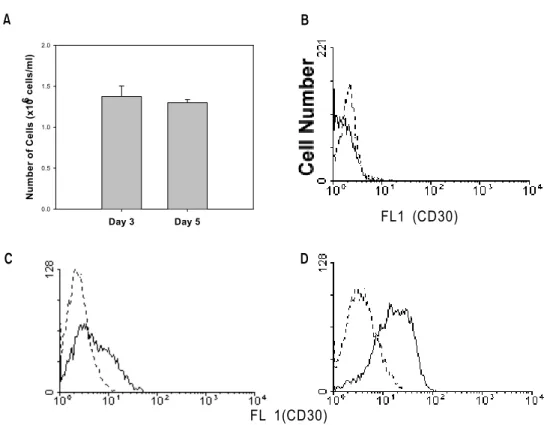
were stimulated with immobilized anti-CD3 at 1×10⁶ cells/ml for 3 days or 5×10⁵ cells/ml for 5 days for comparison. As shown in Fig. 1A, 3 day stimulation at high concentration (1×10⁶ cells/ml) and 5 day stimulation at low concentration (5 days, 5×10⁵ cells/ml) resulted in an equivalent level of cell proliferation (1.3-1.4×10⁶ cells/ml). However, at day 3 of stimulation, CD30 expression was much lower than at day 5 (MFI, 5 vs 15, Fig. 1B and 1C).
It suggests that relatively prolonged stimulation is required for CD30 expression, when compared to cell proliferation.
Change of T cell adhesion by cross-linking of CD30.
Cross-linking of CD30 did not modulate cell proliferation of normal activated T cells in this study (data not shown). Instead, cells were likely to adhere so firmly to the surface that adherence was tested following cross-linking of CD30. Stimulated cells were harvested and CD30 was cross-linked with immobilized anti-CD30 antibody for 2 days and then their adherence to surface was examined. As shown in Fig. 2, immobilized hamster IgG did not affect T cell adhesion whereas anti-CD30 drastically enhanced the adherence from 0.22 to 0.72∼0.88 of OD by
Figure 1. Cell Proliferation and CD30 expression on T cells following CD3 activation. Purified T cells were stimulated with immobilized anti-CD3 at 1×10⁶ cells/ml for 3 days or 4×10⁵ cells/ml for 5 days. Activated cells were harvested and viable cells were counted (A). Non-activated (B) and activated cells (C, D) were also stained with FITC-conjugated anti-CD30 (solid lines) and isotype control (dotted lines) on day 3 (B, C) and 5 (D).
A B
Day 3 Day 5
Number of Cells (x10
6 ce
lls/ml)
0.0 0.5 1.0 1.5 2.0
FL1 (CD30)
FL 1(CD30)
C D
Figure 2. Adherence of activated T cells following CD30 cross-linking. At day 5, cells were incubated with immobilized control hamster IgG or anti-CD30 of indicated concentration for further 2 days. Cell adherence was determined as described in Materials and Methods. Representative data from 2 separate experiments were shown.
Ab Concentrations (µg/ml)
0 5 10 15 20
O.D at 550nm
0.0 0.2 0.4 0.6 0.8 1.0
Anti-CD30 Hamster IgG
10∼20μg/ml of antibody.
Regulation of cell adhesion molecule expression by CD30 signaling. Above result is clearly indicative that CD30 signals can regulate cell adhesion molecule expression.
Therefore, expressional change of cell adhesion molecules expressed on T cells was observed. Of which, CD54 (ICAM-1) was firstly examined in the process of stimulation and cross-linking of CD30.
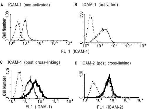
Non-stimulated T cells showed the constitutive but low expression of ICAM-1 as on naive T cells (Fig. 3A).
However, its expression was markedly upregulated by CD3 stimulation (Fig. 3B). Following induction of CD30 expression by stimulation of T cells for 5 days, CD30 on the activated T cells was cross-linked by anti-CD30 antibody or control hamster IgG for further 1 day. Those cells were harvested and stained with antibodies to several adhesion molecules that might be expressed on T cells. As shown in Fig. 3C, and 3d, CD30 signaling considerably enhanced ICAM-1 (Fig. 3C) and, to a lesser extent, CD102 (ICAM-2) expression (Fig. 3D). Repeated experiments showed that these expressional regulations were statistically significant (p<0.01 or p<0.05, respec- tively; Fig. 4). The regulatory function of CD30 was also investigated for other adhesion molecules including CD2, and CD18. However, the expression of CD2 was not affected by CD30 signaling, and slightly lower expression of CD18 was observed after cross-linking of CD30, although the decrement was not significant (Fig. 4).
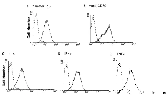
Inability of cytokines to enhance ICAM-1 expression. Several cytokines have been documented as regulators for ICAM-1 expression like IL 4, TNFα and IFNγ (31).
Therefore, these cytokines were tested for their regulatory functions on ICAM-1 expression of lymph node T cells. As shown in Fig. 5, the results showed
Figure 3. Expressional change of ICAM-1 and its regulation by cross-linking of CD30. Purified T cells were incubated in medium (A) or stimulated with immobilized anti-CD3 (B) for 5 days and then stained with FITC-anti-CD54. Harvested cells were incubated with hamster IgG (narrow lines) or anti-CD30 (bold lines) (C, D) for further 1 day and subsequently stained with FITC-anti-CD54 (C) or anti-102 (D) plus FITC-anti-rat IgG or isotype control (dotted lines).
A ICAM-1 (non-activated) B ICAM-1 (activated)
FL 1 (ICAM-1)
D ICAM-2 (post cross-linking) C ICAM-1 (post cross-linking)
FL1 (ICAM-1) FL1 (ICAM-2)
Figure 4. Regulation of cell adhesion molecule expression by CD30. Mouse T cells were stimulated with anti-CD3 for 5 days and then CD30 was cross-linked for further 2 days. *P<0.05,
**P<0.01.
CD2 CD18 CD54 CD102
Mean Fluorescence Intensity
0 20 40 60 80 100 120 140
Hamster IgG Anti-CD30
**
*
that all of these cytokines could not enhance ICAM-1 expression (Fig. 5B-D) and culture supernatant prepared by CD30 signaling also contained no regulatory factors (data not shown).
Contribution of NF-κB activation to CD30-mediated upregulation of ICAM-1 expression. It has been reported that CD30 signals activates NF-κB (20) and which is related to ICAM-1 expression (32). These previous results thus leaded to a hypothesis that CD30 activates NF-κB and which in turn, upregulates ICAM-1 expression. In this study, NF-κB inhibitor, PDTC was added to confirm whether it might block CD30-mediated enhancement of ICAM-1 expression.
As summarized in Table I, 0.1 mM PDTC signi- ficantly reduced ICMA-1 expression induced by immobilized anti-CD30 (MFI, 44 →24) to a control level (MFI, 22) with control IgG. In other words, CD30-mediated enhancement of ICAM-1 expression was mostly abrogated by NF-κB inhibitor, whereas it did not affect both of CD2 and CD102 expression.
This differential outcome strongly supports that NF- κB activation contributes to CD30-mediated ICAM-1 regulation and additionally CD30-mediated ICAM-1 regulation is by no means a default response of cellular activation.
Discussion
CD30 expression requires prior activation like TCR ligation or mitogenic stimulation. In this study, CD30 function was investigated focusing on T cells using purified lymph node T cells and anti-CD3 antibody. Firstly, it was observed that primary stimulation for 3 days induces low level of CD30 even with strong mitogen, plate-bound anti-CD3 in combination with exogenous IL 4 added during primary stimulation, which leaded to high cell proliferation from 1×10⁶ cells/ml to 1.6×10⁶ cells/
ml for 3 days. In this study, prolonged stimulation
Figure 5. Inability of cytokines to enhance ICAM-1 expression on T cells. At day 5 of stimulation, cells were incubated with medium (A), immobilized anti-CD30 (B), or 10 ng/ml of IL 4 (C), IFNγ (D), and TNFα (E) for further 1 day and then stained with FITC-conjugated anti-ICAM-1 (solid lines) and isotype control (dotted lines).
A hamster IgG B +anti-CD30
E TNFα D IFNγ
C IL 4
Table I. Inhibition of CD30-mediated enhancement of ICAM-1 expression by NF-κB inhibitor, PDTC
ꠚꠚꠚꠚꠚꠚꠚꠚꠚꠚꠚꠚꠚꠚꠚꠚꠚꠚꠚꠚꠚꠚꠚꠚꠚꠚꠚꠚꠚꠚꠚꠚꠚꠚꠚꠚꠚꠚꠚꠚꠚꠚꠚꠚꠚꠚ adhesion molecule expression (MFI)**
anti-CD30 PDTC* ꠏꠏꠏꠏꠏꠏꠏꠏꠏꠏꠏꠏꠏꠏꠏꠏꠏꠏꠏꠏꠏꠏꠏꠏꠏꠏꠏꠏꠏꠏ
CD54 CD2 CD102
ꠏꠏꠏꠏꠏꠏꠏꠏꠏꠏꠏꠏꠏꠏꠏꠏꠏꠏꠏꠏꠏꠏꠏꠏꠏꠏꠏꠏꠏꠏꠏꠏꠏꠏꠏꠏꠏꠏꠏꠏꠏꠏꠏꠏꠏꠏ
- - 22 59 28
+ - 44 62 37
+ + 24 64 43
ꠏꠏꠏꠏꠏꠏꠏꠏꠏꠏꠏꠏꠏꠏꠏꠏꠏꠏꠏꠏꠏꠏꠏꠏꠏꠏꠏꠏꠏꠏꠏꠏꠏꠏꠏꠏꠏꠏꠏꠏꠏꠏꠏꠏꠏꠏ
*: added at 0.1 mM, **: representative data were presented from 2 separate experiments with similar results
protocol for 5 days was also evaluated for induction of CD30. The data shown in Fig. 1B, and 1C clearly indicate that longer time period may require for CD30 expression than only for cell proliferation. One possible explanation for this finding is that CD30 expression is not induced only by antigenic stimulation but also require another factors such as, IL 4 (9,10) or CD28 costimulatory signal (10) and which would be provided more abundantly at later stage of stimulation.
Using cultured cell lines, several investigators reported that CD30 enhances cell proliferation or induces apoptosis (14-17). However, in this study, ligation of CD30 alone altered neither cell pro- liferation nor apoptosis of normal activated LN cells (data not shown). It suggests that CD30 signaling effect is variable depending on cell types, antigenic stimulation or other undefined factor(s). Instead, cross-linking of CD30 appeared to induce stronger adherence to the surface on which anti-CD30 was immobilized. This observation was followed by inves- tigation of expressional change of cell adhesion molecules expressed on T cells. CD18 highly ex- pressed on T cells was not altered. In contrast, the data presented here apparently showed that CD30 signaling upregulates ICAM-1 and, to a lesser extent, ICAM-2 expression (Fig. 3). It is also supportive to above results that cross-linking of CD30 induces much higher level of soluble ICAM-1 secretion than control antibody (data in preparation). Expression of ICAM-1 on T cells is constitutive but relatively low (Fig. 3A). By CD3 stimulation, its expression con- siderably increased and subsequently diminished again without stimulating signal (data not shown). CD30 signaling is likely somehow to provide the activation state enough to sustain ICAM-1 production. However, identification of the CAM directly responsible for CD30-mediated adhesion awaits more studies because ICAM-1 expression is unlikely to participate in T cell adhesion to substrate.
Proinflammatory cytokines such as TNFα, and IFNγ and T-cell derived IL 4 are well known re- gulatory molecules for activation (33-35) and ICAM-1 expression (31,36,37) in endothelial cells, and other types of cells including fibroblasts (38), mast cells (39), glioblastoma cells (40), and astrocytes (41). Thus, it was examined if these cytokines might be engaged in CD30-mediated enhancement of ICAM-1 expression.
The results showed that activated T cells did not respond to none of these exogenously added cytokines in ICAM-1 expression (Fig. 5). These results are not likely to be due to absence of cytokine receptors.
Instead, these results suggest that lymphoid cells have a distinct pathway from endothelial cells at the down- stream of cytokine binding with cell surface cytokine
receptor. Piela-Smith et al. (42) have also suggested that those cytokines exert variable effects on mo- dulation of ICAM-1 expression depending on target cell types. In this study, direct assay for CD30-mediated NF-κB activation was not repeated because it was already observed (20). However, inhi- bition of CD30-mediated enhancement of ICAM-1 expression by NF-κB inhibitor (Fig. 6) clearly demonstrated that CD30 upregulates ICAM-1 expression through NF-B activation. Furthermore, addition of PDTC did not affect the CD2 and CD102 expression, indicting that such PDTC effect did not result from non-specific inhibition of T cell activation. NF-κB is involved in T cell activity such as proliferation, synthesis of cytokines and its receptors (43), and replication of human immuno- deficiency virus (HIV) (44). Upregulaton of ICAM-1 expression might be one of the several phenomena resulted from NF-κB activation by CD30 signaling.
Besides ICAM-1 regulation, CD30 may exert many other functions through ubiquitous transcription factor, NF-κB pathway.
Functional significance of CD30 regulation for ICAM-1 expression on T cells was not fully elu- cidated in this study. ICAM-1 is a cell surface gly- coprotein of immunoglobulin superfamily that has been known to mediate various cell-cell interactions, including leukocyte adhesion to vascular endothelial cells, as well as cytotoxicity, tumor progression, and metastasis (reviewed in Ref. 45). Moreover, ICAM-1 itself involves in signal transduction between interacting cells. In particular, the expression and function of LFA-1, the counterpart of ICAM-1, on T cells has been extensively studied. Previous studies on the expression and functions of ICAM-1, however, have mostly focused on antigen-presenting cells or endothelial cells, but not on T cells (reviewed in Refs. 31 and 45). In other words, ICAM-1 is not widely noted as a molecule expressed on T cells and receiving signals, but existing on counterpart cells and giving signals to T cells. It may be due to relatively low expression of ICAM-1 on T cells. However, several reports have demonstrated that ICAM-1 on T cells functionally mediate T-T interaction (46), T-B interaction or mixed lymphocyte reaction (47). These results may implicate that CD30 can exert various functions in certain steps of immune responses through regulation of ICAM-1 expression.
Finally, cell adhesion molecules can help the primary signaling between two interacting cells by augmentation of contact. Accordingly, the effects of CD30 signal may appear variably depending upon the feature of primary signals, although we also cannot rule out the involvement of the signals directly transduced by counterpart molecules.
References
1. Falini B, Pileri S, Pizzolo G, Durkop H, Flenghi L, Stirpe F, Martelli MF, Stein H: CD30 (Ki-1) molecule: a new cytokine receptor of the tumor necrosis factor receptor super- family as a tool for diagnosis and immunotherapy. Blood 85;1-14, 1995
2. Smith CA, Gruss HJ, Davis T, Anderson D, Farrah T, Baker E, Sutherland GR, Brannan CI, Copeland NG, Jenkins NA:
CD30 antigen, a marker for Hodgkin's lymphoma, is a receptor whose ligand defines an emerging family of cyto- kines with homology to TNF. Cell 73; 1349-1360, 1993 3. Schwab U, Stein H, Gerdes J, Lemke H, Kirchner M, Schaadt
M, Diehll V: Production of a monoclonal antibodyspecific for Hodgkin and Sternberg-Reed cells of Hodgkin's disease and a subset of normal lymphoid cells. Nature (Lond.) 299;65-67, 1982
4. Del Prete G, De Carli M, Almerigogna F, Daniel CK, D'Ellios MM, Zanguoghi G, Vinante F, Pizzolo G, Romag- nani S: Preferential expression of CD30 by human CD4+
T cells producing Th2-type cytokines. FASEB J 9; 81-86, 1995
5. Manetti R, Annunziato F, Biagiotti R, Giudizi MG, Piccinni MP, Giannarini L, Sampognaro S, Parronchi P, Vinante F, Pizzolo G, Romagnani S: CD30 expression by CD8+ T cells producing type 2 helper cytokines. Evidence for large num- bers of CD8+CD30+ T cell clones in human immuno- deficiency virus infection. J Exp Med 180;2407-2411, 1994 6. Del Prete G, De Carli M, D'Ellios MM, Daniel CK,
Almerigogna F, Aldeson M, Smith CA, Thomas E, Romag- nani S: CD30-mediated signaling promotes the development of human T helper type 2-like T cells. J Exp Med 182;
1655-1661, 1995
7. Bengtsson A, Johansson C, Linder MT, Hallden G, van der Ploeg I, Scheynius A: Not only Th2 cells but also Th1 and Th0 cells express CD30 after activation. J Leukoc Biol 58;
683-689, 1995
8. Hamann D, Hilkens CMU, Grogan JL, Lens SMA, Kapsenberg MI, Yazdanbakhsh M, van Lier RAW: CD30 expression does not discriminate between human Th1- and Th2-type T cells. J Immunol 156;1387-1391, 1996
9. Nakamura T, Lee RK, Nam SY, Al-Ramadi BK, Koni PA, Bottomly K, Podack ER, Flavell RA: Reciprical regulation of CD30 expression on CD4+ T cells by IL-4 and IFN- gamma. J Immunol 158;2090-2098, 1997
10. Gilfillan MC, Noel PJ, Podack ER, Reiner SL, Thompson CB: Expression of the costimulatory receptor CD30 is regulated by both CD28 and cytokines. J Immunol 160;
2180-2187, 1998
11. Shahinian A, Pfeffer K, Lee KP, Kundig TM, Kishihara K, Wakeham A, Kawai K, Ohashi PS, Thompson CB, Mak TW:
Differential T cell costmulatory requirements in CD28-defi- cient mice. Science 261;609-612, 1993
12. Cory DB, Reiner SL, Linsley PS, Locksley RM: Differential effects of blockade of CD28-B7 on the development of Th1 or Th2 effector cells in experimental leishmaniasis. J Immunol 153;4142-4148, 1994
13. Kubo M, Yamashita M, Abe R, Tada T, Okumura K, Ransom JT, Nakayama T: CD28 costimulation accelerates IL-4 rece- ptor sensitivity and IL-4 mediated Th2 differentiation. J Immunol 163;2432-2442, 1999
14. Gruss HJ, Boiani N, Williams DE, Armitage RJ, Smith CA, Goodwin RG: Pleiotropic effects of the CD30 ligand on CD30-expressing cells and lymphoma cell lines. Blood 83;2045-2056, 1994
15. Telford WG, Nam SY, Podack ER, Miller RA: CD30-
regulated apoptosis in murine CD8 T cells after cessation of TCR signals. Cell Immunol 182;125-136, 1997
16. Masuda M, Ishida C, Arai Y, Okamura T, Ohsawa M, Shimakage M, Mizoguchi H: Dual action of CD30 antigen:
Anti-CD30 antibody induced apoptosis and interleukin-8 se- cretion in Ki-1 lymphoma cells. Int J Hematol 67;257-265, 1998 17. Leca G, Vita N, Maiza H, Fasseu M, Bensussan A: A mono- clonal antibody to the Hodgkin's disease-associated antigen CD30 induces activation and long-term growth of human autoreactive T cell clone. Cell Immunol 156;230-239, 1994 18. Shanebeck KD, Maliszewski CR, Kennedy MK, Picha KS,
Smith CA, Goodwin RG, Grabstein KH: Regulation of murine B cell growth and differentiation by CD30 ligand.
Eur J Immunol 25;2147-2153, 1995
19. Jumper MD, Nishioka Y, Davis LS, Lipsky PE, Meek K:
Regulation of human B cell function by recombinant CD40 ligand and other TNF-related ligands. J Immunol 155;2369- 2378, 1995
20. McDonald P, Cassatella MA, Bald A, Maggi E, Romagnani S, Gruss HJ, Pizzolo G: CD30 ligation induces nuclear factor-kB activation in human T cell lines. Eur J Immunol 25;2870-2876, 1995
21. Bowen MA, Lee RK, Miragliotta G, Nam SY, Podack ER:
Structure and expression of murine CD30 and its role in cytokine production. J Immunol 156;442-449, 1996
22. Bowen MA, Olsen KJ, Cheng L, Avila D, Podack ER:
Functional effects of CD30 on a large granular lymphoma cell line, YT. J Immunol 151;5896-5906, 1993
23. Amakawa R, Hakem A, Kundig TM, Matsuyama T, Simard JJL, Timmers E, Wakeham A, Mittruecker H, Griesser H, Takimoto H, Schmits R, Shahinian A, Ohashi PS, Penninger JM, Mak TW: Impaired negative seletion of T cells in Hodgkin's disease antigen CD30-deficient mice. Cell 84;551-562, 1996 24. Chiarle R, Podda A, Prolla G, Podack ER, Thorbecke GJ,
Inghirami G: CD30 overexpression enhances negative sele- ction in the thymus and mediates programmed cell death via a bcl-2-sensitive pathway. J Immunol 163;194-205, 1999 25. Kurts C, Kosaka H, Carbone FR, Miller JF, Heath WR: Sig-
nalling through CD30 protects against autoimmune diabetes mediated by CD8 T cells. Nature 398;341-344, 1999 26. de Bruin PC, Gruss HJ, van der Valk P, Willemze R, Meijer
CJ: CD30 expression in normal and neoplastic lymphoid tissue:
biological aspects and clinical implications. Leukemia 9;1620- 1627, 1995
27. Sandlund JT, Pui C, Santana VM, Mahmoud H, Robers WM, Morris S, Raimond S, Ribeiro R, Crist WM, Lin J, Mao L, Berard CW, Hutchison RE: Clinical features and treatment outcome for children with CD30+ large-cell non-Hodgkin's lymphoma. J Clinic Oncol 12;895-898, 1994
28. Nadali G, Vinante F, Ambrosetti A, Todeschini G, Veneri D, Zanotti R, Meneghini V, Ricetti M, Benedetti F, Vassanelli G, Perona G, Chilosi M, Menestrina F, Stein H, Pizzolo G:
Serum levels of soluble CD30 are elevated in the majority of untreated patients with Hodgkin's disease and correlate with clinical features and prognosis. J Clinic Oncol 12;
793-797,1994
29. Del Prete G, Maggi E, Pizzolo G, Romagnani S: CD30, Th2 cytokines and HIV infection: a complex and fascinating link.
Immunol Today 16;76-80, 1995
30. Gruss HJ, Sheffrahn I, Hubinger G, Duyster J, Hermann F:
The CD30 ligand and CD40 ligand regulate CD54 surface expression and release of its soluble form by cultured Hodgkin and Reed-Sternberg cells. Leukemia 10;829-835 1996 31. Dustin ML, Springer TA: Role of lymphocyte adhesion re-
ceptors in transient interactions and cell locomotion. Ann Rev Immunol 9;27-66, 1991
32. Baeuerle PA, Henkel T: Function and activation of NF-B in the immune system. Ann Rev Immunol 12;141-179, 1994 33. Hajjar KA, Hajjar DP, Silverstein RL, Nachman RL: Tumor
necrosis factor-mediated release of plate-derived growth factor from cultured endothelial cells. J Exp Med 166;235-245, 1987 34. Yu CL, Haskard DO, Cavender D, Johnson AR, Ziff M:
Human gamma interferon increase the binding of T lymphocytes to endothelial cells. Clin Exp Immunol 62;
554-560, 1985
35. Thornhill MH, Kyan-Aung U, Haskhard DO: IL-4 increase human endothelial cell adhesiveness for T cells but not neutrophils. J Immunol 144;3060-3065, 1990
36. Pober JS, Gimbrone Jr MA, Lapierre LA, Mendrick DL, Fiers W, Rothlein R, Springer TA: Overlapping patterns of acti- vation of human endothelial cells by interleukin 1, tumor necrosis factor and immune interferon. J Immunol 137;1893- 1896, 1986
37. Thornhill MH, Wellicome SM, Mahiouz DL, Lanchbury JSS, Kyan-Aung U, Haskhard DO: Tumor necrosis factor combines with IL-4 or IFN- to selectively enhance endothelial cell adhesiveness for T cells. J Immunol 146;592-598, 1991 38. Dustin ML, Rothlein R, Bhan AK, Dinarello CA, Springer
TA: Induction by IL 1 and interferon-γ: tissue distribution, biochemistry, and function of a natural adherence molecule (ICAM-1). J Immunol 137;245-254, 1986
39. Valent P, Bevec D, Maurer D, Besenser J, DiPadova F, Butterfield JH, Speiser W, Majdic O, Lechner K, Betterheim P: Interleukin 4 promotes expression of mast cell ICAM-1 antigen. Proc. Natl Acad Sci USA 88;3339-3342, 1991 40. Kuppner MC, Van Meir E, Hamou MF, DeTribolet N:
Cytokine regulation of intercellular adhesion molecule-1 (ICAM-1) expression on human glioblastoma cells. Clin Exp Immunol 81;142-148, 1990
41. Frohman EM, Frohman TC, Dustin ML, Vayuvegula B, Choi B, Gupta A, van den Noort S, Gupta S: The induction of intercellular adhesion molecule 1 (ICAM-1) expression on human fetal astrocytes by interferon-gamma, tumor nerosis factor alpha, lymphotoxin, and interleukin-1: relevance to intraceebral antigen presentation. J Neuroimmunol 23;117- 124, 1989
42. Piela-Smith TH, Broketa G, Hand A, Korn JH: Regulation of ICAM-1 expression and function in human dermal fibroblasts by IL-4. J Immunol 148;1375-1381, 1992 43. Bours V, Franzoso G, Brown K, Park S, Azarenko V, Tomita
YM, Kelly K, Siebenlist U: Lymphocyte activation and the family of NF-B transcription factor complexes. Curr Top Mi- crobiol Immunol 182;411-420, 1992
44. Nabel G, Baltimore D: An inducible transcription factor activates expression of human immunodeficiency virus in T cells. Nature 326;711-713, 1987
45. Springer TA: Traffic signals for lymphocyte recirculation and leukocyte emigration: the multistep paradigm. Cell 76;301- 314, 1994
46. Brod SA, Purvee M, Benjamin D, Hafler DA: T-T cell interactions are mediated by adhesion molecules. Eur J Immunol 20;2259-2268, 1990
47. Sanders VM, Vitetta ES: B cell-associated LFA-1 and T cell-associated ICAM-1 transiently cluster in the area of contact between interacting cells. Cell Immunol 132;45-55, 1991