202 http://www.ecevr.org/
CLINICAL
EXPERIMENTAL VACCINE
RESEARCH
Introduction
Human and animal mortality from rabies remains a major burden in many countries around the world. Global estimates reveal about 50,000 human deaths from rabies ev- ery year, and higher mortality among the livestock [1]. Diverse species of warm-blood- ed animals transmit the causative agent, a single stranded RNA virus of the genus Lys- savirus of the viral family Rhabdoviridae. Natural exposure to rabies virus through ani- mal bites is very common in the endemic countries of Asia and Africa where compan- ion animals, mainly dogs, are the main vectors of viral transmission to humans [2]. Ra- bies is 100% fatal but preventable by timely administration of effective pre- or post-ex- posure vaccination.
At present, mainly cell-culture derived vaccines are used across the world to provide immunity against rabies. However, these expensive, temperature-sensitive biologicals are often not available in many endemic areas. Successful immunization of about 70%
© Korean Vaccine Society.
This is an Open Access article distributed under the terms of the Creative Commons Attribution Non-Com- mercial License (http://creativecommons.org/licenses/
by-nc/3.0) which permits unrestricted non-commercial use, distribution, and reproduction in any medium, pro- vided the original work is properly cited.
K O R E A N V A C C I N E S O C I E T Y
K O R E A N K O R E A N A C C I N E O C I E T Y V
S
Clin Exp Vaccine Res 2014;3:202-211 http://dx.doi.org/10.7774/cevr.2014.3.2.202 pISSN 2287-3651 • eISSN 2287-366X
Padinjaremattathil Thankappan Ullas*, Anita Desai, Shampur Narayan Madhusudana
Department of Neurovirology, National Institute of Mental Health and Neurosciences, Bangalore, Karnataka, India
Received: March 25, 2014 Revised: April 29, 2014 Accepted: May 1, 2014 Corresponding author:
Shampur Narayan Madhusudana, MD Department of Neurovirology, National Institute of Mental Health and Neurosciences, Bangalore, Karnataka 560029, India
Tel: +91-80-26995129, Fax: +91-80-26564830 E-mail: mshampur@gmail.com
*Present address: School of Chemical and Bio- technology, SASTRA University, Thanjavur, Tamil Nadu, India
No potential conflict of interest relevant to this article was reported.
The authors gratefully acknowledge the Indian Council for Medical Research, New Delhi, for a research fellowship to PTU.
Purpose: Myeloid differentiation factor 88 (Myd88), a ubiquitous Toll-like receptor adaptor molecule, has been reported to play important roles in B cell responses to infections and vac- cination. The present study evaluated the effects of genetic adjuvanting with Myd88 on the immune responses to a plasmid DNA rabies vaccine.
Materials and Methods: Plasmids encoding rabies glycoprotein alone (pIRES-Rgp) or a frag- ment of Myd88 gene in addition (pIRES-Rgp-Myd) were constructed and administered intra- muscularly or intrademally in Swiss albino mice (on days 0, 7, and 21). Rabies virus neutralizing antibody (RVNA) titres were estimated in the mice sera on days 14 and 28 by rapid fluorescent focus inhibition test. The protective efficacy of the constructs was evaluated by an intracere- bral challenge with challenge virus standard virus on day 35.
Results: Co-expression of Myd88 increased RVNA responses to pIRES-Rgp by 3- and 2-folds, following intramuscular and intradermal immunization, respectively. pIRES-Rgp protected 80%
of the mice following intramuscular and intradermal immunizations, while pIRES-Rgp-Myd af- forded 100% protection following similar administrations.
Conclusion: Genetic adjuvanting with Myd88 enhanced the RVNA responses and protective efficacy of a plasmid DNA rabies vaccine. This strategy might be useful for rabies vaccination of canines in the field, and needs further evaluation.
Keywords: Rabies, DNA vaccines, Adjuvants, Myeloid differentiation factor 88
Immunogenicity and efficacy of
a plasmid DNA rabies vaccine
incorporating Myd88 as a genetic
adjuvant
of susceptible canine population could prevent rabies trans- mission among canines [3], but this faces technical and logis- tic challenges [4]. Canines require annual rabies vaccination from 3 months of age, and often do not develop adequate pro- tective immunity due to poor immunogenicity of the vaccines used, or co-existent malnutrition and diseases. Huge num- bers and rapid turnover of free-roaming canines and difficul- ties in retrieving them for booster vaccinations introduce ad- ditional layers of complexity into canine vaccination progra- mmes in the developing countries. Clearly, potent, inexpen- sive vaccines and efficient mass vaccination strategies are needs of the hour in canine rabies control in the rabies-en- demic countries.
Plasmid-based vaccination has been explored as an alter- native to cell culture-based rabies vaccines for animal pro- phylaxis. A eukaryotic expression vector encoding full-length glycoprotein gene of rabies virus represents the simplest de- sign of an anti-rabies vaccine, capable of generating protec- tive neutralizing antibodies upon in vivo delivery. Such con- structs have been shown to mediate efficient prophylaxis in small animal models, but are observed to be poorly immuno- genic in larger animals ([5], and references therein). In gen- eral, systemic lability, poor cellular uptake and low immuno- genicity remain major hurdles limiting the utility of plasmids for in vivo applications [6]. Attempts to improve plasmid-based vaccination are currently focused on improved vector design, gene modifications and the use of efficient delivery vehicles and molecular adjuvants [7,8].
Delivery approaches employing gene gun, electroporation, cationic lipids and microparticles, and nanopolymers have been evaluated in improving plasmid-raised immune respon- ses [9-15]. Molecular adjuvants in the form of gene fragments coding immunomodulatory molecules have also been em- ployed in plasmid vaccination to enhance its immunogenici- ty and efficacy [16]. Such attempts have generally employed co-administration of discrete plasmids encoding the immu- nogenic gene and the adjuvant, or constructs designed as fu- sions of the two. These molecules could be advantageous in achieving site-specific adjuvanting, and limiting adjuvant toxicity [17]. A variety of cytokine, chemokine, pro-apoptotic and other genes have been reported to be effective adjuvants for plasmid-based vaccines [18-24].
Toll-like receptors (TLRs) are a group of evolutionarily con- served pattern recognition receptors expressed on a wide va- riety of immune and non-immune cells, that sense specific pathogenic ligands and initiate inflammatory and immune
signaling cascades [25]. Their ligands and signaling interme- diaries hold considerable promise as immunomodulatory agents [26-28].
TLR ligands need to be present extracellularly to bind their cognate receptors, a requirement which increases the risk for their non-specific interactions, systemic toxicity and other adverse events. TLR adaptor molecules, however, act within the cell, limiting the possible toxicity, and quickly achieve threshold levels and faster kinetics. Myeloid differentiation factor 88 (MyD88) is an adaptor molecule essential in signal- ing through all TLRs except TLR3, and also has roles in sig- naling through interleukin (IL)-1R1, IL-18R1 and interferon-γ receptor 1 pathways [17]. Studies have reported critical roles for signaling through TLR and MyD88 pathways in the gener- ation of vaccine-generated humoral immunity [27,29,30]. Tak- eshita et al. [30] reported the enhancement of immunogenic- ity and protective efficacy of a plasmid-based influenza vac- cine, upon the use of Myd88 as a genetic adjuvant.
TLR adaptor molecules have not been investigated previ- ously for their adjuvanting potential in plasmid vaccines against rabies. In the present work, we evaluated Myd88 as a genetic adjuvant in a candidate plasmid rabies vaccine, and report that its effects on the immunogenicity and protective efficacy of the vaccine in Swiss albino mice.
Materials and Methods
Cell lines, culture media and supplements
Baby Hamster Kidney (BHK-21) (ATCC CCL-10) was obtained from National Centre for Cell Sciences, Pune, India and main- tained at 37°C under 5% CO2 in Eagle’s minimum essential medium supplemented with 10% fetal bovine serum (Euro- pean Grade FBS, Biological Industries, Beit Haemek, Israel) and antibiotics (100 U/mL of penicillin and 100 µg/mL of strep- tomycin) (Sigma Aldrich, St. Louis, MO, USA).
Plasmids
The bicistronic eukaryotic expression vector pIRES was pro- vided by Dr. Praveen K. Gupta (Indian Veterinary Research institute, Izatnagar, Uttar Pradesh, India). pFLAG-CMV4- hMyd88, containing an 891 bp fragment of human myeloid differentiation factor primary response gene was a kind gift from Dr. Fumihiko Takeshita, Yokohama City University School of Medicine, Japan. The development of pIRES-Rgp encod- ing the glycoprotein gene of rabies virus has been reported by us earlier [8]. Large-scale, endotoxin-free plasmid prepara-
tions were made using a commercial kit (EndoFree Plasmid Purification Giga Kit, Qiagen, Hilden, Germany).
Enzymes, polymerase chain reaction reagents, and chemicals Polymerase chain reaction (PCR) reagents [Pfu DNA polymer- ase (Cat. No. EP0571), dNTP mix (Cat. No. R1121)], T4 DNA ligase (Cat. No. EL0014), TurboFect in vitro Transfection Re- agent (Cat. No. R0531), Lambda DNA/EcoRI+HindIII marker (Cat. No. SM0193), GeneRuler 100 bp DNA ladder (Cat. No.
SM0241) and 6× DNA loading dye (Cat. No. R0611) were pur- chased from Thermo Scientific (Waltham, MA, USA). The re- stricton enzymes NheI (Cat. No. R0131), MluI (Cat. No. R0- 198S), XbaI (Cat. No. R0145S), and SalI (Cat. No. R0138S) and Quick-Load 1 kb DNA ladder (Cat. No. N0486L) were pro- cured from New England Biolabs (Ipswich, MA, USA). Prim- ers to amplify glycoprotein and Myd88 gene fragment were designed from sequences available at NCBI and the plasmid sequence, respectively, using PrimeGen Software (V2 ver- sion), and were synthesized at Eurofins Genomics India Pvt.
Ltd. (Bangalore, India).
Antibodies and antibody conjugates
A murine monoclonal antibody against rabies virus glycopro- tein, produced as part of an earlier project, was used in indi- rect immunofluorescent staining procedure for glycoprotein expression. Myd88 expression was probed with a polyclonal rabbit MyD88 antibody (Cat. No. 3699, Cell Signaling Tech- nology, Danveres, MA, USA). The secondary antibody conju- gates (goat anti-mouse IgG-FITC [Cat. No. 621120380011730], goat anti-rabbit IgG-TRITC [Cat. No. 621150280011730]) were purchased from Merck Biosciences (Mumbai, India). An in- house anti-rabies serum, calibrated against the Second Inter- national Reference Serum (obtained from the National Insti-
tute of Biological Standards, Hertfordshire, UK) was employed as a reference serum in the rapid fluorescent focus inhibition test (RFFIT).
Rabies virus and rabies vaccine
A standard laboratory ‘fixed’ strain (Challenge Virus Standard, CVS-11) of rabies virus, obtained as a freeze-dried mouse brain preparation from the Central Research Institute, Kasauli, In- dia, and maintained by passage in BHK-21 cells and suckling mice, was employed in the in vitro and animal experiments.
A commercial rabies vaccine Rabipur (PCEC vaccine, B.No.
1894, Chiron Behring Vaccines Pvt. Ltd., Ankleshwar, India) having a potency ≥2.5 IU/mL was used as a control immunogen.
Animals
The plan for animal experiments was approved by the Institu- tional Animal Ethics Committee, NIMHANS (AEC/35/193(B)/
N.V. dated 27.04.2009). Swiss albino mice (4-6 weeks of age and of either sex) were used for the in vivo studies. Care of the an- imals and the experimental procedures were performed under the guidelines of the Institutional Animal Ethics Committee.
Cloning experiments
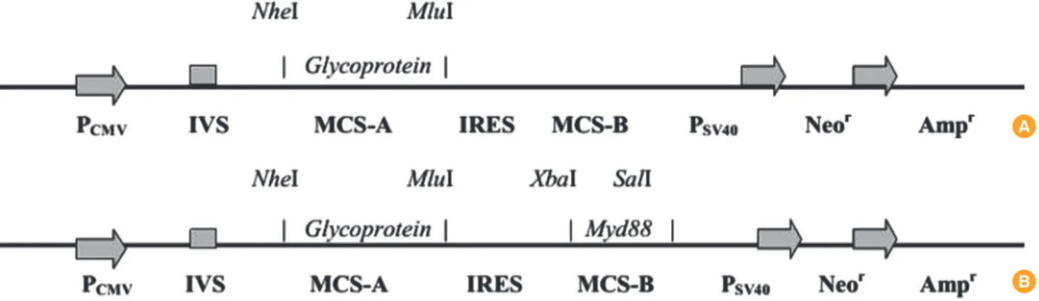
Fig. 1 shows a schematic representation of the proposed plas- mid vaccine constructs.
Construction of pIRES-Rgp
The full-length glycoprotein gene (G) was PCR amplified from pBacPak-GRC9 vector using the primers GFor (ATTAGCTAG- CATGGTTCCTCAGGCTCTCC) and GRev (ATGTAGAATTC- TCAC AGTCTGGTCTGAC) bearing NheI and MluI sites, re- spectively, and subcloned between these restriction sites in the multiple cloning site-A of pIRES vector, as reported earlier
Fig. 1. Scheme of plasmid constructs (linearized). (A) pIRES-Rgp. (B) pIRES-Rgp-Myd.
A
B
[8]. The construct was named pIRES-Rgp.
Construction of pIRES-Rgp-Myd
An 891 bp fragment of the human Myd88 gene was similarly subcloned into the multiple cloning site-B of pIRES-Rgp. Brie- fly, the gene fragment was PCR amplified using MydFor (AT- TATCTACAATGGCTGCAGGAGGTCCC) and MydRev prim- ers (TAAAGTCGACTCAGGGCAGGGACAAGGC) (bearing XbaI and SalI sites, respectively), using pFLAG-CMV4-hMyd88 as template. The reaction was set up in a volume of 50 µL, em- ploying 10 pmol each of MydFor and MydRev, 2.5 units of Pfu DNA polymerase and 250 ng of the template. The amplifica- tion conditions were as follows: denaturation at 95°C for 1 minute, followed by 35 cycles of 95°C for 30 seconds, 68°C for 30 seconds and 72°C for 45 seconds, and final extension at 72°C for 3 minutes. The amplicon was gel-purified using Gen- Elute gel extraction kit (Sigma Aldrich) and quantitated. Se- quential restriction digestion of pIRES vector and the ampli- con was performed with XbaI and SalI enzymes. The digests were again gel-purified, and a ligation reaction was set up with a vector:insert ratio of 1:3 using T4 DNA ligase (Thermo Sci- entific), in a volume of 20 µL. After overnight incubation at 16°C, 10 µL of the reaction mixture was used to transform com- petent Escherichia coli DH5α cells by a standard heat-shock procedure. Transformants were subsequently identified on a selective Luria Bertani (LB)-ampicillin agar plate, and the identity of the recombinants was verified by colony PCR us- ing MydFor and MydRev primers. Glycerol stocks of the re- combinants were made and stored at -80°C till further use.
The identities of the recombinant constructs were verified by restriction enzyme analysis and by nucleotide sequencing (data not shown).
Studying in vitro expression of the glycoprotein and Myd88 genes
Expression of genes encoded in pIRES-Rgp and pIRES-Rgp- Myd was evaluated by indirect immunofluorescent staining of cells transiently transfected with each.
Expression of glycoprotein gene from pIRES-Rgp
The evaluation of G expression from pIRES-Rgp has been re- ported earlier [8].
Expression of glycoprotein and Myd88 genes from pIRES-Rgp- Myd
To evaluate expression of G and Myd88 genes from pIRES-
Rgp-Myd, transient transfection experiment was set up in BHK-21 cells as before, using 2 µg of the plasmid. Fourty-eight hours post-transfection, the cells were acetone-fixed and subjected to indirect immunofluorescent staining for each protein. Staining for glycoprotein expression was performed as done previously. For verifying MyD88 expression, the cells were probed with a rabbit anti-MyD88 polyclonal antibody (1:100 diluted in phosphate buffered saline [PBS] pH 7.4), fol- lowed by goat anti-rabbit IgG-TRITC conjugate (1:100 diluted in PBS pH 7.4). The coverslips were examined under an in- verted fluorescence microscope (TS120, Nikon, Tokyo, Japan) and images obtained with the help of a digital camera.
Large-scale plasmid purification
Glycerol stocks of the plasmid constructs were thawed and streaked on to LB ampicillin agar plates. After overnight incu- bation at 37°C, a single colony was transferred with a sterile inoculating loop into 5 mL of LB ampicillin broth and incu- bated at 37°C for 6-8 hours. The starter culture was subsequen- tly inoculated into 50 mL of fresh LB ampicillin broth and grown at 37°C with vigorous shaking (500 rpm) overnight, which was further inoculated into 2.5 L of fresh LB ampicillin broth and incubated under similar conditions. After 16 hours of growth, the bacteria were pelleted by centrifugation at 5,000 rpm at 4°C. The plasmid identity was once again verified by restriction enzyme analysis of a plasmid miniprep made from 1.5 mL of the overnight culture. Large-scale purification of each plasmid was then performed from the bacterial pellets using a commercial kit (EndoFree Plasmid Purification Giga Kit, Qiagen), as per manufacturer instructions. The quality and concentration of the plasmid preparations were evaluat- ed by spectrophotometry and agarose electrophoresis, and the preparations were stored in aliquots at -20°C till further use.
Evaluation of the immunogenicity and protective ability of plasmid DNA constructs
Groups (n=10 each) of 6-8-week-old Swiss albino mice of ei- ther sex were employed for the experiments. Ninety micro- grams of each plasmid, contained in 100 µL or 50 µL of sterile normal saline, was inoculated intramuscularly or intrader- mally, respectively, into each animal. The intramuscular im- munizations were administered into quadriceps muscle on the left leg, and the intradermal inoculations were done at a point about 1 cm below the base of the tail. The animals re- ceived one dose of the immunogen on days 0, 7, and 21. Con- trol groups were inoculated with a cell culture rabies vaccine
(Rabipur), sterile normal saline, or the empty pIRES vector.
Estimation of rabies virus neutralizing antibody titre by RFFIT Blood samples were collected from the immunized mice by retro-orbital venous plexus puncture under halothane anes- thesia, 14 and 28 days after the first immunization. The sam- ples were allowed to clot, and sera were separated by centrif- ugation at 6,000 rpm for 5 minutes. Sera were heat-inactivat- ed in a water bath at 56°C for 30 minutes, and stored at -20°C till tested.
Rabies virus neutralizing antibody (RVNA) titres in the sera were assayed by a standard RFFIT, with some modifications [31].
Evaluation of protective efficacy of the vaccine formulations against a rabies virus challenge
The mice were subjected to an intracerebral challenge with 50 LD50 of CVS-11 strain of rabies virus, on day 35. The ani- mals were observed for a period of 28 days for rabies-specific symptoms. At the end of the observation period, the percent survival in each group was recorded. The animals showing symptoms of rabies were euthanized, and infection confirm-
ed by direct fluorescent antibody testing on impression smears prepared from extracted brains.
Statistical analysis
The statistical analyses were done using GraphPad PRISM software version 5.00 (GraphPad Software, San Diego, CA, USA). Comparison of antibody titres in the different groups was performed with non-parametric ANOVA test, followed by post-hoc analysis by Tukey’s multiple comparison test. p<
0.05 were considered to be statistically significant.
Results
Cloning experiments Construction of pIRES-Rgp
The development and evaluation of the construct pIRES-Rgp encoding the full-length rabies glycoprotein gene has been reported earlier [8].
Construction of pIRES-Rgp-Myd
The Myd88 fragment (891 bp) was successfully amplified by PCR and cloned into the MCS-B of pIRES-Rgp, between the
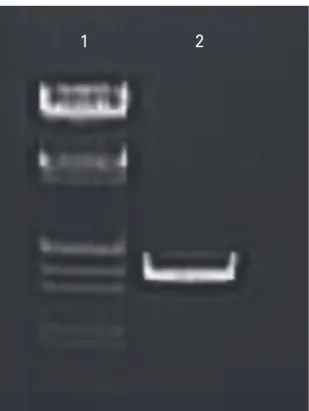
Fig. 2. Gel image showing polymerase chain reaction amplification of rabies virus glycoprotein gene. Lane 1, lambda DNA/EcoRI+HindIII marker; lane 2, ~1.6 kb band indicative of full-length glycoprotein gene.
1 2
Fig. 3. Gel image showing polymerase chain reaction amplification of Myd88 gene fragment. Lane 1, GeneRuler 100 bp DNA ladder; lane 2, 891 bp band indicative of Myd88 gene fragment.
1 2
restriction sites XbaI and SalI, yielding the construct pIRES- Rgp-Myd. Presence of G and Myd88 gene inserts in the con- struct was checked by PCR using gene-specific primers (Figs.
2, 3), and further verified by restriction enzyme analysis and nucleotide sequencing (data not shown).
Immunofluorescence staining for expression of G and MyD88 Expression of G from pIRES-Rgp
BHK-21 cells transiently transfected with pIRES-Rgp revealed expression of the glycoprotein upon indirect immunofluores- cent staining using a glycoprotein-specific monoclonal anti- body, as reported earlier [8].
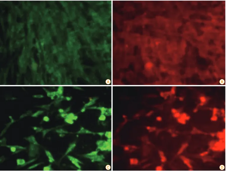
Expression of G and Myd88 genes from pIRES-Rgp-Myd Indirect immunofluorescent staining using specific antibod- ies revealed the expression of both G and MyD88 proteins in BHK-21 cells transfected with pIRES-Rgp-Myd. The trans- fected cells showed typical apple-green, membrane and cy- toplasmic fluorescence, indicative of glycoprotein expression.
Orange-red TRITC fluorescence, indicating MyD88 expres- sion, was observed on the cell membranes and in the cyto- plasm, in the same fields, indicating probable co-localization of both the proteins (Fig. 4).
Large scale plasmid purification
In order to prepare the milligram amounts of plasmids requir-
Fig. 4. Images of BHK-21 cells fluorescently stained for expression of G and MyD88. Cells were grown in 24-well plates and mock-transfected (A, B) or transfected with a liposomal complex of pIRES-Rgp-Myd (C, D). Forty-eight hours later, the cells were stained sequentially with a murine anti-G antibody and a rabbit polyclonal anti-Myd88 antibody, followed by species-specific secondary IgG conjugated with FITC or TRITC, respec- tively. Note the lack of specific fluorescence in mock-transfected cells, and the apple-green fluorescence (indicative of expression of G) and orange-red fluorescence (indicative of MyD88 expression) in pIRES-Rgp-Myd transfected cells, following staining (images at a total magnifica- tion of ×400).
A
C
B
D
ed for immunization experiments, we used a commercial large-scale, endotoxin-free plasmid purification kit, as per manufacturer instructions.
Evaluation of immunogenicity and protective efficacy of the plasmid constructs
Neutralizing antibody response in the immunized animals We collected sera from the mice on days 14 and 28 post-im- munization and estimated their RVNA titres by a RFFIT. The results from a single representative experiment are summa- rized in Table 1.
Results of intramuscular immunization
As seen from Table 1, RVNA titres>0.5 IU/mL, indicative of sero-conversion for rabies, developed by day 14 in all groups of mice immunized with pIRES-Rgp or pIRES-Rgp-Myd. We observed the highest RVNA titres in the group that received two doses of Rabipur (8 IU/mL), followed by the group im- munized with pIRES-Rgp-Myd (3.2 IU/mL). Notably, pIRES- Rgp-Myd induced ~3.2 fold higher levels of antibodies than pIRES-Rgp (p<0.05) at this time point.
The RVNA titres increased further in the groups, after the booster dose on day 21 (Table 1). On day 28, the highest titres of neutralizing antibodies were still seen in the Rabipur group (14.4 IU/mL). Mice immunized with pIRES-Rgp-Myd showed a mean titre of 11.2 IU/mL. The mean titre observed in groups immunized with pIRES-Rgp at this point was 3.6 IU/mL. The RVNA titres increased >3-fold in these groups, following the booster dose on day 21. The RVNA titres following intramus-
cular immunization with pIRES-Rgp-Myd were 3.1-fold high- er than those induced by pIRES-Rgp at this point (p<0.05).
Results of intradermal immunization
Among the intradermally immunized groups, the highest RVNA titres at day 14 were detected in the group immunized with Rabipur (8 IU/mL) (Table 1). pIRES-Rgp-Myd and pIRES- Rgp induced titres of 3.0 and 0.96 IU/mL respectively, at this time point. We observed a 2.5-fold increase in RVNA titre fol- lowing a booster with pIRES-Rgp-Myd, whereas the pIRES- Rgp booster increas ed it by 3.6-fold.
Evaluation of protective efficacy
We evaluated the protective efficacy of the plasmid constructs by an intracerebral challenge study with 50 LD50 of CVS-11 strain of rabies virus, on day 35. The inoculated animals were observed for a period of 21 days for clinical symptoms of ra- bies, and the percentage survival was recorded in each group.
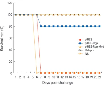
Mice inoculated with the empty vector pIRES and normal sa- line developed clinical symptoms of rabies by day 6 and were euthanized. We observed a survival rate of 80% in the group immunized intramuscularly with pIRES-Rgp (Fig. 5). Nota- bly, pIRES-Rgp-Myd protected 100% of the animals following intramuscular and intradermal immunization. pIRES-Rgp, however, protected only 80% of the mice following immuni- zation via either route. All the animals immunized with Rabi- pur survived the challenge.
Table 1. Rabies virus neutralizing antibody (RVNA) titres observed in the experimental groups following immunization
Sl. No. Immunogen Route RVNA titre (IU/mL) Day 14 Day 28
I pIRES Intramuscular
Intradermal <0.5a)
<0.5a) <0.5a)
<0.5a) II pIRES-Rgp Intramuscular
Intradermal 1.0±0.61
0.96±0.67 3.6±0.89b) 3.6±0.89b) III pIRES-Rgp-Myd Intramuscular
Intradermal
3.2±1.09b) 3.0±1.41
11.2±4.38b) 7.6±5.36b)
IV Rabipur Intramuscular
Intradermal 8.0±0.0
8.0±0.0 14.4±3.5 16.0±0.0 V Normal saline Intramuscular
Intradermal <0.5a)
<0.5a) <0.5a)
<0.5a) Values are presented as mean±standard deviation.
a)The RVNA titres were below the limit of detection by rapid fluorescent focus inhibition test in these groups.
b)p<0.05.
Fig. 5. Graph showing the survival rates of immunized mice following an intracerebral challenge with CVS-11 strain of rabies virus. Results show data compiled from challenge study in the intramuscular and intradermal immunization groups. NS, normal saline.
120
100
80
60
40
20
0 1 2 3 4 5 6 7 8 9 10 11 12 13 14 15 16 17 18 19 20 21 Days post-challenge
Survival rate (%) pIRESpIRES-Rgp
pIRES-Rgp-Myd Rabipur NS
Discussion
Despite successful demonstrations of efficacy of plasmid DNA- based vaccination strategy for rabies in a number of animal models, the technology is still constrained by poor immuno- genicity observed in larger animals and the delay in onset of antibody responses. A number of physical, chemical and bio- logical methods have been developed and evaluated for en- hancing DNA vaccine efficacy, of which, in vivo electropora- tion and gene gun delivery have been found to yield the high- est efficacy [32].
Neutralizing antibodies directed against surface proteins of the pathogen represent crucial correlates of protective im- munity in a number of infections, including rabies. Genera- tion and proliferation of antibody-secreting plasma B-cells and the maintenance of antigen-specific memory B-cells un- derlie efficient and long-lasting humoral immune responses.
The signaling molecules of the TLR pathway offer opportuni- ties to influence these phenomena, in view of their ubiqui- tous expression, and ability to modulate multiple signaling pathways [33].
Experimental evidence abounds on the ability of Myd88 to influence immune and inflammatory signaling through mul- tiple pathways, and protective immunity against many patho- gens [26,28,34-39]. In view of reports suggesting the impor- tant roles played by Myd88 signaling in the generation of hu- moral immune responses, we explored the potential utility of Myd88 as a genetic adjuvant in plasmid vaccination against rabies.
Our study revealed that co-expression of Myd88 in a plas- mid rabies vaccine enhanced the RVNA titres in Swiss albino mice upon intramuscular and intradermal immunization (Table 1). Notably, the mean RVNA titre at day 28 was 3-fold higher in the group immunized intramuscularly with pIRES- Rgp-Myd than in the group immunized with pIRES-Rgp (p<0.05).
This is in agreement with Takeshita et al. [30] who reported enhancement in antibody titres upon Myd88 adjuvanting in a plasmid vaccine against influenza. pIRES-Rgp-Myd increas- ed the RVNA titres > 2-fold upon intradermal immunization, compared to pIRES-Rgp (p<0.05).
We observed comparable antibody titres in groups receiv- ing pIRES-Rgp intramuscularly and intradermally. This ob- servation is intriguing, as it is known that intradermal immu- nization generally induces better immune responses than the intramuscular immunization.
In a previous study, Pinto et al. [23] studied the effect of co-
administration of plasmids encoding chemokine and cyto- kine genes (RANTES, MCP-1, MIP-1-β, and TRANCE) in plas- mid vaccines against rabies, and observed a modest adjuvant effect to a secreted and truncated form of the protein. The authors commented that genetic adjuvants may be unable to influence early events of immune response, due to temporal differences in expression of the immunogen and the adjuvant genes. The present study, in contrast, evaluated immune re- sponses to plasmids co-expressing viral glycoprotein and Myd88 genes. Co-expression of both the genes was apprecia- ble in fluorescently-stained transfected cells, and it would seem likely that MyD88 expression could have influenced ac- tivation and maturation of antigen presenting cells at the site of inoculation.
An early onset and a high initial titre of RVNA would be ideal in the context of rabies vaccination. Whereas the former holds critical importance in post-exposure prophylaxis, the decline of antibody responses to non-protective levels can be delayed by high initial titres of RVNA. This would be benefi- cial in maintaining protective immunity over long periods in immunized animals, especially in canines. Findings from the study appear to suggest the ability of Myd88 adjuvanting in influencing these two relevant attributes of protective immu- nity against rabies.
We evaluated the protective efficacy of the vaccine formu- lations by a viral challenge study performed on the immuniz- ed animals on day 35. pIRES-Rgp protected 80% of the immu- nized animals against the challenge, following intramuscular and intradermal routes of administration. Notably, 100% of the animals immunized with pIRES-Rgp-Myd (in either route) survived the challenge (Fig. 5).
Kaur et al. [40] reported variation in protective efficacy aga- inst a viral challenge despite similar magnitude of immune response to different plasmid vaccine constructs, suggesting the probable contribution of cellular immune factors in pro- tective immunity against rabies. Findings from the present study seem to support this, as complete protection against the lethal virus challenge was not observed in the groups im- munized with pIRES-Rgp. We propose that the enhanced RVNA responses and protective efficacy following immuniza- tion with pIRES-Rgp-Myd might have resulted from stimula- tion of the cellular immune responses.
In conclusion, the study reveals that genetic adjuvanting with Myd88 considerably enhances the protective immune responses to plasmid-based rabies vaccines. Plasmid vaccines encoding Myd88 in addition to the protective immunogen
might represent efficacious vaccine candidates for generation of long-lasting protective immunity against rabies and other infectious diseases, especially for immunization of animals, and needs to be evaluated further in field conditions.
References
1. Knobel DL, Cleaveland S, Coleman PG, et al. Re-evaluating the burden of rabies in Africa and Asia. Bull World Health Organ 2005;83:360-8.
2. Rupprecht CE, Hanlon CA, Hemachudha T. Rabies re-ex- amined. Lancet Infect Dis 2002;2:327-43.
3. World Health Organization. Guidelines for dog rabies con- trol. VH/83.43 Rev. 1. Geneva: WHO; 1986.
4. Bahloul C, Taieb D, Diouani MF, et al. Field trials of a very potent rabies DNA vaccine which induced long lasting vi- rus neutralizing antibodies and protection in dogs in ex- perimental conditions. Vaccine 2006;24:1063-72.
5. Ullas PT, Desai A, Madhusudana SN. Rabies DNA vaccines:
current status and future. World J Vaccines 2012;2:36-45.
6. Barry ME, Pinto-Gonzalez D, Orson FM, McKenzie GJ, Petry GR, Barry MA. Role of endogenous endonucleases and tissue site in transfection and CpG-mediated immune activation after naked DNA injection. Hum Gene Ther 1999;
10:2461-80.
7. Ulmer JB, Wahren B, Liu MA. Gene-based vaccines: re- cent technical and clinical advances. Trends Mol Med 2006;
12:216-22.
8. Ullas PT, Madhusudana SN, Desai A, et al. Enhancement of immunogenicity and efficacy of a plasmid DNA rabies vaccine by nanoformulation with a fourth-generation ami- ne-terminated poly (ether imine) dendrimer. Int J Nano- medicine 2014;9:627-34.
9. Singh M, Briones M, Ott G, O’Hagan D. Cationic micro- particles: a potent delivery system for DNA vaccines. Proc Natl Acad Sci U S A 2000;97:811-6.
10. Lodmell DL, Parnell MJ, Bailey JR, Ewalt LC, Hanlon CA.
Rabies DNA vaccination of non-human primates: post- exposure studies using gene gun methodology that accel- erates induction of neutralizing antibody and enhances neutralizing antibody titers. Vaccine 2002;20:2221-8.
11. Margalith M, Vilalta A. Sustained protective rabies neu- tralizing antibody titers after administration of cationic lipid-formulated pDNA vaccine. Genet Vaccines Ther 2006;
4:2.
12. Bodles-Brakhop AM, Heller R, Draghia-Akli R. Electro-
poration for the delivery of DNA-based vaccines and im- munotherapeutics: current clinical developments. Mol Ther 2009;17:585-92.
13. Kaur M, Saxena A, Rai A, Bhatnagar R. Rabies DNA vac- cine encoding lysosome-targeted glycoprotein supple- mented with Emulsigen-D confers complete protection in preexposure and postexposure studies in BALB/c mice.
FASEB J 2010;24:173-83.
14. Xiang SD, Selomulya C, Ho J, Apostolopoulos V, Plebanski M. Delivery of DNA vaccines: an overview on the use of biodegradable polymeric and magnetic nanoparticles.
Wiley Interdiscip Rev Nanomed Nanobiotechnol 2010;2:
205-18.
15. Nawwab Al-Deen FM, Selomulya C, Kong YY, et al. De- sign of magnetic polyplexes taken up efficiently by den- dritic cell for enhanced DNA vaccine delivery. Gene Ther 2014;21:212-8.
16. Kobiyama K, Jounai N, Aoshi T, et al. Innate immune sig- naling by, and genetic adjuvants for DNA vaccination. Vac- cines 2013;1:278-92.
17. Wales J, Andreakos E, Feldmann M, Foxwell B. Targeting intracellular mediators of pattern-recognition receptor signalling to adjuvant vaccination. Biochem Soc Trans 2007;35(Pt 6):1501-3.
18. Xiang Z, Ertl HC. Manipulation of the immune response to a plasmid-encoded viral antigen by coinoculation with plasmids expressing cytokines. Immunity 1995;2:129-35.
19. Geissler M, Gesien A, Tokushige K, Wands JR. Enhance- ment of cellular and humoral immune responses to hepa- titis C virus core protein using DNA-based vaccines aug- mented with cytokine-expressing plasmids. J Immunol 1997;158:1231-7.
20. Tsuji T, Hamajima K, Ishii N, et al. Immunomodulatory effects of a plasmid expressing B7-2 on human immuno- deficiency virus-1-specific cell-mediated immunity induc- ed by a plasmid encoding the viral antigen. Eur J Immu- nol 1997;27:782-7.
21. Xiang ZQ, He Z, Wang Y, Ertl HC. The effect of interferon- gamma on genetic immunization. Vaccine 1997;15:896-8.
22. Sin JI, Kim JJ, Ugen KE, Ciccarelli RB, Higgins TJ, Weiner DB. Enhancement of protective humoral (Th2) and cell- mediated (Th1) immune responses against herpes sim- plex virus-2 through co-delivery of granulocyte-macro- phage colony-stimulating factor expression cassettes. Eur J Immunol 1998;28:3530-40.
23. Pinto AR, Reyes-Sandoval A, Ertl HC. Chemokines and
TRANCE as genetic adjuvants for a DNA vaccine to rabies virus. Cell Immunol 2003;224:106-13.
24. Bramson JL, Dayball K, Hall JR, et al. Super-activated in- terferon-regulatory factors can enhance plasmid immu- nization. Vaccine 2003;21:1363-70.
25. O’Neill LA, Golenbock D, Bowie AG. The history of Toll- like receptors: redefining innate immunity. Nat Rev Im- munol 2013;13:453-60.
26. Genestier L, Taillardet M, Mondiere P, Gheit H, Bella C, Defrance T. TLR agonists selectively promote terminal plasma cell differentiation of B cell subsets specialized in thymus-independent responses. J Immunol 2007;178:7779- 86.
27. O’Neill LA, Bowie AG. The family of five: TIR-domain-con- taining adaptors in Toll-like receptor signalling. Nat Rev Immunol 2007;7:353-64.
28. Rubtsov AV, Swanson CL, Troy S, Strauch P, Pelanda R, Tor- res RM. TLR agonists promote marginal zone B cell acti- vation and facilitate T-dependent IgM responses. J Immu- nol 2008;180:3882-8.
29. Krieg AM. Toll-free vaccines? Nat Biotechnol 2007;25:303-5.
30. Takeshita F, Tanaka T, Matsuda T, et al. Toll-like receptor adaptor molecules enhance DNA-raised adaptive immune responses against influenza and tumors through activa- tion of innate immunity. J Virol 2006;80:6218-24.
31. Smith JS, Yager PA, Baer GM. A rapid reproducible test for determining rabies virus-neutralizing antibody. In: Mes- lin FX, Kaplan MM, Koprowski H, editors. Laboratory tech- niques in rabies. 4th ed. Geneva: World Health Organiza- tion; 1996. p.181-92.
32. Mintzer MA, Simanek EE. Nonviral vectors for gene deliv- ery. Chem Rev 2009;109:259-302.
33. Gururajan M, Jacob J, Pulendran B. Toll-like receptor ex- pression and responsiveness of distinct murine splenic and mucosal B-cell subsets. PLoS One 2007;2:e863.
34. Ha SA, Tsuji M, Suzuki K, et al. Regulation of B1 cell mi- gration by signals through Toll-like receptors. J Exp Med 2006;203:2541-50.
35. Guay HM, Andreyeva TA, Garcea RL, Welsh RM, Szomo- lanyi-Tsuda E. MyD88 is required for the formation of long- term humoral immunity to virus infection. J Immunol 2007;
178:5124-31.
36. von Bernuth H, Picard C, Jin Z, et al. Pyogenic bacterial infections in humans with MyD88 deficiency. Science 2008;
321:691-6.
37. Sheahan T, Morrison TE, Funkhouser W, et al. MyD88 is required for protection from lethal infection with a mouse- adapted SARS-CoV. PLoS Pathog 2008;4:e1000240.
38. Neves P, Lampropoulou V, Calderon-Gomez E, et al. Sig- naling via the MyD88 adaptor protein in B cells suppress- es protective immunity during Salmonella typhimurium infection. Immunity 2010;33:777-90.
39. Kang SM, Yoo DG, Kim MC, et al. MyD88 plays an essen- tial role in inducing B cells capable of differentiating into antibody-secreting cells after vaccination. J Virol 2011;85:
11391-400.
40. Kaur M, Rai A, Bhatnagar R. Rabies DNA vaccine: no im- pact of MHC class I and class II targeting sequences on immune response and protection against lethal challenge.
Vaccine 2009;27:2128-37.