1)
Introduction
Hyperphosphatemia is a common biochemical alte- ration in patients with renal failure, hypoparathyroidism, pseudohypoparathyroidism, metabolic acidosis, acrome- galy, and tumor lysis syndrome1). Serum phosphate is elevated occasionally in patients with multiple myeloma.
However, hyperphosphatemia is rare in patients with multiple myeloma unless renal failure is present, and most cases of multiple myeloma that present with hyper- phosphatemia are accompanied by significant reduction of renal function below 30 mL/min of glomerular filtra- tion rate (GFR)2, 3). We report a case of pseudohyper- phosphatemia from a patient with multiple myeloma whose serum phosphate was constantly elevated without any symptom or sign of hyperphosphatemia.
Case Report
A 69-year-old woman suffered from dyspnea on exertion for a week. Severe anemia (hemoglobin 5.8 g/
dL) and aortic stenoinsufficiency was revealed on her blood test and echocardiogram. She was admitted for evaluation of anemia. The albumin-globulin ratio was reversed on her blood chemistry test, and Bence- Jones proteinuria was detected by routine urinalysis.
The bone marrow biopsy and serum and urine protein electrophoreses were performed, and she was diag- nosed as the immunoglobulin G (IgG) kappa type of multiple myeloma. The sequential investigation showed that the stage of multiple myeloma was IIIA. There were no skin lesions such as calcified nodules or eczema, and deep tendon reflexes were normal at both upper and lower extremities. Neither lytic bone lesion nor soft tissue calcification was found on her simple radiograms. Serum creatinine was 1.6 mg/dL, and estimated GFR calculated by the abbreviated Modifi- cation of Diet in Renal Disease (MDRD) study equ- ation4)was 29.7 mL/min/1.73 m2. Serum calcium was
Pseudohyperphosphatemia in a Patient with Multiple Myeloma
Yonggu Lee, M.D.1, Taiyon Koo, M.D.1, Joo-Hark Yi, M.D.1, Jung-Hye Choi, M.D.1 Sang-Woong Han, M.D.1, Ile-Kyu Park, M.D.2 and Ho-Jung Kim, M.D.1
Departments of1Internal Medicine and 2Laboratory Medicine Hanyang University Guri Hospital, Guri, Korea
Hyperphosphatemia is an unusual manifestation in patients with multiple myeloma without a significantly reduced glomerular filtration rate. Serum phosphate may be falsely elevated when a large amount of paraproteins is present in the serum, because ultraviolet light absorbance is elevated with the phospho- molybdate ultraviolet assay, which is most commonly used for serum phosphate measurement. This pseudohyperphosphatemia can be confirmed by deproteinization of the serum of patients. We report a case of multiple myeloma presenting with spurious hyperphosphatemia revealing pseudohyperphos- phatemia by deproteinization of serum using sulfosalicylic acid.
Paraproteinemia, Multiple myeloma, Pseudohyperphosphatemia Case report
8.3 mg/dL, and intact parathyroid hormone was 55.9 pg/mL. However, serum phosphate was elevated to 12.7 mg/dL. She was given mephalan 8 mg/m2per day, prednisolone 60 mg/m2 per day for 7 days every month, and oral CaCO3500 mg three times a day in the middle of her meal. After three months when she was admitted for her 3rd cycle of chemotherapy, serum phosphate was still elevated to 13.1 mg/dL. Serum creatinine was 1.2 mg/dL, and estimated GFR was improved to 40.9 mL/min/1.73 m2. Urinary phosphate collected for 24 hours was normal. Other laboratory findings at the initial presentation and admission for the 3rd cycle of chemotherapy are shown in Table 1.
The hyperphosphatemia was suspected to be spuri- ous because intact parathyroid hormone was normal and estimated GFR was not remarkably decreased.
Therefore, we took a single blood sample from her, and split it into two. One of the samples was treated with 20% sulfosalicylate to remove the paraprotein, and the serum phosphate, calcium, and albumin concentrations were compared between the two samples. The phos- phate level was decreased to 3.5 mg/dL in the depro- teinized sample, but it was 10.5 mg/dL in the other (Table 2).
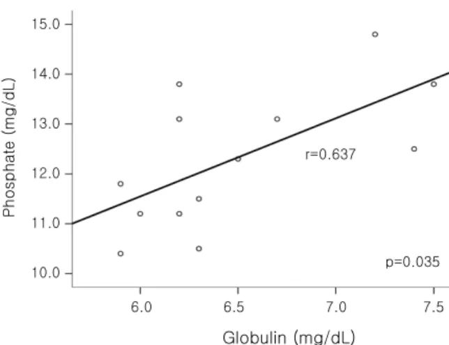
We analyzed the relationship between serum phos- phate and serum globulin of the patient. Thirteen mea- surements were selected among all of the 21 simul- taneous measurements of serum phosphate, calcium, total protein, and albumin, where the corrected calcium [= serum calcium 0.8(4 - serum albumin)] was nor- mal (8.2-10.5 mg/dL). Serum globulin was defined as the difference between serum protein and serum albumin. Spearman s correlation of SPSS 13.0 was used’ for statistic analysis. The concentration of serum phos- phate was correlated positively with that of serum globulin (r=0.637, p=0.035, Fig. 1).
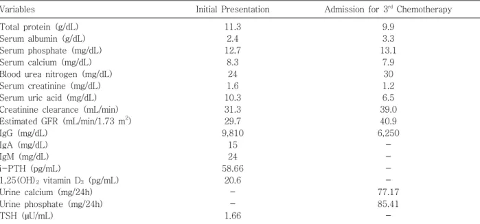
Finally, she was diagnosed as pseudohyperphospha- temia occurring due to the interference of serum para- protein with phosphate measurement, and she stopped Table 1. Biochemical Data of the Patient with Multiple Myeloma and Spurious Hyperphosphatemia
Variables Initial Presentation Admission for 3rd Chemotherapy
Total protein (g/dL) Serum albumin (g/dL) Serum phosphate (mg/dL) Serum calcium (mg/dL) Blood urea nitrogen (mg/dL) Serum creatinine (mg/dL) Serum uric acid (mg/dL) Creatinine clearance (mL/min) Estimated GFR (mL/min/1.73 m2) IgG (mg/dL)
IgA (mg/dL) IgM (mg/dL) i-PTH (pg/mL)
1,25(OH)2vitamin D3(pg/mL) Urine calcium (mg/24h) Urine phosphate (mg/24h) TSH ( U/mL)µ
11.3 2.4 12.7
8.3 24 1.6 10.3 31.3 29.7 9,810
15 24 58.66
20.6 - - 1.66
9.9 3.3 13.1
7.9 30 1.2 6.5 39.0 40.9 6,250
- - - - 77.17 85.41 -
GFR, glomerular filtration rate; Estimated GFR was calculated by the abbreviated Modification of Diet in Renal Disease study equation; iPTH, intact parathyroid hormone; TSH, thyroid-stimulating hormnone.
Table 2. Serum Concentrations of Calcium, Phos- phate, and Protein in the Patient before and after Deproteinization with Sulfosalicylic Acid
Variables Before
Deproteinization
After Deproteinization Total protein (g/dL)
Serum albumin (g/dL) Serum phosphate
(mg/dL)
Serum calcium (mg/dL)
9.3 3.0 10.5
7.8
0.4 - 3.5
9.0
her CaCO3medication.
Discussion
Pseudohyperphosphatemia is characterized by fal- sely elevated serum phosphate due to serum parapro- tein in multiple myeloma patients without impairment of renal function and clinical manifestations of hyper- phosphatemia. Phosphomolybdate ultraviolet (UV) assay is most commonly used for measurement of serum phosphate5). It relies on the formation of a UV-absorbing complex between phosphate and molybdate. An inor- ganic phosphate reacts with an ammonium molybdate in the presence of sulphuric acid to form an ammonium phosphomolybdate complex, of which absorbance is measured at 340 nm3). Serum of a patient with multiple myeloma has falsely elevated absorbance because paraproteins react with ammonium molybdate to make the serum cloudy5-8). The level of serum phosphate returns to normal when it is measured after depro- teinization with sulfosalicylate5-7, 9).
We searched the PubMed and Korean medical li- terature database (KMbase and KoreaMed) for similar case reports. Keyword search under pseudo“ hyperpho- sphatemia and multiple myeloma was per” “ ” formed.
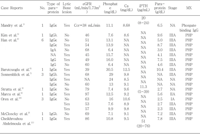
Eight case reports from PubMed and 2 case reports from Korean databases were found1, 5-12) (Table 3).
Han et al. reported that pseudohyperphosphatemia was identified in 6 of 67 (9%) patients with multiple myelo- ma8). Clinical characteristics of these cases are sum- marized in Table 3. Most cases were of the IgG type myeloma, stage IIIA, but other types and stages have been also reported. Existence of osteolytic lesion and serum calcium levels were inconsistent, but parathyroid hormone levels were all normal when measured. Serum creatinine levels was various, but there were no cases in which estimated GFR from the abbreviated MDRD study equation was below 30 mL/min/1.73 m2.
The positive correlation between serum phosphate and globulin (or immunoglobulin) has been reported in several case reports of pseudohyperphosphatemia as in ours2, 3, 5-8). It suggests that the absorbance of UV light at 340 nm increases in parallel with the concentration of immunoglobulin13). We think that the larger the tumor burden of multiple myeloma is, the greater the chances for pseudohyperphosphatemia to occur are. This could explain why the most pseudohyperphosphatemia cases were of IgG type and stage IIIA.
Interestingly, Mandry et al. reported a case of hy- perphosphatemia due to phosphate-binding immuno- globulin from a patient with multiple myeloma1). The increased serum phosphate bound with immunoglo- bulin in the patient was demonstrated by comparison with serum phosphate in a normal control using a radioisotope method with32P. In this case, differentia- tion from the pseudohyperphosphatemia was possible because hyperphosphatemia still existed when the se- rum was treated with sulfosalicylate because of weak phosphate binding capacity of immunoglobulin not enough to precipitate with phosphate11). Pseudohyper- phosphatemia does not have any necessity of treat- ment. In contrast to the cases of pseudohyperphos- phatemia, this case of phosphate-binding immunoglo- bulin had a decreased level of 1,25(OH)2vitamin D3, suggesting a role in induction of bone diseases such as osteomalacia.
In conclusion, pseudohyperphosphatemia should be considered when serum phosphate increases in a Globulin (mg/dL)
6.0 6.5 7.0 7.5 r=0.637
p=0.035
Phosphate (mg/dL)
15.0
14.0
13.0
12.0
11.0
10.0
Fig. 1. Relationship between serum globulin and phos- phate in the multiple myeloma patient with pseudohyper- phosphatemia.
patient with multiple myeloma whose renal function is not remarkably decreased. Analysis of the relationship between serum globulin and serum phosphate, and measurement of 1,25(OH)2vitamin D3and parathyroid hormone may be helpful for the identification and dif- ferentiation of hyperphosphatemia. When pseudohy- perphosphatemia is suspected, measurement of serum phosphate after deproteinization with sulfosalicylate will prevent unnecessary phosphate-lowering treat- ments.
References
1) Mandry JM, Posner MR, Tucci JR, Eil C: Hyper- phosphatemia in multiple myeloma due to a phos- phate-binding immunoglobulin. Cancer 68:1092- 1094, 1991
2) McCloskey EV, Galloway J, Morgan MA, Kanis JA:
Pseudohyperphosphataemia in multiple myeloma.BMJ 299:1381-1382, 1989
3) Stratta P, Canavese C, Quaglia M, Lazzarich E, Morellini V, Brustia M, Bardone B, Bellomo G: A patient with unexplained hyperphosphataemia. Nephrol Dial Transplant21:2664-2666, 2006
4) Levey AS, Greene T, Kusek JW, Beck GJ, MDRD Study Group: A simplified equation to predict GFR from serum creatinine. [Abstract]J Am Soc Nephrol11:155, 2000 5) Kim KS, Kim DU, Lee WS, Chung WS: Pseudohy- perphosphatemia in multiple myeloma. Korean J Hematol29:351-355, 1994
6) Barutcuoglu B, Parildar Z, Mutaf I, Habif S, Bayindir O:
Spuriously elevated inorganic phosphate level in a multiple myeloma patient. Clin Lab Haematol 25:
271-274, 2003
7) Sonnenblick M, Eylath U, Brisk R, Eldad C, Hershko C:
Paraprotein interference with colorimetry of phos- phate in Serum of some patients with multiple myeloma.
Clin Chem32:1537-1539, 1986
8) Han JY, Lee JH, Choi BG, Moon YS, Jin SW, Han WH, Kim YG, Hong YS, Kim HK, Kim BK, Lee KS, Kim DJ:
Table 3. Case Reports of Pseudohyperphosphatemia in Patients with Multiple Myeloma
Case Reports No.
Type of para- protein
Lytic Bone lesion
eGFR (mL/min/1.73m2
)
Phosphat e (mg/dL)
Ca (mg/dL)
iPTH (pg/mL)
Para- protein (g/dL)
Stage MX
Mandry et al.1)
Kim et al.5) Han et al.8)
Barutcuoglu et al.6) Sonnenblick et al.7)
Stratta et al.3) Marcu et al.9) Oren et al.10)
McCloscky et al.2) Cheikhrouhou
Abdelmoula et al.11) 1
1 6
1 3
1 1 3
1 1
IgGκ IgGλ IgGκ IgGκ IgA NA IgG IgG IgGκ IgGλ IgGκ IgGκ IgGκ IgGκ IgGκ
IgGλ IgGκ
Yes
No No Yes
No Yes Yes No Yes Yes Yes No No Yes
No Yes Yes No Yes
Ccr=38 mL/min
46 51 54 68 45 49 60 39 68 NA 60 79 97 63 53 57 69 86
11.1
7.6 13.1 13.9 6.4 15.7 16.0 6.4 30.5 29 24 13 7.4 12.5 16 7.6 9.9 7.1 10.8
8.68
8.6 NA NA NA NA NA NA 12.5
9.8 8.5 9.1 9.6 9.2 10.6
8.9 9.8 9.1 9.5
20 (8-24)
NA NA NA NA NA NA NA NA NA NA NA 11.3 (5-39)
NA NA NA NA NA 51 (20-76)
6.5
9.6 5.0 8.7 3.0 4.1 7.5 4.6 10.4
NA NA NA 2.7 5.6 2.5 2.7 2.3 7.2 7.8
NA
IIIA IIIA IIIA IIIA IIIA IIIA IIIA IIIA IIIA NA NA NA IIA IA IIIA IIIA IIIA IIIA
Phospate binding IgG
PHP PHP PHP PHP PHP PHP PHP PHP PHP PHP PHP PHP PHP PHP PHP PHP PHP PHP
No, number of case; Ccr, Creatinine clearance. Serum creatinine was not available in the paper; NA, variable that was not available in the paper; eGFR, estimated glomerular filtration rate calculated by the abbreviated Modification of Diet in Renal Disease study equation; MX, mechanism of hyperphosphatemia; PHP, pseudohyperphosphatemia; iPTH, intact parathyroid hormone; Ca, serum calcium; IgG , immunoglobulin G kappa; IgG , immunoglobulin G lambda; IgA,κ λ immunoglobulin A.
Clinical Spectrums of Pseudohyperphosphatemia in Multiple Myeloma.Korean J Med 51: 157-159, 1996 9) Marcu CB, Hotchkiss M: Pseudohyperphosphatemia in a patient with multiple myeloma.Conn Med 68: 71-72, 2004
10) Oren S, Feldman A, Turkot S, Lugassy G: Hyper- phosphatemia in multiple myeloma.Ann Hematol 69:
41-43, 1994
11) Cheikhrouhou Abdelmoula L, Amira C, Chaabouni L,
Kchir MM, Zouari R: Hyperphosphatemia in multiple myeloma. Joint Bone Spine70:541-542, 2003 12) Adler SG, Laidlaw SA, Lubran MM, Kopple JD:
Hyperglobulinemia may spuriously elevate measured serum inorganic phosphate levels. Am J Kidney Dis 11:260-263, 1988
13) Teppo AM: Immunoturbidimetry of albumin and im- munoglobulin G in urine. Clin Chem 28:1359-1361, 1982