Current Approaches for Carpal Tunnel Syndrome
Poong-Taek Kim, MD, Hyun-Joo Lee, MD, Tae-Gong Kim, MD*, In-Ho Jeon, MD
†Department of Orthopaedic Surgery, Kyungpook National University School of Medicine, Daegu,
*Department of Orthopaedic Surgery, Park Hospital, Daegu,
†Department of Orthopaedic Surgery, Asan Medical Center, University of Ulsan College of Medicine, Seoul, Korea
Received October 4, 2013; Accepted November 27, 2013 Correspondence to: In-Ho Jeon, MD
Department of Orthopaedic Surgery, Asan Medical Center, University of Ulsan College of Medicine, 88 Olympic-ro 43-gil, Songpa-gu, Seoul 138- 736, Korea
Tel: +82-2-3010-3036, Fax: +82-2-488-7877 E-mail: [email protected]
Carpal tunnel syndrome (CTS), the most common form of entrapment neuropathy in upper extremity, is estimated to occur in 3.8% of the general population. On the basis of clinical examinations and nerve conduction studies, it has been approximated that one in every five subjects who complain of symptoms such as pain, numbness, and a tin- gling sensation in the hands could have CTS.1)
According to the biomechanical and histological findings, the most characteristic histological finding is noninflammatory fibrosis and thickening of the subsyno- vial connective tissue. The hypothesis says surgical trauma to the synovium and the flexor tendons within the carpal tunnel associated with aging process or repetitive/forceful movement could lead to degeneration, thus as the volume of the carpal tunnel contents increases, median nerve within the tunnel is compressed and eventually resulted in CTS.
With advancement in biomechanical and biological research on idiopathic carpal tunnel syndrome, the insight on the pathophysiol- ogy of carpal tunnel syndrome has gained much clinical relevance. Open carpal tunnel release is still a gold standard procedure for carpal tunnel syndrome, which has evolved into mini-open procedure with development of new devices. Endoscopic carpal tunnel release has become popular in recent practice of hand surgery with an advantage of early recovery of hand function with minimal morbidity. However, endoscopic carpal tunnel release has its own limitation such as long learning curve with obvious surgical risk reported in the literature. In this review article, various treatment protocols for idiopathic carpal tunnel syndrome are presented with special highlight on endoscopic carpal tunnel release, which is gaining popularity in current practice.
Keywords: Carpal tunnel syndrome, Open, Endoscopic, Release
CONSERVATIVE TREATMENT
When the pathophysiology of idiopathic CTS remained unclear, definitive treatment strategies have not been es- tablished. However, in recent practice, treatment should be selected considering the various factors, such as the stage of the disease, the severity of the symptoms, or the patient's preference. Splinting, local injection of corticoste- roids into the carpal tunnel, and oral corticosteroid treat- ment have proven effective in some cases.2)
The rationale for wrist splint is based on observa- tions that CTS symptoms improve with rest and aggravate with activity. Subsequent research has suggested that the therapeutic effect of wrist splinting arises from minimiz- ing carpal tunnel pressure.3)
Corticosteroid treatment is effective in reducing inflammation and edema of synovium and tendons, it also has harmful effect on tenocyte function by reducing col- lagen and proteoglycan synthesis. This eventually reduces the mechanical strength of the tendon and leading to fur- ther degeneration.4) The effect of corticosteroids or local anesthetic agents on the peripheral nerve fibers or epin- erium cells reported traumatic epineural injury, axonal degeneration and intrafascial syrinx created by hydrostatic pressure.5) Further research is required to clarify the ap- propriate injection method and the optimum preparation,
Copyright © 2014 by The Korean Orthopaedic Association
This is an Open Access article distributed under the terms of the Creative Commons Attribution Non-Commercial License (http://creativecommons.org/licenses/by-nc/3.0) which permits unrestricted non-commercial use, distribution, and reproduction in any medium, provided the original work is properly cited.
Clinics in Orthopedic Surgery • pISSN 2005-291X eISSN 2005-4408
dose, and volume of corticosteroid.6)
SURGICAL TREATMENT
Open Carpal Tunnel Release
When conservative treatment fails, surgical treatment is considered. Despite the equivocal nature of CTS etiology, simple decompression of the median nerve by division of the transverse carpal ligament (TCL) is the treatment of choice and is considered to yield excellent results in 75%
of the patients.7)
In the recent literature, surgical treatment has been reported to be more effective than splinting7) and other conservative treatment.8) Carpal tunnel release (CTR) with TCL division is accepted as the most reliable procedure for relieving symptoms. The TCL can be divided by various surgical techniques, including conventional open carpal tunnel release (OCTR), mini-OCTR, and endoscopic car- pal tunnel release (ECTR). OCTR is generally accepted method,9) and reported high success rate with minimal
complication, although in worst scenario, some wound- related symptoms may persist for 2 years postoperatively.10) This surgery is indicated and feasible for CTS with any type of pathology, such as CTS due to any space occupying lesion, deformity or even in revision surgeries.
Endoscopic Carpal Tunnel Release
ECTR has gained popularity since their introduction two decades ago as single portal surgery by Okutsu et al.11) and two portal technique by Chow.12) Okutsu et al.13) developed their procedure with special tube like instrument to visu- alize whole TCL and it was transected under endoscopic assistance. At first, visualizing the dorsal aspect of the TCL by using endoscopy seemed highly innovative, attractive approach (Table 1, Fig. 1).14)
Authors’ preferred technique: anatomy principle based ECTR procedure
1. Local anesthetics for instant feedback from the patient during surgery: We use Ropivacaine 7.5 mg/mL, 10 mL
Table 1. Reported Complications Related to First-Time ECTR Complications Trans-bursal Extra-bursal Total
Incomplete release 59 124 183
SPA injury 0 7 7
Ulnar nerve injury 1 0 1
Median nerve injury 0 2 2
Complication rate (%) 48 33 36
ECTR: endoscopic carpal tunnel release, SPA: superficial palmar arch.
Reprint from Makowiec et al.14) with permission from Elsevier.
Table 2. Principles to Prevent Complications during ECTR Topographic anatomy of entry and exit portals
Manipulation of instrument outside of tendon sheath Clear space between TCL & cannula
Conversion to open in case of pain and difficulty
ECTR: endoscopic carpal tunnel release, TCL: transverse carpal ligament.
Fig. 1. Introducing the cannula to the exit portal. HH: hook of hamate, KL:
Kaplan line, TCL: transverse carpal ligament.
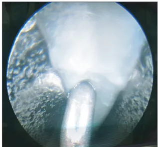
Fig. 2. Undersurface of transverse carpal ligament with washboard appearance.
in total: 4 mL was administered in the proximal direction under the subcutaneous fascia, 2 mL subcutaneoulsy in the distal wrist crease, and 4 mL subcutaneously in the palm.15)
2. Robust anatomic land marks to avoid poten- tial damage of normal structure: A line is drawn for the planned incision that is a transverse incision on the distal wrist crease starting from the radial border of the palmaris longus (PL) extending ulnarly for 1.5 cm. If the PL is ab- sent, a 1.5-cm incision centered on the line drawn from the ulnar border of the middle finger is used. The PL ten- don is the dissected and retracted radially to protect the palmar cutaneous branch of the median nerve exposing the deep fascia of the forearm (Table 2, Fig. 2).16)
3. Good endoscopic visualization and tapping the TCL with probe: Blunt dissection is performed to the proximal edge of the TCL. A elevator is used to clean the undersurface of the ligament to provide a good view with the endoscope. By palpating palm and watching it with endoscopy or inserting a needle from the palm into the canal, 4 cm distal to the transverse wrist crease, the end of the TCL is marked (Fig. 3).
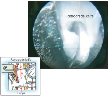
4. Transecting the TCL: The probe knife is used to make the first cut, distally to proximally. The distal bor- der of the TCL should be saved at this stage. The scope is switched into proximally and retrograde knife is brought in and the second cut to release complete TCL (Fig. 4).
Clinical results of ECTR
ECTR, either by single or dual-portal technique, can yield almost same clinical results comparable with those of OCTR. Unless iatrogenic nerve injury is counted, ECTR provides good symptom resolution, less scar and pillar pain, providing better physical findings and electrophysi- ologic recovery.17-19)
Comparison with OCTR
ECTR is useful for achieving median nerve decompres- sion, its effectiveness in comparison with the minimally invasive OCTR for restoring function of the affected hand early after treatment is still under debate. The grip strength temporarily decreases after ECTR because of postoperative pain over the incision site, which is the case after any CTR procedure. Because of the ambiguity in the definition of
‘resuming daily activities,’ the average time required to re- sume daily activities or work after ECTR could not be ad- dressed and compared directly. However, by using limited skin incision for ECTR, we expect the intact subcutaneous fatty tissue and palmar fascia will help for decreasing post- operative pain.
The benefit of ECTR over OCTR is that by dividing the TCL from below, the overlying skin and muscle are preserved, potentially improving postoperative morbidity, facilitating an earlier return to work, and preserving grip strength. The rate of major nerve, vessel, or tendon injury was shown to be lower in ECTR group, at 0.19% compared with 0.49% for the OCTR group. Similarly, Boecksyns and Sorensen20) reported a rate of 0.3% for irreversible Fig. 3. Triangular blade cutting the middle of the transverse carpal
ligament in retrograde fashion.
Fig. 4. Transection of the transverse carpal ligament by using a retrograde knife.
nerve damage in the ECTR group, and 0.2% for the CTR group.21)
On the other hand, CTR had more wound problems such as infection, hypertrophic scar, and scar tenderness.
Thoma and colleagues demonstrated a benefit of ECTR with respect to scar tenderness. ECTR also provides a faster recovery to operated patients for the first 2 weeks, with faster relief of pain and faster improvement in func- tional abilities. But at 1 year, both open and endoscopic techniques seem to be equivalently efficient (Table 3).22,23)
PEARLS AND PITFALLS TO AVOID COMPLICATION
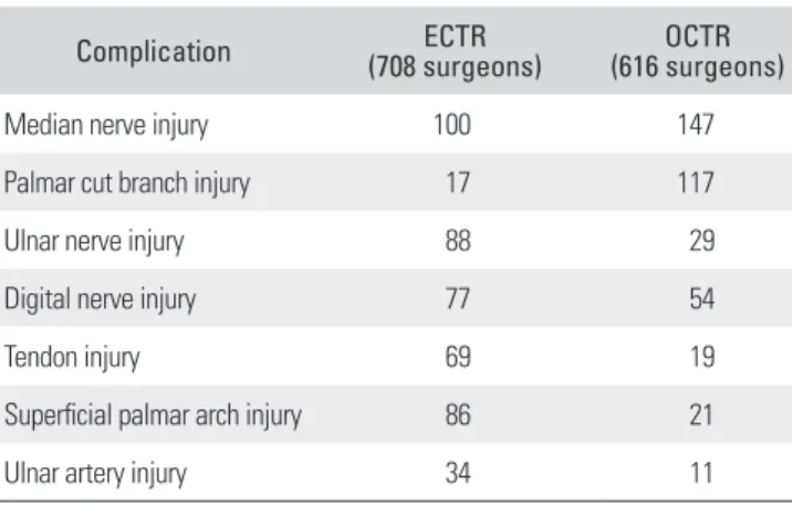
Practicing ECTR could result in a few disastrous compli- cations such as transection of the median nerve, flexor tendons, superficial palmar arch or even the ulnar nerve, mostly because of technical errors (Table 4).
These complications usually could be avoided by adhering some principles to the recommended procedure:
(1) the TCL should not be cut if soft tissue obstructs surgi- cal view; and (2) very low threshold to conversion to open procedure if the surgeon encounters any difficulty intro- ducing the cannula or achieving proper working space and good visualization just as like any endoscopic surgery (Table 2). Careful physical examination and preoperative ultrasonography or magnetic resonance imaging may be useful to detect space-occupying lesion for CTS.
Anatomic variations should be always kept in mind considering ECTR. Reported prevalence of anatomic variations encountered in elective CTR was 31 anomalies in total out of 526 CTR (5.7%). These included aberrant
muscle/tendon variation, anomalies of the median nerve or its palmar cutaneous or motor branches, and anomaly of the ulnar nerve crossing the carpal tunnel incision.24)
Another potential limitation of ECTR-namely is related to the cannula (diameter 5.5 mm in Chow's tech- nique) within the limited space of carpal tunnel which has already preceding pathologies in. As stated earlier, the pressure in the carpal tunnel was measured 5–85 mmHg in idiopathic CTS , which is greater than non-symptomatic population measured 3–6 mmHg.25)
ECTR is advised not to attempt without clear un- derstanding of the anatomy of the target space because potential complications are mostly related to the technical pitfalls and anatomic variations.
CONCLUSIONS
The current concepts of CTS and the role of ECTR in cur- rent surgical procedures were reviewed. ECTR is a useful technique for achieving median nerve decompression.
However, there is lack of evidence-based clinical data to support ECTR provides superior clinical results to OCTR in terms of early recovery of hand function. Principle- based ECTR procedure, however provides few complica- tions with successful clinical outcome so far. New treat- ment strategies can be established for CTS of different pathologies based on these clinical experiences.
CONFLICT OF INTEREST
No potential conflict of interest relevant to this article was reported.
Table 3. Advantages and Disadvantages of Endoscopic Carpal Tunnel Release
Advantage Disadvantage
Less pain Unable to see all computed tomography contents
Earlier mobility Higher risk in the presence of aberrant motor branch
Bilateral easier Higher skill/training requirement Earlier return to work Cost of endoscopic equipment Minimal skin/retinacular scar
Nerve not ‘scarred’
Reduced ‘pillar’ pain
Table 4. Reported Complication after ECTR and OCTR
Complication ECTR
(708 surgeons) OCTR
(616 surgeons)
Median nerve injury 100 147
Palmar cut branch injury 17 117
Ulnar nerve injury 88 29
Digital nerve injury 77 54
Tendon injury 69 19
Superficial palmar arch injury 86 21
Ulnar artery injury 34 11
ECTR: endoscopic carpal tunnel release, OCTR: open carpal tunnel release.
Modified from Palmer and Toivonen23) with permission from Elsevier.
REFERENCES
1. Atroshi I, Gummesson C, Johnsson R, Ornstein E, Ranstam J, Rosen I. Prevalence of carpal tunnel syndrome in a gen- eral population. JAMA. 1999;282(2):153-8.
2. Piazzini DB, Aprile I, Ferrara PE, et al. A systematic review of conservative treatment of carpal tunnel syndrome. Clin Rehabil. 2007;21(4):299-314.
3. Walker WC, Metzler M, Cifu DX, Swartz Z. Neutral wrist splinting in carpal tunnel syndrome: a comparison of night- only versus full-time wear instructions. Arch Phys Med Re- habil. 2000;81(4):424-9.
4. Scutt N, Rolf CG, Scutt A. Glucocorticoids inhibit tenocyte proliferation and Tendon progenitor cell recruitment. J Or- thop Res. 2006;24(2):173-82.
5. Cassuto J, Sinclair R, Bonderovic M. Anti-inflammatory properties of local anesthetics and their present and po- tential clinical implications. Acta Anaesthesiol Scand.
2006;50(3):265-82.
6. Davies T. Injection with methylprednisolone for carpal tun- nel syndrome: study is needed to determine best treatment for this syndrome. BMJ. 2000;320(7235):646.
7. Bland JD. Treatment of carpal tunnel syndrome. Muscle Nerve. 2007;36(2):167-71.
8. Verdugo RJ, Salinas RA, Castillo JL, Cea JG. Surgical versus non-surgical treatment for carpal tunnel syndrome. Co- chrane Database Syst Rev. 2008;(4):CD001552.
9. Badger SA, O'Donnell ME, Sherigar JM, Connolly P, Spence RA. Open carpal tunnel release: still a safe and effective op- eration. Ulster Med J. 2008;77(1):22-4.
10. Boya H, Ozcan O, Oztekin HH. Long-term complications of open carpal tunnel release. Muscle Nerve. 2008;38(5):1443- 6.
11. Okutsu I, Ninomiya S, Takatori Y, Ugawa Y. Endoscopic management of carpal tunnel syndrome. Arthroscopy.
1989;5(1):11-8.
12. Chow JC. Endoscopic release of the carpal ligament: a new technique for carpal tunnel syndrome. Arthroscopy.
1989;5(1):19-24.
13. Okutsu I, Ninomiya S, Hamanaka I, Kuroshima N, Inanami H. Measurement of pressure in the carpal canal before and after endoscopic management of carpal tunnel syndrome. J Bone Joint Surg Am. 1989;71(5):679-83.
14. Makowiec RL, Nagle DJ, Chow JC. Outcome of first-time
endoscopic carpal tunnel release in a teaching environment.
Arthroscopy. 2002;18(1):27-31.
15. Sorensen AM, Dalsgaard J, Hansen TB. Local anaesthesia versus intravenous regional anaesthesia in endoscopic car- pal tunnel release: a randomized controlled trial. J Hand Surg Eur Vol. 2013;38(5):481-4.
16. Ip WY, Sweed TA, Fung KK, Tipoe GL, Pun TS. A new technique of single portal endoscopic carpal tunnel release.
Tech Hand Up Extrem Surg. 2012;16(1):27-9.
17. Pajardi G, Pegoli L, Pivato G, Zerbinati P. Endoscopic carpal tunnel release: our experience with 12,702 cases. Hand Surg.
2008;13(1):21-6.
18. Hankins CL, Brown MG, Lopez RA, Lee AK, Dang J, Harper RD. A 12-year experience using the Brown two- portal endoscopic procedure of transverse carpal ligament release in 14,722 patients: defining a new paradigm in the treatment of carpal tunnel syndrome. Plast Reconstr Surg.
2007;120(7):1911-21.
19. Oertel J, Schroeder HW, Gaab MR. Dual-portal endo- scopic release of the transverse ligament in carpal tunnel syndrome: results of 411 procedures with special reference to technique, efficacy, and complications. Neurosurgery.
2006;59(2):333-40.
20. Boeckstyns ME, Sorensen AI. Does endoscopic carpal tun- nel release have a higher rate of complications than open carpal tunnel release? An analysis of published series. J Hand Surg Br. 1999;24(1):9-15.
21. Mintalucci DJ, Leinberry CF Jr. Open versus endoscopic car- pal tunnel release. Orthop Clin North Am. 2012;43(4):431-7.
22. Vasiliadis HS, Xenakis TA, Mitsionis G, Paschos N, Geor- goulis A. Endoscopic versus open carpal tunnel release. Ar- throscopy. 2010;26(1):26-33.
23. Palmer AK, Toivonen DA. Complications of endoscopic and open carpal tunnel release. J Hand Surg Am. 1999;24(3):
561-5.
24. Lindley SG, Kleinert JM. Prevalence of anatomic variations encountered in elective carpal tunnel release. J Hand Surg Am. 2003;28(5):849-55.
25. Werner R, Armstrong TJ, Bir C, Aylard MK. Intracarpal canal pressures: the role of finger, hand, wrist and forearm position. Clin Biomech (Bristol, Avon). 1997;12(1):44-51.