Introduction
Although there has been a raising awareness of the impor- tance of stroke as aging is increasing, mechanism of stroke is less clear. However, it has been assured that cerebrovascular ac- cident due to rupture of a vulnerable atherosclerotic plaque is the most common cause of stroke.1,2 Approximately 87% of st- roke in the United States are ischemic, about 10% are intracere- bral hemorrhages.3 In Korea, from 70% to 80% of stroke are isc- hemic and most of the population aged 50 and over have ath- erosclerotic plaque.4 Those facts of Korea and the ultrasound (US) sustained the assumption that vulnerable plaque rupture triggers stroke.
So far, stroke has been depended on the primary treatment5 but lots of clinical trials supporting the efficacy of HMG-CoA re- ductase inhibitors (statins) have been conducted since its laun- ch.6-10 As a result, numerous findings reported that statin thera- py plays a remarkable role to reduce the accumulation of athero- ma11-20 and the level of circulating inflammatory marker.21-27 Nevertheless, there is little data and guideline on efficiency of st- atin treatment in patient with calcified or advanced plaques. Also, little is known about the natural course of an advanced plaque.
In reality, patients with calcified or hyperechoic carotid ath- eroma came to the tertiary hospital, so most clinical doctors are confused what is the best treatment between medication and operation. Korea Food and Drug Administration recommended
Vascular Neurology 2011;3:45-50 ISSN 2092-6855
Evaluation of Regression of Advanced Carotid Atherosclerotic Plaques
Hye Kyoung Pae, Kyoung Joo Cho, Su Kyung Lee, Gyung Whan Kim
Department of Neurology, College of Medicine, Yonsei University, Seoul, Korea
Received August 31, 2011 Revised September 6, 2011 Accepted September 7, 2011 Correspondence Gyung Whan Kim, MD, PhD Department of Neurology,
College of Medicine, Yonsei University, 50 Yonsei-ro, Seodaemun-gu, Seoul 120-752, Korea Tel +82-2-2228-1600 Fax +82-2-393-0705 E-mail gyungkim@yuhs.ac
ObjectiveaaRecent studies have determined evidences of effects of hypolipidemic drug on athero- sclerotic plaque as well as marked change in composition that affects plaque stability. In particular, high dose statin treatment plays an important role in regression of advanced atherosclerotic plaque.
In this study, we aimed to investigate the relationship between the disruption of atherosclerotic plaque and inflammatory molecules and propose the reliable guideline of therapy of advanced (cal- cified) carotid atheroma.
MethodsaaThe outpatients of 23 who had advanced carotid atherosclerotic plaque were selected from the neurology outpatient clinic and were treated statin, aspirin, and clopidogrel for secondary prevention for a year. All participants were measured carotid artery intima-media thickness, plaque morphology, and thickness of carotid plaque by an external B-mode ultrasound and were assessed express P-selectin expression (CD62p), platelet-leukocyte aggregation, CD11b and CD40L by flow cytometry and by the platelet function analyzer.
ResultsaaThe outpatients of 23 were more than 60 years old (69.8±6.9, male=16, female=7). The average of carotid artery intima-media thickness was 0.93 mm at left and 0.91 at right. We found the significant fact that the number of total plaques in all subjects was 83. The distribution of plaques in different arterial segments was: common carotid artery, 21.7%; bulb, 54.2%; and internal carotid artery, 24.1. 77.1% (n=64) of total plaques were heterogeneous with hyperechoic morphology and 14.5% (n=12) of those were hypoechoic plaques in echogenicity. High sensitivity C-reactive protein of participants was 1.9±2.3. The difference of carotid atherosclerotic plaque thickness before and after intensive statin treatment was 2.6±0.8 and 2.4±0.7 (p=0.001). The level of CD11b and CD40L in monocyte decreased significantly after high dose statin therapy.
ConclusionsaaThese findings support the central role of statin to reduce plaque thickness and the level of inflammatory markers. Intensive statin therapy is crucial for patients with advanced athero- sclerotic plaques in carotid artery. Thus, it is urgent to establish optimal dose of stain for each patient without undergoing adverse reactions. Vascular Neurology 2011;3:45-50 Key Wordsaa Atherosclerosis, Calcified carotid artery atheroma,
Circulation inflammatory molecules, Stroke.
statin use when high sensitivity C-reactive protein (CRP) is more than 0.2 mg/dL in serum without approved dose. Food and Drug Administration (FDA) of the US has warned about high- dose statin because of side-effects such as severe myopathy, mu- scle pain, kidney damage, etc.28 Thus, FDA strictly has approv- ed statin of 10 to 20 mg as the general does.
Therefore, this study performed to evaluate the hypothesis that thickness of plaque and the level of circulating inflamma- tory molecules in patients with carotid atherosclerosis would highly decrease under receiving statin therapy and to propose the reliable guideline of treatment of calcified carotid atheroma.
We also analyzed the regression of calcified carotid atheroma monthly under statin medication.
Materials and Methods
Study design and subjects
This study performed a prospective observational study and enrolled 27 patients who had carotid atherosclerotic plaque was selected from the neurology outpatient clinic of Yonsei Univer- sity hospital and was treated with statin, aspirin, and clopidogrel for secondary prevention for a year. Participants were detected advanced or calcified plaques that ranged more than 3 mm in plaque thickness.
Exclusion criteria included operation or angioplasty for symp- tomatic stenosis performed within 3 months or planned for the future, known allergy against or previous treatment with aspirin, clopidogrel, and statin therapy, bleeding history, severe kidney or liver disease, cancer, acute myocardial infarct, and thyroid disease. Twenty-seven subjects were identified by clinical data re- cord and were willing to participate. Of these, 4 patients were excluded because of terminal stage cancer (n=2) and end stage renal disease (n=2). Therefore, 23 subjects were entered into the study. Characteristics of the participants were shown in Table 1. All participants provided written informed consent and the study protocol was approved by the Institutional Review Board of the Yonsei University Hospital.
Basic investigation
All participants were measured carotid artery intima-media thickness (CIMT), plaque morphology, and thickness of carotid plaque by B-mode ultrasonography and were assessed the level of circulating inflammatory markers such as express P-selectin (CD62p), platelet-monocyte complexes, CD40 ligand (CD40L) and Mac1 every month by flow cytometry and by the platelet function analyzer. All subjects answered a questionnaire of their medical history, smoking habits and medication. Recordings of height and weight were performed and sitting brachial blood pressure (BP) was measured using a validated automatic BP device (JMW160KA, Seoul, Korea).
Serum glucose, total and high density lipoprotein cholesterol, triglycerides and low density lipoprotein cholesterol were mea-
sured with fasting. Subjects visited to the neurology outpatient clinic monthly and were inquired about potential reverse effects and received a physical examination.
Pre-study for determination the maximum therapeutic dosage of a statin
FDA of South Korea recommended statin use when high sen- sitivity CRP is more than 0.2 mg/dL in serum but there is no ap- proved statin dose and no data for the maximum dose of that.
Therefore, we had to perform an observational study to deter- mine the maximum therapeutic dosage of a statin. We rando- mized outpatients with atherosclerosis who visited neurology outpatient. They provided written informed consent and the study protocol was approved by the Institutional Review Board of the Yonsei University Hospital. We prescribed randomized outpatients statin of 20 mg/day (mg/d) at first. They were asked about potential side effects and received a physical examination Table 1. Clinical and demographic characteristic of the study popu- lation
Variables Case group
(n=23)
Age (y) 69.8±6.9
Male % (n) 69.6 (16)
Female % (n) 30.4 (7)
Body mass index 24.3±2.7
Current smoking, % (n) 8.7 (2)
Current drinking, % (n) 17.3 (4)0
History of diabetes mellitus, % (n) 8.3 (2) History of hypertension, % (n) 16.7 (4)0
History of stroke, % (n) 16.7 (4)0
History of transient ischemic attack, % (n) 66.7 (16) History of carotid artery atherosclerosis, % (n) 83.3 (20)
Total carotid plaques, (n) 83
Carotid artery plaques, (n) 03.8±1.3
Common carotid plaque, % (n) 21.7 (18)
Bifurcation (bulb), % (n) 54.2 (45)
Internal carotid artery, % (n) 24.1 (20)
Lt carotid artery ID, mm 08.1±0.8
Rt carotid artery ID, mm 08.2±1.0
Lt carotid artery IMT, mm 00.9±0.2
Rt carotid artery IMT, mm 00.9±0.2
Systolic blood pressure 126.3±10.9
Diastolic blood pressure 075.3±10.4
Fasting glucose, mg/dL 104.0±27.2
Total cholesterol, mg/dL 144.7±32.5
High density lipoprotein, mg/dL 046.4±11.6 Low density lipoprotein, mg/dL 074.4±28.1
Triglyceride, mg/dL 117.5±31.5
High sensitivity CRP, mg/dL 01.9±2.3
Data are shown as mean±SD or number of subjects, frequency (%). ID: inner diameter, IMT: intima-media thickness.
including laboratory tests. If they had no side-effects or pro- gressed the thickness of atheroma for 2 months, we increased gradually statin dose of 20 mg/d. We maintained statin of 60 mg/d in women and 80 mg/d in men as a maximum dose. They did not undergo any adverse effects and we decided to provide men statin of 80 mg/d and women statin of 60 mg as the maxi- mum therapeutic dosage.
In this study, men in subjects took aspirin (100 mg/d), clopi- dogrel (75 mg/d), and statin (80 mg/d) and women took aspirin (100 mg/d), clopidogrel (75 mg/d), and statin (60 mg/d) for a year. Intake of study drugs was controlled by counting of deliv- ered tablets.
B-mode ultrasound examination
Carotid artery plaque was assessed by high-resolution B-mo- de ultrasound (Accuvix V10, Seoul, Korea) using a system with a multi-frequency 5 to 10 MHz linear-array transducer. All mea- surements were performed by a technologist trained in ultra- sound research according to a standard scanning and reading protocol. The CIMT was measured offline on the far wall of the common carotid artery (CCA) in a longitudinal view in a region free of plaque using a computerized system. The upper limit of normal for the intima-media thickness (IMT) was defined as 1.0 mm, and lesions with an IMT ≥1.1 mm were defined as ath- eromatous plaques, which were measured everywhere.
Plaque was also defined as normal with 1.0 mm ≥plaque th- ickness, mild to moderate with that of 1.0 mm to 3.0 mm, and moderate to severe with that more than 3 mm.
Maximum CIMT measures were obtained from walls of th- ree arterial segments of both carotid arteries; the near and far wall of the proximal 10 mm of the internal carotid artery (ICA), the near and far wall of the carotid bifurcation beginning at the tip of the flow divider and extending 10 mm proximally, and the near and far wall of the arterial segment extending 10 to 20 mm proximally to the tip the flow divider into the CCA.29 Thick- ness of calcified carotid atheroma was measured in a longitudi- nal and vertical view for screening accurate plaque. Color Dop- pler and pulsed Doppler were also used to assist in confirmation of possible plaques.
Plaque echogenicity was assessed by the gray scale median (GSM) using the histogram function. Plaque morphology was classified by GSM value, which was calculated from pixel anal- ysis within in the range 0-256. Plaques were defined as hypo- echoic if GSM value was lower than 70, as hyperechoic or calci- fied if GSM was higher than 70, and as heterogenous if echoge- nicity was mixed hypoechoic with hyperechoic.30
Assessment of platelet and leukocyte activation A trained nurse collected blood samples twelve times throu- ghout the study. Blood samples were collected into Vacutainer tubes containing 0.5 mL of 3.2% buffered sodium citrate. Imme- diately after that the citrated blood sample was added to a fixa-
tion solution (4% formaldehyde) to minimize ex-vivo platelet activation and then was prepared as below for flow cytometry.
The whole blood samples were used to assess platelet activity.
Whole blood resuspended in Tyrode’s solution was incubated with phycoerythrin (PE)-conjugated anti-CD41a to immunolo- gically identify platelets. The samples were simultaneously in- cubated with fluorescein isothiocyanate (FITC)-conjugated an- ti-CD62P at saturating concentrations for 15 min at room tem- perature in the dark. Platelet-bound anti-CD62P was determin- ed by analyzing 5,000 platelets for FITC fluorescence. Results were expressed as a percentage of antibody-positive platelets.
The red blood cell-lysed blood samples were used to deter- mine Mac-1 expression in leukocytes and evaluate platelet-leu- kocyte interaction. Granulocytes were identified by forward and sideward scatter properties of PE-CD45 fluorescence posi- tive leukocytes. Monocytes and lymphocytes were identified by strong expression of PE-CD14 and PE-CD162, respectively. To observe the Mac-1 expression in leukocyte subsets, the samples were simultaneously incubated with CD11b-FITC for 20 min at room temperature in the dark. Platelet-leukocyte subset ag- gregates (platelet-granulocyte, -monocyte, and -lymphocyte ag- gregates) were recorded with simultaneous detection of CD42b- FITC labeled platelets. As isotype control experiments, FITC- conjugated IgG antibody was used. A minimum of 5,000 cell ev- ents were analyzed in each assay. The results of Mac-1 were ex- pressed as the mean fluorescence intensity of antibody-positive leukocytes for each leukocyte subgroup. The percentage of pla- telet-granulocyte, -monocyte, and -lymphocyte complexes were calculated.
The blood samples were analyzed on the flow cytometer wi- thin three hours of venepuncture. Flow cytometric specimens were kept before analysis at 0-4°C and analyzed using FACS- Calibur (Becton Dickinson Biosciences, San Jose, CA, USA).
Statistical analysis
Characteristics and plaque thickness of participants are pre- sented as means±SD and results as means. The regression of ca- rotid artery plaque was compared using the paired t-test or Wilcoxon signed rank test. Probability values of p<0.05 were considered statistically significant (Statistical Package for the Social Sciences, Windows version 15.0, Chicago, IL, USA).
Results
Twenty-three subjects were included in this study. We analyz- ed plaque regression monthly between Dec 2009 and Nov 2010.
The population of subjects were over aged 60 (69.8±6.9, male=
16, female=7). In terms of laboratory data, triglyceride and high sensitivity CRP was 117.5±31.5 and 1.9±2.3. The average of ca- rotid artery IMT was 0.93 mm at left and 0.91 at right. The num- ber of total plaques in all subjects was 83 (3.8±1.3). The distribu- tion of plaques in different arterial segments was: CCA, 21.7%;
bulb, 54.2%; and ICA, 24.1. Subject characteristics are detailed in Table 1.
There were no significant differences between before and af- ter statin treated in carotid plaque thickness and echogenicity.
The difference of carotid atherosclerotic plaque thickness be- tween before and after intensive statin therapy was 2.6±0.8 and 2.4±0.7 (p=0.001) (Table 2 and 3). The number of echogenicity was: echolucent, 14.5%; hyperechoic, 8.4%; and heterogeneous with hyperechoic morphology, 77.1 in baseline and echolucent, 15.7%; hyperechoic, 12.0%; and heterogeneous with hyperechoic morphology, 72.3 in statin use.
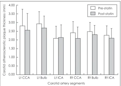
Table 3 showed that plaque thickness of pre-statin is contra- sted with that of post-statin. The change of plaque thickness in pre and post-statin therapy was shown in Fig. 1. There was a con- siderable downward trend in plaque thickness between before and after statin treatment. Plaque thickness of all carotid artery segments except Lt ICA decreased significantly after statin therapy. The reduced width of plaque thickness in CCA and
bulb is much higher than that in ICA.
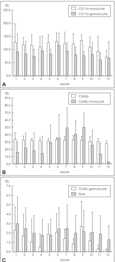
The level of inflammatory molecules monthly was shown in Fig. 2. The level of CD11b-monocyte, CD40L-monocyte and CD40L-granulocyte fluctuated for early 9 months. From Sep- tember to December, there were significant reductions in the level of CD11b-monocyte, CD40L-monocyte and CD40L-gran- ulocyte. There was no considerable difference in the level of CD11b-granulocyte, platelet-leukocyte aggregates (PMA) from Table 2. Comparison of carotid plaque thickness and echogenicity before and after statin treated
Variables Total plaques (n=83) % (n) Maximum (mm) Mean (mm) p value
Before statin treatment (baseline) 2.6±0.8 0.001
Lt CCA 16.9 (14) 4.9 2.8±1.0
Lt Bulb 27.7 (23) 3.9 2.9±0.7
Lt ICA 15.7 (13) 2.8 2.1±0.6
Rt CCA 4.8 (4) 3.1 2.4±0.5
Rt Bulb 26.5 (22) 3.8 2.5±0.7
Rt ICA 8.4 (7) 3.4 2.3±0.6
Echogenicity
Hypoechoic (echolucent) 14.5 (12)
Hyperechoic 8.4 (7)
Heterogenous, mixed 77.1 (64)
After statin treatment (follow-up) 2.4±0.7 0.001
Lt CCA 16.9 (14) 4.5 2.6±1.0
Lt Bulb 27.7 (23) 3.9 2.7±0.7
Lt ICA 15.7 (13) 3.5 2.1±0.7
Rt CCA 4.8 (4) 3.0 2.1±0.7
Rt Bulb 26.5 (22) 3.4 2.3±0.5
Rt ICA 8.4 (7) 3.3 2.1±0.6
Echogenicity
Hypoechoic (echolucent) 15.7 (13)
Hyperechoic 12.0 (10)
Heterogenous, mixed 72.3 (60)
Data are shown as mean±SD or number of plaques, n (%); thickness of plaques, mm. p<0.05 by Wilcoxon signed rank test for all com- parisons between before and after statin. CCA: common carotid artery, Bulb: bifurcation, ICA: internal carotid artery, ECA: external carotid artery.
Table 3. Difference of plaque thickness between pre and post- statin treatment
t Dif Sig. (2-tailed) Before statin-After statin 3.601 82 0.001 p values by paired t-test, p values<0.05 were considered as sig- nificant.
Figure 1. The change of plaque thickness before and after statin th- erapy. CCA: common carotid artery, Bulb: bifurcation, ICA: internal carotid artery, ECA: external carotid artery.
44.00 3.50 3.00 2.50 2.00 1.50 1.00 0.50
0.00 Lt CCA Lt Bulb Lt ICA
Carotid artery segments
Rt CCA Rt Bulb Rt ICA
Carotid atherosclerotic plaque thickness (mm)
Pre-statin Post-statin
January to December. The level of express P-selectin (CD62p) showed steady pattern during the experiment.
Discussion
There is a well recognized need for clinically applicable me-
dication to monitor plaque thickness and circulation inflamma- tory markers in the management and prevention of stroke. The administration of statin was well tolerated and safe in our pa- tients during the treatment period of one year. Serious adverse reactions were not occurred related to statin use. The main hy- pothesis of this study was that statin may reduce the level of cir- culating inflammatory biomarkers and plaque thickness in pa- tients with calcified (advanced) atherosclerotic plaque.
We monitored the thickness of calcified plaque in pre-statin treatment and in post-statin treatment during one year (Fig. 1).
There was a remarkable decrease in calcified plaque thickness unlike the previous studies,21,22 although which is different from the subjects of our study, found no difference in Coronary ar- tery Calcification progression in 80 subjects treated with 80 mg of simvastatin versus placebo and in 366 asymptomatic pa- tients randomized to either 10 or 80 mg of atovastatin over 12 months. We need more follow time to affect the plaque thick- ness and the inflammatory biomarkers for support this study convincingly.
In addition, the levels of circulating inflammatory molecules, such as CD11b-monocyte, CD40L-monocyte and CD40L-gran- ulocyte decreased significantly, which corresponded to earlier studies23-25 that did not coincide with the subject of this study. So, further researches aimed at patients with calcified plaque need to support this study convincingly.
There was non-considerable reduction in the level of CD62p and PMA unlike previous studies which reported that statin therapy reduces the level of CD62p expression and platelet- leukocyte adhesion in hypercholesterolemia subjects.26,27 There is no evidence to explain why CD62p expression and PMA did not decrease in our patients. We speculated that the difference between subjects with calcified plaque and without would exist in the mechanism of the circulating inflammatory molecules.
Thus, further investigation to subjects with calcified plaque re- quires. The problem also remains how best to treat patients with calcified atherosclerotic plaque without adverse effects. The cli- nical trials mentioned in this study provide substantial support for the institution of intensive statin treatment in diverse clinical setting. Nevertheless, the FDA has warned safety and adverse effects of intensive statin therapy and has recommended to pre- scribe low dose or moderate dose. If new solutions to solve kn- own problems of high dose statin therapy are reconfirmed by the majority of clinical trials, the management for patients with calcified atherosclerotic plaque will be established.
Conclusion
These findings support the central role of statin to reduce plaque thickness and the level of inflammatory markers. Inten- sive statin therapy is crucial for patients with advanced athero- sclerotic plaques in carotid artery. Thus, it is urgent to establish optimal dose of stain for each patient without undergoing ad- verse reactions.
Figure 2. The level of CD11b-monocyte and CD11b-granulocyte (A).
The level of CD62p and CD40L-monocyte (B). The level of CD40L- granulocyte and PMA (C). PMA: platelet-leukocyte aggregates. CD 62p: P-selectin expression.
250.0 200.0 150.0 100.0 50.0
0.0 1 2 3 4 5 6 7 8 9 10 11 12 Month
(%) CD11b-monocyte
CD11b-granulocyte
A
90.0 80.0 70.0 60.0 50.0 40.0 30.0 20.0 10.0
0.0 1 2 3 4 5 6 7 8 9 10 11 12 Month
(%) CD62p
CD40L-monocyte
B
7.0 6.0 5.0 4.0 3.0 2.0 1.0
0.0 1 2 3 4 5 6 7 8 9 10 11 12 Month
(%)
CD40L-granulocyte PMA
C
REFERENCES
1. Elkind MS. Inflammatory mechanisms of stroke. Stroke 2010;41:S3-S8.
2. Yazdani SK, Vorpahl M, Ladich E, Virmani R. Pathology and vulnera- bility of atherosclerotic plaque: identification, treatment options, and in- dividual patient differences for prevention of stroke. Curr Treat Options Cardiovasc Med 2010;12:297-314.
3. American Heart Association. Heart Disease and Stroke Statistics-2008 Update. Dallas: American Heart Association, 2008.
4. KNSO. Statistical survey of death causes in 2005-2009. Seoul: Korea National Statistical Office, 2007.
5. Sacco RL, Adams R, Albers G, Alberts MJ, Benavente O, Furie K, et al.
Guidelines for prevention of stroke in patients with ischemic stroke or transient ischemic attack: a statement for healthcare professionals from the American Heart Association/American Stroke Association Council on Stroke: co-sponsored by the Council on Cardiovascular Radiology and Intervention: the American Academy of Neurology af- firms the value of this guideline. Stroke 2006;37:577-617.
6. Baigent C, Keech A, Kearney PM, Blackwell L, Buck G, Pollicino C, et al. Efficacy and safety of cholesterol-lowering treatment: prospective meta-analysis of data from 90,056 participants in 14 randomised tri- als of statins. Lancet 2005;366:1267-1278.
7. Law MR, Wald NJ, Rudnicka AR. Quantifying effect of statins on low density lipoprotein cholesterol, ischaemic heart disease, and stroke:
systematic review and meta-analysis. BMJ 2003;326:1423.
8. Paciaroni M, Hennerici M, Agnelli G, Bogousslavsky J. Statins and st- roke prevention. Cerebrovasc Dis 2007;24:170-182.
9. Amarenco P, Labreuche J. Lipid management in the prevention of st- roke: review and updated meta-analysis of statins for stroke prevention.
Lancet Neurol 2009;8:453-463.
10. Amarenco P. Effect of statins in stroke prevention. Curr Opin Lipidol 2005;16:614-618.
11. Cha JK, Jo WS, Shin HC, Bae HR, Ho JM, Kim JW. Increased platelet CD63 and P-selectin expression persist in atherosclerotic ischemic stroke. Platelets 2004;15:3-7.
12. Marquardt L, Ruf A, Mansmann U, Winter R, Schuler M, Buggle F, et al. Course of platelet activation markers after ischemic stroke. Stroke 2002;33:2570-2574.
13. Koyama H, Maeno T, Fukumoto S, Shoji T, Yamane T, Yokoyama H, et al. Platelet P-selectin expression is associated with atherosclerotic wall thickness in carotid artery in humans. Circulation 2003;108:524-529.
14. Shoji T, Koyama H, Fukumoto S, Maeno T, Yokoyama H, Shinohara K, et al. Platelet activation is associated with hypoadiponectinemia and carotid atherosclerosis. Atherosclerosis 2006;188:190-195.
15. Lutgens E, Gorelik L, Daemen MJ, de Muinck ED, Grewal IS, Kotelian- sky VE, et al. Requirement for CD154 in the progression of atheroscle- rosis. Nat Med 1999;5:1313-1316.
16. Mathiesen EB, Bønaa KH, Joakimsen O. Low levels of high-density li- poprotein cholesterol are associated with echolucent carotid artery pla- ques: the tromsø study. Stroke 2001;32:1960-1965.
17. Mathiesen EB, Bønaa KH, Joakimsen O. Echolucent plaques are as- sociated with high risk of ischemic cerebrovascular events in carotid ste- nosis: the tromsø study. Circulation 2001;103:2171-2175.
18. Sterpetti AV, Schultz RD, Feldhaus RJ, Davenport KL, Richardson M, Farina C, et al. Ultrasonographic features of carotid plaque and the risk of subsequent neurologic deficits. Surgery 1988;104:652-660.
19. Polak JF, Shemanski L, O’Leary DH, Lefkowitz D, Price TR, Savage PJ, et al. Hypoechoic plaque at US of the carotid artery: an independent risk factor for incident stroke in adults aged 65 years or older. Cardio- vascular Health Study. Radiology 1998;208:649-654.
20. Dotsenko O, Chaturvedi N, Thom SA, Wright AR, Mayet J, Shore A, et al. Platelet and leukocyte activation, atherosclerosis and inflamma- tion in European and South Asian men. J Thromb Haemost 2007;5:
2036-2042.
21. Schmermund A, Achenbach S, Budde T, Buziashvili Y, Förster A, Friedrich G, et al. Effect of intensive versus standard lipid-lowering treatment with atorvastatin on the progression of calcified coronary atherosclerosis over 12 months: a multicenter, randomized, double- blind trial. Circulation 2006;113:427-437.
22. Terry JG, Carr JJ, Kouba EO, Davis DH, Menon L, Bender K, et al. Ef- fect of simvastatin (80 mg) on coronary and abdominal aortic arterial calcium (from the coronary artery calcification treatment with zocor [CATZ] study). Am J Cardiol 2007;99:1714-1717.
23. Türk U, Alioğlu E, Tengiz I, Ercan E, Mahmudov R, Duygu H, et al.
Statin use is associated with decreased CD-40 ligand expression on T lymphocytes of coronary atheroma plaque in patients with stable cor- onary artery disease. Anadolu Kardiyol Derg 2008;8:99-103.
24. Sanguigni V, Pignatelli P, Lenti L, Ferro D, Bellia A, Carnevale R, et al.
Short-term treatment with atorvastatin reduces platelet CD40 ligand and thrombin generation in hypercholesterolemic patients. Circulation 2005;111:412-419.
25. Weber C, Erl W, Weber KS, Weber PC. HMG-CoA reductase inhibi- tors decrease CD11b expression and CD11b-dependent adhesion of monocytes to endothelium and reduce increased adhesiveness of mo- nocytes isolated from patients with hypercholesterolemia. J Am Coll Cardiol 1997;30:1212-1217.
26. Blann AD, Nadar SK, Lip GY. The adhesion molecule P-selectin and cardiovascular disease. Eur Heart J 2003;24:2166-2179.
27. Romano M, Mezzetti A, Marulli C, Ciabattoni G, Febo F, Di Ienno S, et al. Fluvastatin reduces soluble P-selectin and ICAM-1 levels in hy- percholesterolemic patients: role of nitric oxide. J Investig Med 2000;
48:183-189.
28. Gaist D, Rodríguez LA, Huerta C, Hallas J, Sindrup SH. Lipid-lower- ing drugs and risk of myopathy: a population-based follow-up study.
Epidemiology 2001;12:565-569.
29. Espeland MA, Craven TE, Riley WA, Corson J, Romont A, Furberg CD.
Reliability of longitudinal ultrasonographic measurements of carotid intimal-medial thicknesses. Asymptomatic Carotid Artery Progression Study Research Group. Stroke 1996;27:480-485.
30. Andersson J, Sundström J, Kurland L, Gustavsson T, Hulthe J, El- mgren A, et al. The carotid artery plaque size and echogenicity are re- lated to different cardiovascular risk factors in the elderly: the Prospec- tive Investigation of the Vasculature in Uppsala Seniors (PIVUS) study. Lipids 2009;44:397-403.