Original Article
Received: Jan 4, 2017; Revised: Feb 13, 2017; Accepted: Feb 28, 2017 Correspondence to: Syed Omair Adil
Department of Research, Dow University of Health Sciences, Gulzar-e-Hijri, Ojha Campus, Suparco Road, KDA Scheme-33, Karachi, Pakistan.
Tel: +92-3322319119, Fax: +92-21-99215763, E-mail: [email protected]
Evaluation of the Causes of Erectile Dysfunction in Patients Undergoing Penile Doppler Ultrasonography in Pakistan
Usman Khanzada1, Sohail Ahmed Khan1, Munawar Hussain1, Hatem Adel1, Kamran Masood1, Syed Omair Adil2, Murli Manohar1
1Department of Radiology, Dow Institute of Radiology, 2Department of Research, Dow University of Health Sciences, Karachi, Pakistan
Purpose: In patients with erectile dysfunction, it is important to differentiate psychogenic from organic causes. Penile Doppler ultrasonography is a relatively inexpensive and minimally invasive tool for this purpose. This study was conducted to evaluate the causes of erectile dysfunction in an adult male population, using penile Doppler ultrasonography.
Materials and Methods: A retrospective study was conducted in a single center. All patients who presented with complaints of erectile dysfunction and underwent penile Doppler ultrasonography between July 2014 and June 2016 were included in this study. All examinations were performed using GE Voluson S6 and GE Logiq P5 devices. Following baseline scans, an intracavernosal injection of 20 μg of prostaglandin E1 was given. Peak systolic and end diastolic velocities were measured in each cavernosal artery. Patients with a peak systolic velocity of <25 cm/s were considered to have arterial insufficiency, while an end diastolic velocity of >5 cm/s was considered to indicate venous incompetence.
Results: Out of 97 patients (mean age, 37.09±11.59 years; range, 19∼69 years), 50 patients (51.5%) had normal findings, 24 patients (24.7%) had arterial insufficiency, 15 patients (15.5%) had a venous leak, and 8 patients (8.2%) patients had arterial insufficiency with a venous leak. Psychogenic erectile dysfunction was significantly higher among patients aged ≤40 years, while arterial insufficiency with or without a venous leak was significantly higher among patients aged >40 years (p=0.022).
Conclusions: A majority of the studied individuals demonstrated no organic cause of erectile dysfunction, thus confirming a high prevalence of the psychogenic etiology, particularly in relatively young individuals.
Key Words: Doppler, color; Impotence, vasculogenic; Ultrasonography
INTRODUCTION
Inadequate penile erection, also known as erectile dys- function, is the most common sexual disorder in men. It is defined as the inability to achieve and maintain penile
erection of adequate value to perform satisfactory sexual activity [1]. Erectile dysfunction can occur due to etiol- ogies that are related to endocrinology, neurology, phar- macology, or vascular pathology, or can be psychogenic.
Hemodynamic dysfunction is responsible for most of the
https://doi.org/10.5534/wjmh.2017.35.1.22
www.wjmh.org
cases due to venous incompetency or arterial in- sufficiency, with a relatively small number of patients suf- fering from a psychological etiology alone [2,3].
Although the exact prevalence of this disease in the male population is still not completely known, many stud- ies have been conducted, with the Massachusetts Male Aging Study being the first large-scale community-based study of this pathology. This study reported the prevalence of erectile dysfunction to be 2.6% [4]. Another study re- ported the prevalence to be 52% among non-institu- tionalized males aged between 40 years and 70 years [5].
In patients having erectile dysfunction, it is important to differentiate a neurological or psychological cause from an organic cause. In addition to a comprehensive clinical history, physical examination, and appropriate laboratory investigations, color Doppler ultrasonography of the penis has become valuable in the evaluation of erectile dysfunc- tion. It is a relatively inexpensive and minimally invasive tool that allows a good view of the penile anatomy [6-8], as well as the flow patterns in the vessels, in the diagnosis of erectile dysfunction [9]. This study was conducted with the aim of determining the frequency of organic and psy- chogenic causes of erectile dysfunction.
MATERIALS AND METHODS
This retrospective cross-sectional study was conducted at the Department of Radiology of Dow University of Health Sciences from July 2014 to June 2016. The con- dition of Institutional review board approval was waived as this was a retrospective study and all data were re- trieved from electronic medical record. All patients who presented with erectile dysfunction and underwent a pen- ile color Doppler examination with the injection of 20 μg of prostaglandin E1 during the study period were included in this study. The patients’ previous medical records, de- tailed history, and other laboratory investigations were al- so reviewed at the time of the Doppler examination.
1. Penile Doppler procedure
A precise history was taken from every patient, and a peaceful surrounding with appropriate privacy was pro- vided to relieve the patient’s anxiety. Informed consent was obtained prior to the procedure. The examination was
performed by a radiologist with more than 3 years of expe- rience in ultrasonography. All the examinations were per- formed on GE Voluson S6 and GE Logiq P5 devices (GE Medical Systems, Milwaukee, WI, USA), with a high-fre- quency probe and the availability of color Doppler ultrasonography. Grayscale ultrasonography was per- formed in the transverse and the longitudinal sections to look for any abnormality. Following this baseline, the ve- locities of the right and the left cavernosal arteries were re- corded before the injection. Spectral waveforms from the cavernosal arteries were measured at the base of the penis because it is the location with the highest velocities and the optimum angle correction.
On the basis of the baseline values, an intracavernosal injection of 20 μg of prostaglandin E1 was given close to the base of the penis. The peak systolic velocity and the end diastolic velocity were measured in each cavernosal artery at intervals of 5 minutes for a total of 30 minutes.
Patients with a peak systolic velocity of less than 25 cm/s were considered to have arterial insufficiency. An end dia- stolic velocity of more than 5 cm/s was considered to in- dicate venous incompetence. Images were saved and printed on paper.
Erection grading of the penis at an interval of 10 minutes was done using the following erection hardness grading score [10]: 1, increase in the penis size but no hardness; 2, slightly hard erection; 3, sufficiently hard erection for pen- etration, but not fully hard; and 4, fully hard and rigid erection.
2. Statistical analysis
IBM SPSS ver. 20 (IBM Co., Armonk, NY, USA) was used for the statistical analysis. The mean and the standard deviation were calculated for quantitative variables, such as age, maximum velocity in the right cavernosal artery, and maximum velocity in the left cavernosal artery. The frequency and percentages were calculated for the causes of erectile dysfunction. An association of the cause of erec- tile dysfunction with the patient’s age was observed. The chi-squared test was applied, and p-values <0.05 were considered significant.
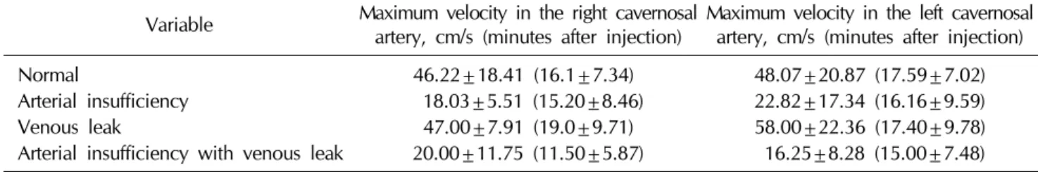
Table 1. Maximum velocities in the cavernosal arteries among patients with normal and abnormal penile Doppler findings (n=97)
Variable Maximum velocity in the right cavernosal
artery, cm/s (minutes after injection) Maximum velocity in the left cavernosal artery, cm/s (minutes after injection)
Normal 46.22±18.41 (16.1±7.34) 48.07±20.87 (17.59±7.02)
Arterial insufficiency 18.03±5.51 (15.20±8.46) 22.82±17.34 (16.16±9.59)
Venous leak 47.00±7.91 (19.0±9.71) 58.00±22.36 (17.40±9.78)
Arterial insufficiency with venous leak 20.00±11.75 (11.50±5.87) 16.25±8.28 (15.00±7.48) Values are presented as mean±standard deviation (mean±standard deviation).
Fig. 1. Causes of erectile dysfunction (n=97).
Fig. 2. Causes of erectile dysfunction with respect to age group.
RESULTS
Out of 97 patients (mean age, 37.09±11.59 years;
range, 19∼69 years), 50 patients (51.5%) had normal penile Doppler ultrasonographic findings (maximum ve- locity in the right cavernosal artery: 46.22±18.41 cm/s at 16.1±7.34 minutes after the injection; maximum velocity in the left cavernosal artery: 48.07±20.87 cm/s at 17.59±
7.02 minutes after the injection), 24 patients (24.7%) had arterial insufficiency (maximum velocity in the right cav- ernosal artery: 18.03±5.51 cm/s at 15.20±8.46 minutes after the injection; maximum velocity in the left cav- ernosal artery: 22.82±17.34 cm/s at 16.16±9.59 minutes after the injection), 15 patients (15.5%) had a venous leak (maximum velocity in the right cavernosal artery: 47.00±
7.91 cm/s at 19.0±9.71 minutes after the injection; max- imum velocity in the left cavernosal artery: 58.00±22.36 cm/s at 17.40±9.78 minutes after the injection), while 8 patients (8.2%) patients had arterial insufficiency with a venous leak (maximum velocity in the right cavernosal ar- tery: 20.00±11.75 cm/s at 11.50±5.87 minutes after the injection; maximum velocity in the left cavernosal artery:
Age was significantly associated with the cause of erec- tile dysfunction (p=0.022) with a higher frequency of psy- chogenic causes in patients aged ≤40 years, while arterial insufficiency with or without a venous leak was higher among patients aged >40 years (Fig. 2).
DISCUSSION
www.wjmh.org
organic from psychological causes. With the advance- ment of the treatment options of male sexual dysfunction, this imaging modality will become important for the eval- uation of the success rates of the treatment.
In this study, penile Doppler ultrasonography revealed a surprisingly high prevalence of psychogenic erectile dysfunction, as indicated by normal penile Doppler ultra- sonography; this was found in the majority of the patients.
This finding is similar to the findings of previous studies that reported a high rate of psychogenic erectile dysfunc- tion using both similar and different techniques [11,12].
In the present study, most of the patients suffering from psychogenic causes were younger than 40 years of age and had no known comorbidity. Upon taking a detailed history, we identified relationship problems after marriage and family pressures as the most important precipitating and maintaining factors for psychogenic erectile dysfunc- tion. The demand for a premarital complete sexual check-up has now increased in our population, and this has led to a significant amount of stress in young men regarding failure to perform intercourse after marriage. In the existing liter- ature, performance anxiety, that is, fear of failure during intercourse, has also been reported as a potential factor re- lated to psychogenic erectile dysfunction. Further, multi- ple developmental, cognitive, affective, and interpersonal factors can predispose men to sexual dysfunction [2].
Among all organic causes, arterial insufficiency was found to be the most common in our study population.
Organic causes of erectile dysfunction were more com- mon in patients aged >40 years. The existing literature al- so states that the various risk factors of erectile dysfunction are associated with penile arterial insufficiency, including hypertension, hyperlipidemia, cigarette smoking, and dia- betes mellitus [13-16]. Such disorders are highly prevalent in our community [17-19]. Moreover, a relatively large number of cases of arterial insufficiency have been re- ported by other researchers [20,21]. Another local study has also reported a relatively large number of cases of arte- rial erectile dysfunction, as compared to venous insuffi- ciency [11].
Further, endothelial dysfunction is the common de- nominator of many vascular risk factors that can lead to ar- teriogenic erectile dysfunction [22,23]. A previous study reported that erectile dysfunction improved when the con-
centrations of the elevated total and low-density lipopro- tein cholesterol, were lowered either by dietary measures or by statin administration [2].
The findings of our study should be noted in the light of the following limitations. First, a psychogenic cause was determined based on the combination of the patient’s his- tory and normal ultrasound findings. We recommend identifying the cause-and-effect relationship of the psy- chogenic causes related to anxiety, depression, and other social factors, and research utilizing standardized ques- tionnaires should be carried out for this purpose. The sec- ond limitation of this study is that the arterial velocities are the maximum near the base of the penis and decrease as they advance; operator dependency may have led to varia- bility in recording the velocities. To minimize this varia- bility, we need to record the velocities near the base of the penis where they are highest. Third, organic causes of erectile dysfunction were identified on the basis of color Doppler findings, and their relationship with risk factors such as hypertension, diabetes, and metabolic syndrome could not be clarified. Various studies have reported dia- betes mellitus, hypertension, hyperlipidemia, metabolic syndrome, depression, and lower urinary tract symptoms to be causes of erectile dysfunction [2,13-16,24-26]. In this study, we were not able to determine the devel- opmental cause of erectile dysfunction in the study population.
However, despite the abovementioned limitations, this study is a significant effort in reporting the outcome of pen- ile Doppler ultrasonography from our developing country.
Further prospective studies are recommended for the eval- uation of causes leading to erectile dysfunction in our population. The higher frequency of psychogenic erectile dysfunction in this study suggests the need of psychosex- ual therapy in our population, particularly in people with significant psychological problems. Moreover, studies should be conducted to monitor the efficacy of psychosex- ual therapy in these patients. A recent systematic review also recommended that while treating erectile dysfunc- tion, clinicians should ascertain the cognitive schemas, personality, sexual abuse history, and sexual expectations of the patients.
CONCLUSIONS
The higher frequency of psychogenic erectile dysfunc- tion in a relatively younger age group suggests the need of psychosexual therapy in our population, particularly in patients with significant psychological problems. In the older age group, the predominant cause of erectile dys- function was vascular problems. Early detection of psy- chogenic and vascular causes can lead to effective treat- ment and will ultimately eventually decrease the rate of in- fertility and depression among the population.
CONFLICT OF INTEREST
No potential conflict of interest relevant to this article was reported.
REFERENCES
1. Juszczak K, Wyczółkowski M, Filipek M, Thor PJ. Patho- physiology of erectile dysfunction in men. Folia Med Cracov 2008;49:67-77.
2. Shamloul R, Ghanem H. Erectile dysfunction. Lancet 2013;
381:153-65.
3. Hatzimouratidis K, Eardley I, Giuliano F, Moncada I, Salonia A. Guidelines on male sexual dysfunction: erectile dysfunction and premature ejaculation. Arnhem: European Association of Urology; 2015.
4. Johannes CB, Araujo AB, Feldman HA, Derby CA, Kleinman KP, McKinlay JB. Incidence of erectile dysfunc- tion in men 40 to 69 years old: longitudinal results from the Massachusetts male aging study. J Urol 2000;163:460-3.
5. Araujo AB, Travison TG, Ganz P, Chiu GR, Kupelian V, Rosen RC, et al. Erectile dysfunction and mortality. J Sex Med 2009;6:2445-54.
6. Golijanin D, Singer E, Davis R, Bhatt S, Seftel A, Dogra V.
Doppler evaluation of erectile dysfunction: part 2. Int J Impot Res 2007;19:43-8.
7. Aversa A, Bruzziches R, Spera G. Diagnosing erectile dys- function: the penile dynamic colour duplex ultrasound revisited. Int J Androl 2005;28 Suppl 2:61-3.
8. Smith JF, Brant WO, Fradet V, Shindel AW, Vittinghoff E, Chi T, et al. Penile sonographic and clinical characteristics in men with Peyronie's disease. J Sex Med 2009;6:2858-67.
9. Golubinski AJ, Sikorski A. Usefulness of power Doppler ul- trasonography in evaluating erectile dysfunction. BJU Int 2002;89:779-82.
11. Bari V, Ahmed MN, Rafique MZ, Ashraf K, Memon WA, Usman MU. Evaluation of erectile dysfunction with color Doppler sonography. J Pak Med Assoc 2006;56:258-61.
12. Yildirim D, Bozkurt IH, Gurses B, Cirakoglu A. A new pa- rameter in the diagnosis of vascular erectile dysfunction with penile Doppler ultrasound: cavernous artery ondula- tion index. Eur Rev Med Pharmacol Sci 2013;17:1382-8.
13. Irekpita E, Salami TA. Erectile dysfunction and its relation- ship with cardiovascular risk factors and disease. Saudi Med J 2009;30:184-90.
14. Hatzichristou D. Understanding individuals' response to erectile dysfunction. Int J Impot Res 2008;20 Suppl 2:
S15-20.
15. Steinke EE, Jaarsma T, Barnason SA, Byrne M, Doherty S, Dougherty CM, et al. Sexual counselling for individuals with cardiovascular disease and their partners: a consensus document from the American Heart Association and the ESC Council on Cardiovascular Nursing and Allied Profes- sions (CCNAP). Eur Heart J 2013;34:3217-35.
16. Phé V, Rouprêt M. Erectile dysfunction and diabetes: a re- view of the current evidence-based medicine and a syn- thesis of the main available therapies. Diabetes Metab 2012;38:1-13.
17. Fawwad A, Siddiqui IA, Basit A, Zeeshan NF, Shahid SM, Nawab SN, et al. Common variant within the FTO gene, rs9939609, obesity and type 2 diabetes in population of Karachi, Pakistan. Diabetes Metab Syndr 2016;10:43-7.
18. Aziz KU. Evolution of systemic hypertension in Pakistani population. J Coll Physicians Surg Pak 2015;25:286-91.
19. Ahmad S, Zhao W, Renström F, Rasheed A, Samuel M, Zaidi M, et al. Physical activity, smoking, and genetic pre- disposition to obesity in people from Pakistan: the PROMIS study. BMC Med Genet 2015;16:114.
20. Coelho MF, Santos PB. Erectile dysfunction of vascular cause: statistical evaluation on the plurimetabolic syndro- me's risk factors and their correlation with penile eco-dop- pler rates. Acta Med Port 2011;24 Suppl 2:379-82.
21. Yavas US, Calisir C, Kaya T, Degirmenci NA. A sign of arte- riogenic insufficiency on penile Doppler sonography: retro- grade flow in penile cavernosal-spongiosal communica- tions. J Ultrasound Med 2007;26:1643-8.
22. Ring JD, Lwin AA, Köhler TS. Endovascular approaches to penile arterial revascularization for vasculogenic erectile dysfunction. In: Köhler TS, McVary KT, editors. Contemporary treatment of erectile dysfunction. New York City: Springer International Publishing; 2016;221-40.
23. Gareri P, Castagna A, Francomano D, Cerminara G, De Fazio P. Erectile dysfunction in the elderly: an old wide- spread issue with novel treatment perspectives. Int J Endocrinol 2014. doi: 10.1155/2014/878670.
24. Brotto L, Atallah S, Johnson-Agbakwu C, Rosenbaum T, Abdo C, Byers ES, et al. Psychological and interpersonal di-
www.wjmh.org J, et al. The Princeton III Consensus recommendations for
the management of erectile dysfunction and cardiovascular disease. Mayo Clin Proc 2012;87:766-78.
26. Boutari C, Doumas M, Manolis AJ. Approach to erectile dys-
function in patients with hypertension and coronary artery disease. In: Andreadis EA, editor. Hypertension and car- diovascular disease. New York City: Springer International Publishing; 2016;309-27.