Received: August 5, 2019 Revised: October 7, 2019 Accepted: November 29, 2019
Address for Correspondence: Byung-Koo Yoon, Department of Obstetrics and Gynecology, Samsung Medical Center, Sungkyunkwan University School of Medicine, 81 Irwon-ro, Gangnam-gu, Seoul 06351, Korea
Tel: 82-2-3410-3519, E-mail: bkyoon@skku.edu, ORCID: https://orcid.org/0000-0002-1326-6102
Address for Correspondence: Chi-Dug Kang, Department of Biochemistry, Pusan National University School of Medicine, 49 Busandaehak-ro, Mulgeum-eup, Yangsan 50612, Korea
Tel: 82-51-510-8082, E-mail: kcdshbw@pusan.ac.kr, ORCID: https://orcid.org/0000-0002-7860-5347 Won-Jong Oh’s current affiliation is Korea Brain Research Institute, Daegu, Korea.
ORIGINAL ARTICLE
INTRODUCTION
Coronary heart disease (CHD) is the leading cause of mortality in women. Although the pathogenesis of CHD has not been fully understood, thrombosis plays a crucial role in the development and progression of
atherosclerosis [1]. Thrombosis is normally balanced by fibrinolysis, which is under refined regulation by tissue-type plasminogen activator (t-PA) and plas- minogen activator inhibitor type 1 (PAI-1) [2]. The t-PA from vascular cells converts plasminogen into plasmin, which degrades the fibrin polymer into small
pISSN: 2288-6478, eISSN: 2288-6761
Effects of 17ββ-Estradiol on the Plasminogen Activator System in Vascular Smooth Muscle Cells Treated with Lysophophatidylcholine
Byung-Koo Yoon1,2, Young-Hee Kang2, Won-Jong Oh2, Dong-Yun Lee1, Duk-Kyung Kim3, Bruce Kessel4, Chi-Dug Kang5
1Department of Obstetrics and Gynecology, Samsung Medical Center, Sungkyunkwan University School of Medicine, Seoul, Korea,
2Samsung Biomedical Research Institute, Samsung Medical Center, Seoul, Korea, 3Department of Medicine, Samsung Medical Center, Sungkyunkwan University School of Medicine, Seoul, Korea, 4Department of Obstetrics and Gynecology, University of Hawaii, Honolulu, HI, USA, 5Department of Biochemistry, Pusan National University School of Medicine, Yangsan, Korea
Objectives: When administered soon after menopause, hormone therapy can prevent coronary heart diseases in women. To explore the mechanism underlying the cardioprotective actions of estrogen, we investigated the effects of 17β-estradiol (17β-E2) on the plasminogen activator system using cultured vascular smooth muscle cells (VSMCs).
Methods: VSMCs were isolated from rat aortas. Protein expression of plasminogen activator inhibitor type 1 (PAI-1) and tissue-type plasminogen activator (t-PA) were evaluated using Western blotting and enzyme-linked immunosorbent assay, respectively. The enzyme activity of PAI-1 in a conditioned medium was assessed via reverse fibrin overlay zymography and that of t-PA was assessed via fibrin overlay zymography. Gene expression was quantified using real-time reverse transcription-polymerase chain reaction.
Results: Following pre-treatment for 24 hours, 17β-E2 suppressed both protein expression and enzyme activity of PAI-1 stimulated by lysophosphatidylcholine (lysoPC) in a significant and dose-dependent manner at a near physiological concentration. Moreover, 17β-E2 (10−7 M) inhibited PAI-1 gene expression, and ICI 182,780—a specific estrogen receptor antagonist—blocked the effects of 17β-E2 on the PAI-1 protein. 17β-E2 did not affect t-PA secretion but significantly enhanced free t-PA activity through reduced binding to PAI-1. Furthermore, 17β-E2 suppressed intracellular reactive oxygen species production and nuclear factor-κB-mediated transcription.
Conclusions: In VSMCs stimulated with lysoPC, 17β-E2 reduced PAI-1 expression through a non-receptor-mediated mechanism via antioxidant activity as well as a receptor-mediated mechanism; however, it did not alter t-PA secretion. Of note, 17β-E2 suppressed PAI- 1 activity and concurrently enhanced t-PA activity, suggesting a beneficial influence on fibrinolysis.
Key Words: Estrogens, Fibrinolysis, Myocytes, smooth muscle, Plasminogen inactivators, Tissue plasminogen activator
fragments, and thus helps to dissolve clots. In contrast, PAI-1 binds to t-PA and inhibits the activation of plas- minogen. Indeed, an increased blood level of PAI-1 is associated with increased risk of CHD [3].
If started early after menopause, menopausal hor- mone therapy (MHT) prevents CHD, one of the late health problems associated with estrogen deficiency af- ter menopause [4,5]. In addition to beneficial systemic impacts on lipid profiles, blood pressure, and glucose metabolism, direct actions of MHT on arteries may be a major mechanism for cardio-protection [6].
Oxidative stress, which induces the oxidative modi- fication of low-density lipoprotein (LDL), is another key component in the development of atherosclerosis.
Oxidized LDL exerts arterial impacts in various ad- verse ways [7]. We reported that lysophosphatidylcho- line (lysoPC), an active component of oxidized LDL, induced PAI-1 expression in vascular smooth muscle cells (VSMCs) [8]. Further lysoPC stimulated the enzyme activity of PAI-1 and inhibited that of t-PA, probably leading to decreased fibrinolysis. The present study was undertaken to investigate the direct effect of 17β-estradiol (E2) on the plasminogen activator (PA) system in cultured VSMCs.
MATERIALS AND METHODS
This study was performed according to materials and methods described previously by our group [8]. Here, the materials and methods will be described briefly.
Materials
Sprague–Dawley rats were purchased from Charles River Japan (Hino, Japan). Dulbeccoʼs modified Eagleʼs medium (DMEM) and DMEM/F-12 without phenol red, fetal bovine serum (FBS), trypsin-ethylenediami- netetraacetic acid (EDTA), and penicillin-streptomycin were purchased from GIBCO BRL (Grand Island, NY, USA). Human plasminogen and bovine fibrinogen were obtained from Enzyme Research Laboratories, Inc. (Swamsea, UK). Human urokinase and bovine thrombin were purchased from Calbiochem (Darm- stadt, Germany). Monoclonal antibody for α-smooth muscle actin was purchased from DAKO (Glostrup, Denmark). 2´,7´-dichlorofluorescin diacetate (DCF- DA) was obtained from Molecular Probes (Eugene, OR, USA) and was dissolved in dimethyl sulfoxide (DMSO). 1,3-bis(4-hydroxyphenyl)-4-methyl-5-[4-(2- piperidinylethoxy)phenol]-1H-pyrazole dihydrochlo-
ride (MPP) and 4-[2-phenyl-5,7-bis(trifluoromethyl) pyrazolo[1,5-a]-pyrimidin-3-yl]phenol (PHTPP) were purchased from Tocris Bioscience (Bristol, UK).
LysoPC and all other chemicals were obtained from Sigma Chemical Co. (St. Louis, MO, USA). E2 was dis- solved in ethanol (EtOH), whereas ICI 182,780, MPP, and PHTPP were dissolved in DMSO. All of the other chemicals were dissolved in water.
Cell culture
VSMCs were isolated from thoracic aortas of 3-month- old Sprague-Dawley rats (160–180 g) using a specific enzyme digestion method and were grown in DMEM/
F-12 (50 : 50) without phenol red, containing antibiot- ics and 10% FBS. The cells were stained positively for α-smooth muscle actin. In order to obtain quiescent cells, the cells were incubated for 48 hours in a defined serum-free (DSF) medium containing insulin (0.5 µM), transferrin (5 mg/mL), and ascorbate (0.2 mM).
This study protocol was reviewed and approved by the Institutional Animal Care and Use Committee (IACUC) of Samsung Biomedical Research Institute (IACUC No.
C-A3-220-2), which has been accredited by the Asso- ciation for Assessment and Accreditation of Laboratory Animal Care International and abides by the guide of the Institute of Laboratory Animal Resources.
Western blot analysis
Cells were lysed for 30 minutes on ice in RIPA buffer (50 mM Tris-HCl [pH, 7.5], 200 mM NaCl, 1% NP-40, 0.5% sodium deoxycholate, and 0.1% sodium dodecyl sulfate [SDS]) containing 1 mM phenylmethanesul- fonyl fluoride. Thirty micrograms of cellular proteins were separated by SDS-PAGE, electrotransferred to a polyvinyldifluoride transfer membrane (Schleicher
& Schuell, Keene, NH, USA), and then blocked and incubated with rabbit polyclonal anti-PAI-1 antibody (American Diagnostica Inc., Stamford, CT, USA). After washing, blots were incubated with anti-rabbit/anti- mouse horseradish peroxidase conjugated secondary antibody (Amersham Biosciences, Buckinghamshire, UK), and the bands were detected with ECL reagents (Amersham Biosciences). β-actin was used as a control.
RNA preparation and real-time reverse transcrip- tase-polymerase chain reaction assay
Total RNA was extracted from cells using the easy- spinTM Total RNA Extraction Kit (Intron Biotechnol- ogy, Seongnam, Korea). Conversion to cDNA was
achieved through the PrimeScriptTM RT Master Mix (Takara Bio USA, Inc., Mountain View, CA, USA).
Real-time reverse transcriptase-polymerase chain re- action (RT-PCR) was carried out using SYBR Premix Ex TaqTM (Takara Bio USA, Inc.). The PCR reactions were run in an Applied Biosystems QuantStudio 6 Flex Real-Time PCR system (Applied Biosystems, Foster City, CA, USA) and the relative expression of PAI-1 was calculated after normalization to glyceraldehyde 3-phosphate dehydrogenase using the difference in the cycle threshold (ΔCt) method. Primers for rat PAI-1 were 5´-CAATGGAAGACCCCCTTCTTAGAG-3´
(forward) and 5´-CATGGGCACGGAGATGGT-3´
(reverse). Primers for rat glyceraldehyde 3-phosphate dehydrogenase were 5´-GTATCGGACGCCTGGT- TACC-3´ (forward) and 5´-TTGATGGCAACAAT- GTCCACTTTG-3´ (reverse).
Enzyme-linked immunosorbent assay
The t-PA levels were quantified using enzyme-linked immunosorbent assay (ELISA; Molecular Innovations Inc., Novi, MI, USA). The color reaction was per- formed with a 3,3´,5,5´-tetramethylbenzidine substrate, and measured at 450 µm by a SmartSpec 3000 spectro- photometer (Bio-Rad, Hercules, CA, USA).
Fibrin overlay zymography/reverse fibrin overlay zymography
Conditioned media (CM) was subjected to electro- phoresis using a 9% polyacrylamide gel containing 0.1%
SDS. After electrophoresis, the gel was washed twice in 2.5% Triton X-100 for 1 hour to remove the SDS.
In order to detect PA activity with the fibrin overlay assay, the gel was placed on an opaque fibrin indica- tor gel containing 1% low-gelling-temperature agarose (FMC Corporation, Philadelphia, PA, USA), human plasminogen (12.5 µg/mL), bovine thrombin (0.5 U/
mL), and bovine fibrinogen (2 mg/mL). Afterwards, this combined gel (polyacrylamide/fibrin indicator) was incubated at 37°C, and PA activity was detected by the appearance of clear bands in the opaque indicator gel. In addition, activity of PA inhibitor was detected by a reverse overlay assay, which is similar to the fibrin overlay assay described above, except that it also con- tained human urokinase (1.5 U/mL). Since the fibrin in the reverse indicator gel was lysed in the absence of PAI, opaque bands on the indicator gel indicated the presence of PAI.
Analysis of intracellular formation of reactive oxy- gen species
Intracellular free radical production was determined using DCF-DA. To analyse the reactive oxygen spe- cies (ROS) formation quantitatively, flow cytometry analysis was used. After incubation of the quiescent cells with 10 µM DCF-DA and 5 µM lysoPC, the cells were trypsinized and resuspended in PBS containing 1 mM EDTA, and then analyzed immediately by flow cy- tometry (10,000 cells/sample) using a beam of 488 nm excitation. The median intensity of fluorescence was determined by CellQuest software (Becton-Dickinson, Franklin Lakes, NJ, USA).
Determination of nuclear factor-κB activation Activation of nuclear factor-κB (NF-κB) was deter- mined in the VSMCs after transfection with a reporter plasmid containing the luciferase reporter gene linked to five repeats of the NF-κB binding sites. The VSMCs (1 × 10⁵ cells/well) were plated in 24-well plates and grown to about 70% confluence. Cells were then tran- siently cotransfected with 1 µg of NF-κB–luciferase reporter plasmid and 1 µg of β-galactosidase plasmid using Lipofectamine plus (Invitrogen, Carlsbad, CA, USA). At 6 hours post-transfection, cells were starved for 48 hours before stimulation with lysoPC. Transfect- ed cells were exposed to 5 µM lysoPC for the indicated time periods. Luciferase activity was measured using a luciferase assay kit (Promega, Madison, WI, USA) with signal detection for 5 seconds in a luminometer (Panomics Inc., Fremont, CA, USA). A β-galactosidase enzyme assay (Promega) was used to determine the β-galactosidase activity with a SmartSpec 3000 spec- trophotometer at 420 nm. The results are expressed relative to the NF-κB activity compared with con- trols after normalizing for β-galactosidase activity and protein concentration.
Data analysis and statistics
Data are presented as mean ± standard error of the mean, and N is the number of performed experiments.
The Kruskal–Wallis test and Wilcoxon rank sum test were carried out for statistical analysis using Statistics Package for Social Sciences Version 20.0 (IBM Corp., Armonk, NY, USA). A two-tailed value of P < 0.05 was considered statistically significant.
0 10
8
6
4
2
Bandintensity(foldoverDSFmedium)
Time (h) 0
4 8 12 24
DSF medium LysoPC
a
b
c
d e
e
e
a,b,c,d
LysoPC (5 M)
PAI-1
-actin
0 4 8 12 24 4 8 12 24 h
n = 5
DSF medium 40
30
20
10
ExpressionlevelofPAI-1mRNA (foldoverDSFmedium)
LysoPC (5 M, 4 h) 0
a
n = 4
Alone EtOH
(0.1%)
Amountofsecretedt-PAprotein(ng/mL)
a
e
0 60
50
40
30
20
10
0
n = 3
4 8 12 24
DSF medium LysoPC (5 M)
Time (h)
A
DSF medium 7 6 5 4 3 2 1
Bandintensity(foldoverDSFmedium)
0
EtOH (0.1%)
PAI-1
-actin 0.1 1 2.5 5 7.5 10
n = 5
Alone EtOH (0.1%)
0.1 1 2.5 5 7.5 10
LysoPC (5 M, 8 h)
LysoPC (5 M)
a
a b,c
b
c
B
C D
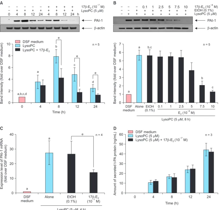
Fig. 1. Effects of 17β-estradiol (E2) on the plasminogen activator system in lysophosphatidylcholine (LysoPC, 5 µM)-treated vascular smooth muscle cells after 24-hour pre-treatment. (A) Time-course effects and (B) dose-response effects of 17β-E2 on plasminogen activator inhibitor type 1 (PAI-1) in cell lysates as determined by Western blot analysis. Representative blots are shown in the upper section of each panel. The intensity of the bands was densitometrically determined and normalized to that of corresponding β-actin bands. Graphic data are depicted in the lower part of the panel. (C) Effects of 10–7 M 17β-E2 on gene expression of PAI-1 as evaluated by real-time reverse transcriptase-polymerase chain reaction. (D) Time-course effects of 10–7 M 17β-E2 on secreted tissue-type plasminogen activator (t-PA) in the conditioned media as measured by enzyme-linked immunosorbent assay. Data are expressed as mean ± standard error of the mean. DSF: defined serum-free, EtOH: ethanol. a, b, c, d, eP < 0.05.
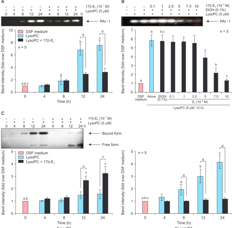
Fig. 2. Effects of 17β-estradiol (E2) on enzymatic activities of the plasminogen activator system in the conditioned media harvested from cultured vascular smooth muscle cells stimulated with lysophosphatidylcholine (LysoPC, 5 µM) after 24-hour pre-treatment. Representative zymographs are shown in the up- per section of each panel. The intensity of the bands was densitometrically determined, and graphic data are depicted in the lower part of the panel. (A) Time- course effects and (B) dose-response effects of 17β-E2 on plasminogen activator inhibitor type 1 (PAI-1) as determined by reverse fibrin overlay zymography.
(C) Time-course effects of 10–7 M 17β-E2 on tissue-type plasminogen activator (t-PA) as examined by fibrin overlay zymography. Data are expressed as mean
± standard error of the mean. DSF: defined serum-free, EtOH: ethanol. a, b, c, dP < 0.05.
0 4 8 12 24
10
8
6
4
2
Bandintensity(foldoverDSFmedium)
Time (h) 0
A B
a,b,c
a
b
d c d
n = 5
LysoPC (5 M)
PAI 1 0
+ 4
+ 8
+ 12
+ 24
++ 4
++ 8
++ 12
++ 24 h
DSF
medium 0.1 1 2.5 5 7.5 10
7 6 5 4 3 2 1
Bandintensity(foldoverDSFmedium)
0 a
n = 5 EtOH (0.1%) LysoPC (5 M)
PAI 1 +
+ +
0.1 + +
1 + +
2.5 + +
5 + +
7.5 + +
10 + +
Alone EtOH (0.1%)
LysoPC (5 M, 12 h)
a b,c
b
c
C
0 4 8 12 24
5
4
3
2
1
Bandintensity(foldoverDSFmedium)
Time (h) 0
a,b
d
b d
LysoPC (5 M)
Bound form 0
+ 4
+ 8
+ 12
+ 24
++ 4
++ 8
++ 12
++ 24 h
Free tPA
a
0 4 8 12 24
5
4
3
2
1
Bandintensity(foldoverDSFmedium)
Time (h) 0
a,b,c
b
c
Bound tPA a n = 5
d
d Free form d
RESULTS
17β-estradiol inhibited lysophophatidylcholine- induced expression of plasminogen activator in- hibitor type 1 protein and mRNA with no change in tissue-type plasminogen activator secretion in the vascular smooth muscle cell
Our group had previously shown that lysoPC signifi- cantly increased PAI-1 expression in the VSMC at 5 µM
or higher concentration of lysoPC in a dose-dependent manner [8]. In Western blot analysis, co-treatment of 17β-E2 did not alter PAI-1 expression stimulated with lysoPC (data not shown). We then tested the effects of 17β-E2 after 24 hours of pre-treatment. In a time- course study, 10–7 M of 17β-E2 decreased PAI-1 expres- sion induced with 5 µM lysoPC, reaching statistical significance after 8 hours (Fig. 1A). EtOH (0.1%), a vehicle, did not change PAI-1 expression. Compared to
DSF medium 4
3
2
1 IntracellularlevelofROS (foldoverDSFmedium)
LysoPC (5 M, 2 h) 0
a n = 5
Alone EtOH
(0.1%) a
A b B
DSF medium 3
2
1
TranscriptionalactivityNF-B (foldoverDSFmedium)
LysoPC (5 M, 2 h) 0
a n = 4
Alone EtOH
(0.1%) a
b
DSF medium 10
8
6
4
2
Bandintensity(foldoverDSFmedium)
0
DMSO (0.1%)
PAI-1
-actin
n = 4 LysoPC (5 M)
a
C
LysoPC alone
DMSO (0.1%) a
b
b b ICI MPP PHTPP
Fig. 3. Mechanisms of 17β-estradiol (E2, 10–7 M) action in lysophosphatidyl- choline (LysoPC, 5 µM)-treated vascular smooth muscle cells after 24-hour pre-treatment. (A) Effects of 17β-E2 treatment for 2 hours on intracellular formation of reactive oxygen species (ROS) as assessed by flow cytometry analysis using 2´,7´-dichlorofluorescin diacetate. (B) Effects of 17β-E2 for 2 hours on nuclear factor-κB (NF-κB)-mediated transcriptional activity as measured by luciferase reporter assay. (C) Effects of estrogen receptor antagonists on 17β-E2 action for 8 hours as determined by Western blot analysis. Representative blots are shown in the upper section. The intensity of the bands was densitometrically determined and normalized to that of corresponding β-actin bands. Graphic data are shown in the lower part.
Data are expressed as mean ± standard error of the mean. DSF: defined serum-free, EtOH: ethanol, DMSO: dimethyl sulfoxide, ICI: ICI 182,780, MPP:
1,3-bis(4-hydroxyphenyl)-4-methyl-5-[4-(2-piperidinylethoxy)phenol]-1H- pyrazole dihydrochloride, THTPP: 4-[2-phenyl-5,7-bis(trifluoromethyl)
vehicle-treated controls, PAI-1 expression was reduced in a dose-dependent manner when checked at 8 hours of treatment with lysoPC, and significant reductions were observed at 7.5 × 10⁻⁸ M or a higher concentration of 17β-E2 (Fig. 1B). The effect of 17β-E2 was also inves- tigated at the level of gene expression after 24 hours of pre-treatment. We had previously reported maximal expression of the PAI-1 gene at 4-hour treatment of lysoPC [8]. EtOH (0.1%) had no influence on PAI-1 gene expression. Compared to vehicle-treated controls, lysoPC-induced expression of PAI-1 mRNA at 4 hours was significantly down-regulated with 10–7 M 17β-E2, as assessed by RT-PCT (Fig. 1C).
In addition, t-PA change in CM was analysed by ELISA. We have reported that neither production nor secretion of t-PA was altered by lysoPC [8]. Constitu- tive secretion of t-PA into the CM was undetectable and increased with time following lysoPC treatment.
t-PA secretion was not affected by 10–7 M 17β-E2 after pre-treatment for 24 hours (Fig. 1D). These results sug- gest that 17β-E2 inhibits lysoPC-induced expression of PAI-1 protein and mRNA without influencing t-PA secretion in the VSMC.
17β-estradiol suppressed enzyme activity of plas- minogen activator inhibitor type 1 and enhanced that of free tissue-type plasminogen activator in the conditioned media
Time-course experiments using reverse fibrin over- lay zymography demonstrated that PAI-1 activity in response to 5 µM lysoPC was significantly reduced after 12 hours by pre-treatment with 17β-E2 (10–7 M) for 24 hours (Fig. 2A). In dose-response experiments performed at 12 hours after lysoPC treatment, EtOH (0.1%) did not affect PAI-1 activity. In comparison to vehicle-treated controls, 17β-E2 treatment suppressed PAI-1 activity in a dose-dependent fashion. Significant reductions were observed at 7.5 × 10⁻⁸ M and higher concentrations of 17β-E2 (Fig. 2B).
Enzyme activity of t-PA was also examined using fibrin overlay zymography (Fig. 2C). As reported previ- ously [8], lysoPC increased higher molecular weight complexes after 8 hours, which are presumed to be inactive t-PA bound to PAI-1. Parallel to the change in bound form, free t-PA activity was barely detected with lysoPC. After 24-hour pre-treatment, 17β-E2 (10–7 M) significantly reversed t-PA responses: bound-form activity was significantly lowered after 8 hours, while free-form activity was elevated after 12 hours. These
results suggest that 17β-E2 suppresses enzymatic activ- ity of PAI-1 possibly through downregulation of PAI-1 levels and consequently leads to an increased free form of t-PA in the CM.
Mechanisms of 17β-estradiol action
LysoPC increases PAI-1 expression via oxidative stress and downstream signaling involving NF-κB-mediated transcriptional activity [8]. Therefore, the influence on intracellular ROS production was explored first. EtOH (0.1%) did not change ROS production in response to 2-hour 5 µM lysoPC treatment. Compared to vehicle- treated controls, the increased ROS production by lysoPC was significantly reduced by pre-treatment with 17β-E2 (10–7 M) for 24 hours (Fig. 3A). While NF- κB activity after 2-hour treatment with 5 µM lysoPC was not altered with EtOH (0.1%), 17β-E2 suppressed the lysoPC-stimulated NF-κB activity, as compared to vehicle-treated control (Fig. 3B).
Estrogen receptors (ERs) α and β are expressed in rat VSMCs [9]. Whether 17β-E2 action is mediated by ERs was also examined (Fig. 3C). DMSO (0.1%) did not affect PAI-1 protein expression in response to 17β- E2. ICI 182,780 (10–6 M), an antagonist against both ERs α and β, significantly abolished 17β-E2 responses as compared to vehicle-treated controls. In particu- lar, MPP (10–6 M), a specific antagonist against ERα, significantly restored PAI-1 expression suppressed by treatment with 17β-E2 (10–7 M). Meanwhile, PHTPP (10–6 M), a specific antagonist against ERβ, showed a similar blocking effect without statistical significance.
These results suggest that 17β-E2 reduces PAI-1 expres- sion via receptor-mediated and non-receptor-mediated mechanisms including inhibition of NF-κB, which can be activated by lysoPC.
DISCUSSION
This study was carried out to examine the direct ef- fects of 17β-E2 on the PA system in cultured VSMCs.
We report that 17β-E2 diminished lysoPC-induced PAI-1 protein and activity levels and concomitantly promoted t-PA activity in VSMCs.
Higher PAI values are found in myocardial infarction and re-infarction patients [3]. Further, elevated blood PAI-1 levels have a causal effect on CHD risk [10]. Es- trogen therapy decreases blood PAI antigen and activity and increases release of active t-PA in postmenopausal women [11]. Estrogen is reported to decrease PAI-
1 production in hepatocytes [12] and adipose tissue [13]. In the present study, we focused on the PA system within the artery.
In CHD, local fibrinolysis systems are critically im- portant for arterial thrombosis in response to endovas- cular injury. As well as in mouse [14], the level of PAI-1 mRNA in human arteries is correlated with the degree of atherosclerosis [15]. Although PAI-1 expression is detected primarily in vascular endothelial cells (VECs) of healthy arteries, PAI-1 is also found in intimal VSMCs in early atherosclerotic lesions and the fibrous cap in advanced atheromatous plaques [16]. Accord- ingly, direct modulation of the PA system in VSMCs might be more clinically relevant than in VECs.
We have reported that lysoPC might negatively affect fibrinolysis in VSMCs by increasing protein expression and activity of PAI-1 and decreasing enzyme activity of t-PA [8]. The present study demonstrated that after 24-hour pre-treatment, 17β-E2 suppressed PAI-1 pro- tein expression and activity in VSMCs stimulated with lysoPC in a dose-dependent fashion. These effects of estrogen occurred at near a physiologic concentration of 7.5 × 10⁻⁸ M. PAI-1 mRNA levels were also down- regulated by 17β-E2, which was restored by ICI 182,780.
These observations support a receptor-mediated effect of estrogen. In the present study, specific antagonists against ERα (MPP) or β (PHTPP) showed a similar trend of inhibition, even though ERα antagonist ef- fects alone reached statistical significance. In contrast, opposing regulation of PAI-1 promoter activity by two ER isoforms was reported in VECs: ERα activated, whereas ERβ suppressed the promoter [17]. Further study is needed to investigate the underlying molecu- lar mechanisms for differential control of PAI-1 gene expression by estrogen in vascular cells. This study also showed that PAI-1 activity in the CM was inhibited in parallel with the reduction of PAI-1 protein levels. On the whole, 17β-E2 suppressed lysoPC-induced PAI-1 in VSMCs at all levels of mRNA, protein and enzyme activity.
LysoPC induces PAI-1 expression in VSMCs via oxi- dative stress [8]. As for further mechanisms of action, 17β-E2 rapidly reduced intracellular ROS production in the present study, which reproduced previous data pub- lished by our group [9]. Moreover, 17β-E2 suppressed NF-κB activity, one of major redox-sensitive signal pathways [18]. These findings support the antioxidant effects of estrogen.
With respect to t-PA, lysoPC does not affect t-PA pro-
duction and secretion in VSMCs [8]. We did not ob- serve a change in t-PA secretion with 17β-E2 but dem- onstrated an increase in free t-PA activity concomitant with a decrease in bound-form activity in the CM. This increase is likely due to inhibited PAI-1 synthesis by 17β-E2 in VSMCs.
To our knowledge, this is the first study to report favorable impacts of 17β-E2 on PA system in VSMCs.
Besides, positive influences of 17β-E2 on PA system in VECs are also published. 17β-E2 upregulated mRNA expression and activity of t-PA [19] and decreased secretion of PAI-1 [20,21]. Taken together, estrogen might enhance vascular fibrinolytic function, which serves as an important mechanism for cardio-protec- tion with postmenopausal estrogen therapy.
On the other hand, PAI-1 plays a crucial role in vas- cular remodeling. PAI-1 is involved in pathological intimal hyperplasia [22]. PAI-1 also reduces VSMC mi- gration, likely resulting in a thinned fibrous atheroma cap [2]. Further, PA system is implicated in vascular in- flammation. Plasmin can amplify the inflammatory re- sponse of monocyte [23]. Beneficial effects of estrogen on PAI-1 in VSMCs demonstrated in this study might contribute to attenuating atherosclerosis progression including neointima formation and plaque instability.
Additional studies are required to better understand how estrogen modulates the PA system in CHD.
Although using VSMCs derived from rat aortas is an established in vitro model, cells from human coronary arteries might be more appropriate for further experi- ments to study the direct vascular effects of estrogen.
In conclusion, 17β-E2 inhibits PAI-1 expression via non-receptor mediated mechanism by antioxidant activity and receptor-mediated mechanism, but does not alter t-PA secretion in VSMCs treated with lysoPC.
Importantly, 17β-E2 suppresses PAI-1 activity and con- currently enhances t-PA activity, suggesting a beneficial influence on fibrinolysis.
ACKNOWLEDGMENTS
This work was supported in part by the Sungkyunk- wan University Foundation for Corporate Collabora- tion (S-2008-0021-000 and S-2010-0228-000), Sam- sung Biomedical Research Institute grant (#SBRI C-95- 036 and C-A3-220-2), and the IN-SUNG Foundation for Medical Research (C-A4-815-1).
CONFLICT OF INTEREST
No potential conflict of interest relevant to this article was reported.
REFERENCES
1. Ibrahim H, Kleiman NS. Platelet pathophysiology, pharmacology, and function in coronary artery disease. Coron Artery Dis 2017;
28: 614-23.
2. Sobel BE, Taatjes DJ, Schneider DJ. Intramural plasminogen acti- vator inhibitor type-1 and coronary atherosclerosis. Arterioscler Thromb Vasc Biol 2003; 23: 1979-89.
3. Ilić M, Majkić-Singh N, Lalić K. Plasminogen activator inhibitor in patients with acute myocardial infarction and re-infarction in syndrome X. Clin Lab 2002; 48: 125-8.
4. Rossouw JE, Prentice RL, Manson JE, Wu L, Barad D, Barnabei VM, et al. Postmenopausal hormone therapy and risk of cardio- vascular disease by age and years since menopause. JAMA 2007;
297: 1465-77.
5. Boardman HM, Hartley L, Eisinga A, Main C, Roqué i Figuls M, Bonfill Cosp X, et al. Hormone therapy for preventing cardiovas- cular disease in post-menopausal women. Cochrane Database Syst Rev 2015; (3): CD002229.
6. Mendelsohn ME, Karas RH. Molecular and cellular basis of car- diovascular gender differences. Science 2005; 308: 1583-7.
7. Koenig W, Karakas M, Zierer A, Herder C, Baumert J, Meisinger C, et al. Oxidized LDL and the risk of coronary heart disease: results from the MONICA/KORA Augsburg Study. Clin Chem 2011; 57:
1196-200.
8. Yoon BK, Kang YH, Oh WJ, Park K, Lee DY, Choi D, et al. Impact of lysophosphatidylcholine on the plasminogen activator system in cultured vascular smooth muscle cells. J Korean Med Sci 2012;
27: 803-10.
9. Yoon BK, Oh WJ, Kessel B, Roh CR, Choi D, Lee JH, et al. 17Beta- estradiol inhibits proliferation of cultured vascular smooth muscle cells induced by lysophosphatidylcholine via a nongenomic anti- oxidant mechanism. Menopause 2001; 8: 58-64.
10. Song C, Burgess S, Eicher JD, O'Donnell CJ, Johnson AD. Causal effect of plasminogen activator inhibitor type 1 on coronary heart disease. J Am Heart Assoc 2017; 6: e004918.
11. Pretorius M, van Guilder GP, Guzman RJ, Luther JM, Brown NJ.
17Beta-estradiol increases basal but not bradykinin-stimulated re- lease of active t-PA in young postmenopausal women. Hyperten- sion 2008; 51: 1190-6.
12. Kilbourne EJ, Scicchitano MS. The activation of plasminogen
activator inhibitor-1 expression by IL-1beta is attenuated by estro- gen in hepatoblastoma HepG2 cells expressing estrogen receptor alpha. Thromb Haemost 1999; 81: 423-7.
13. He G, Pedersen SB, Bruun JM, Richelsen B. Regulation of plas- minogen activitor inhibitor-1 in human adipose tissue: interaction between cytokines, cortisol and estrogen. Horm Metab Res 2000;
32: 515-20.
14. Schafer K, Müller K, Hecke A, Mounier E, Goebel J, Loskutoff DJ, et al. Enhanced thrombosis in atherosclerosis-prone mice is asso- ciated with increased arterial expression of plasminogen activator inhibitor-1. Arterioscler Thromb Vasc Biol 2003; 23: 2097-103.
15. Schneiderman J, Sawdey MS, Keeton MR, Bordin GM, Bernstein EF, Dilley RB, et al. Increased type 1 plasminogen activator inhibi- tor gene expression in atherosclerotic human arteries. Proc Natl Acad Sci U S A 1992; 89: 6998-7002.
16. Lupu F, Bergonzelli GE, Heim DA, Cousin E, Genton CY, Bach- mann F, et al. Localization and production of plasminogen acti- vator inhibitor-1 in human healthy and atherosclerotic arteries.
Arterioscler Thromb 1993; 13: 1090-100.
17. Smith LH, Coats SR, Qin H, Petrie MS, Covington JW, Su M, et al.
Differential and opposing regulation of PAI-1 promoter activity by estrogen receptor alpha and estrogen receptor beta in endothelial cells. Circ Res 2004; 95: 269-75.
18. Kabe Y, Ando K, Hirao S, Yoshida M, Handa H. Redox regulation of NF-kappaB activation: distinct redox regulation between the cytoplasm and the nucleus. Antioxid Redox Signal 2005; 7: 395- 403.
19. Zhang Y, Zhu G, Duan Y. [Effect of 17 beta-estradiol on activity and gene expression of fibrinolytic factor in human umbilical vein endothelial cell]. Zhongguo Yi Xue Ke Xue Yuan Xue Bao 1999;
21: 326-30. Chinese.
20. Sobel MI, Winkel CA, Macy LB, Liao P, Bjornsson TD. The regulation of plasminogen activators and plasminogen activator inhibitor type 1 in endothelial cells by sex hormones. Am J Obstet Gynecol 1995; 173(3 Pt 1): 801-8.
21. Mueck AO, Seeger H, Wallwiener D. Medroxyprogesterone ac- etate versus norethisterone: effect on estradiol-induced changes of markers for endothelial function and atherosclerotic plaque characteristics in human female coronary endothelial cell cultures.
Menopause 2002; 9: 273-81.
22. Ji Y, Weng Z, Fish P, Goyal N, Luo M, Myears SP, et al. Pharma- cological targeting of plasminogen activator inhibitor-1 decreases vascular smooth muscle cell migration and neointima formation.
Arterioscler Thromb Vasc Biol 2016; 36: 2167-75.
23. Foley JH. Plasmin(ogen) at the nexus of fibrinolysis, inflamma- tion, and complement. Semin Thromb Hemost 2017; 43: 135-42.