COMPARISON BETWEEN TIUNITE
TMAND ANOTHER OXIDIZED IMPLANT USING THE RABBIT TIBIA MODEL
In-Sung Yeo1, D.D.S., M.S.D., Jai-Bong Lee2, D.D.S., M.S.D., Ph.D.,
Jung-Suk Han3, D.D.S, M.S.D., Ph.D., Sung-Hun Kim4, D.D.S., M.S.D., Ph.D., Jae-Ho Yang5, D.D.S., M.S.D., Ph.D.
1Graduate student, Department of Prosthodontics, School of Dentistry, Seoul National University
2Professor, Department of Prosthodontics, School of Dentistry, Seoul National University
3Associate Professor, Department of Prosthodontics, School of Dentistry, Seoul National University
4Assistant Professor, Department of Prosthodontics, School of Dentistry, Seoul National University
5Professor, Department of Prosthodontics, School of Dentistry, Seoul National University
Statement of problem. Various anodic oxidation techniques can be applied to dental implant surfaces. But the condition for optimal anodized surfaces has not been described yet.
Purpose.The purpose of this investigation was to compare an implant that was oxidized by another method with TiUniteTMthrough resonance frequency analysis and histomorphome- try.
Material and methods. Turned (control), TiUniteTMand another oxidized fixtures, which used Ca2+solution for anodic oxidation, were placed in the tibiae of 5 New Zealand White rabbits.
The bone responses were evaluated and compared by consecutive resonance frequency analysis once a week for 6 weeks and histomorphometry after a healing period of 6 weeks.
Results.At the first week, both oxidized implants showed significantly higher implant sta- bility quotient (ISQ) values than the control. No significant differences in resonance fre- quency analysis were found between the two oxidized groups for 6 weeks. The means and stan- dard deviations of bone-to-implant contact (BIC) ratios were 71.0 ± 4.2 for TiUniteTM, 67.5 ± 10.3 for the Ca2+-based oxidation fixture, 22.8 ± 6.5 for the control. Both oxidized implants were significantly superior in osseointegration to the turned one. There was, however, no statisti- cally significant difference between the two oxidized implants.
Conclusion.TiUniteTMand the Ca2+-based oxidation fixture showed superior early bone response than the control with respect to resonance frequency analysis and histomorphometry. No sig- nificant differences between the oxidized groups, however, were found in this investigation using the rabbit tibia model.
Key Words
Anodic oxidation, Surface modification, Dental implant, Resonance frequency analysis, Histomorphometry
J Korean Acad Prosthodont : Volume 45, Number 3, 2007
D
ental implants have been an excellent and popular treatment option in restoring singly, partially and fully edentulous areas. However, the dental implant material, commercially pure tita- nium itself requires much time for osseointe- gration. In order to achieve better and more rapid bone response to implant surfaces, various surface modifications have been introduced1-3. Anodic oxidation is one of them.Machined titanium implants with anodized poly-crystalline, thick oxide and a microporous roughness and structures on the submicro- and micrometer level (TiUniteTM, NobelBiocare, Gthenburg, Sweden) have shown good experi- mental results4-6. But the optimum surface chem- istry about anodic oxidation has not been con- firmed and is still under study7-8. In another oxi- dized implant, which used Ca2+solution for anodic oxidation (Osstem Co., Pusan, Korea), the chemical composition of the solution and other conditions for oxidation are known to be slightly different from those of TiUniteTMalthough the exact formulation including voltage and cur- rent is not known. The surface properties of the oxide such as thickness, microstructure, and composition depend on different process para- meters including electrolyte composition, anod- ic potential, electric current, temperature, and electrode geometry9. The purpose of this inves- tigation is to test and compare biocompatibilities between two differently oxidized dental implants using the rabbit tibia model.
MATERIAL AND METHODS
Thirty threaded and turned titanium (grade IV) dental implants with a length of 7 mm and a diameter of 3.75 mm were used in this study. The implants were divided into 3 groups: TiUniteTM, the Ca2+-based oxidation, and turned (control)
implants.
This investigation using animals was approved by the Animal Research Committee of Seoul National University (approved number: SNU- 061003-1) and all experimentation was done in accordance with the Institute of Laboratory Animal Resources (ILAR) guidelines of Seoul National University (SNU). Five New Zealand White rabbits, weighing 3.0 to 3.5 kg, were used in this investigation. Prior to surgery, the shaved skin in the proximal tibial area was washed with Betadine and preoperative antibiotics, 0.12 g kanamycin IM, was administered prophylactically.
Rabbits were anesthetized with a combination of ketamine (28.8 mg/kg) and xylazine (11.7 mg/kg) intramuscularly. Local anesthesia with 1.8 ml of 2% lidocaine was administered in the regions planned for surgery. The proximal aspect of each tibia was surgically exposed via skin incision and the muscles were dissected to allow elevation of the periosteum. The flat surface on the medi- al aspect of the proximal tibia was selected for implant placement. The implantation holes were drilled with a low rotational speed, profuse saline irrigation and with successively increasing diameters, no countersink preparation, and final- ly tapped with a 3.3 mm tap. Three types of implant were installed at each tibia. The implants penetrated the first cortical layer only, with two threads visible above the cortex. The periosteum and fascia were sutured with chromic gut and the skin was sutured with silk. Each rabbit recovered without complications and received 0.06 g kanamycin IM per day for three days postoper- atively.
Huang et al10concluded that resonance fre- quency analysis (RFA) was a reliable and accurate method for early assessment of the osseointe- gration process with respect to implant stability.
For RFA, the rabbits were anesthetized with the same method above once a week for six weeks. The
suture silk was removed and only the skin was dis- sected. Then, implant stability quotient (ISQ) values were measured by OsstellTM(Integration Diagnostics Ltd., Gteborgsvgen, Sweden). The skin was resutured with silk after measurement.
At the sixth week, all the rabbits were anes- thetized, the ISQ values were measured and then the rabbits were sacrificed in a carbon diox- ide chamber. The fixtures were surgically removed en bloc with an adjacent bone collar and imme- diately fixed in 4% neutral formaldehyde. The spec- imens were processed to be embedded in light-cur- ing resin (Technovit 7200 VLC, Kultzer, Wehrheim, Germany). Un-decalcified, cut and ground sections were prepared using the Exakt�system (Exakt Apparatebau, Norderstedt, Germany) based on a method described by Donath11. The specimens were ground to an approximate thickness of 30 μm and stained with hematoxylin and eosin (HE- staining). An IBM personal computer connected to an Olympus BX microscope (Olympus, Tokyo, Japan) and image analysis software (Image Analysis, Bildanalysis, Stockholm, Sweden) was used for calculating the percentage of bone-to- implant contact (BIC). All light microscopic cal- culations were made with a 10 objective and 10 eye- pieces. The highest percentage of BIC in the two consecutive threads was calculated.
The statistical significance of the differences in RFA and BIC ratio among the groups was assessed by one-way ANOVA and Scheffe’s
post hoc analysis. Values of P < .05 were considered to be statistically significant.
RESULTS
All rabbits recovered from anesthesia. Slight inflammation at skin was found during the mea- surement for RFA but neither inflammation nor infection of the bone was noted.
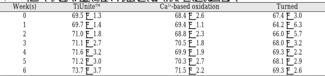
Fig. 1 shows the means of ISQ values for each implant group. The ISQ values for TiUniteTM kept increasing for 6 weeks while those for the con- trol group decreased at the first week of healing (Fig. 1 and Table I). Both the anodized implants showed significantly higher ISQ values than the control at the healing period of the first week (P
< .05). There was almost no significant differ-
Table I. The means and standard deviations of ISQ values for each group
Week(s) TiUniteTM Ca2+-based oxidation Turned
0 69.5 ± 1.3 68.4 ± 2.6 67.4 ± 3.0
1 69.7 ± 1.4 69.4 ± 1.1 64.2 ± 6.3
2 71.0 ± 1.8 68.8 ± 2.3 66.0 ± 5.7
3 71.1 ± 2.7 70.5 ± 1.8 68.0 ± 3.2
4 71.6 ± 3.2 69.9 ± 1.9 69.3 ± 2.2
5 71.2 ± 3.0 70.3 ± 2.7 68.1 ± 2.9
6 73.7 ± 3.7 71.5 ± 2.2 69.3 ± 2.6
Fig. 1. The means of ISQ values for each implant group.
ence between TiUniteTMand the Ca2+-based oxi- dation fixtures. However, because TiUniteTM showed significantly higher ISQ values than the control (P < .05) at the second and sixth weeks, while the Ca2+-based oxidation and the control implants showed no significant difference, TiUniteTM was considered to be slightly superior in RFA to the Ca2+-based oxidation fixture although nei-
ther of the anodized groups was found to be significantly different in resonance frequency measurements from each other.
The means and standard deviations of bone-to- implant contact (BIC) ratios were 71.0 ± 4.2 for TiUniteTM, 67.5 ± 10.3 for the Ca2+-based oxidation group, and 22.8 ± 6.5 for the control (Fig. 2 and Table II). TiUniteTM and the Ca2+-based oxida- tion fixtures showed better osseointegration (P <
.05) than the turned implant (Fig. 3). There was no significant difference in BIC between the two anodized groups.
DISCUSSION
The resonance frequency analysis told that TiUniteTMwas superior in early implant stability and maintenance of it. The Ca2+-based oxidation implant showed similar but more various results than TiUniteTM although both the anodized implants reported better resonance frequency values than the control group. Slightly fluctuat- Table II. The means and standard deviations of BIC ratios for each group
TiUniteTM Ca2+-based oxidation Turned
BIC ratio (%) 71.0 ± 4.2 67.5 ± 10.3 22.8 ± 6.5
Fig. 3. The means and standard deviations of bone-to- implant contact ratio for each implant group.
(a) (b) (c)
Fig. 2. Histologic views at 100 magnification for each implant group: (a) TiUniteTM(b) Ca2+-based oxidation (c) Turned.
ed ISQ values were reported in the Ca2+-based oxi- dation fixture, which indicated such an implant system can have a problem in early bone response and achieving proper stability. More investigations about biocompatibility, implant designs and sur- face characteristics seem to be required.
Although the means and standard deviations of BIC ratios were slightly different maybe because of the differences in experimental conditions and healing periods, this investigation reported the results that were similar to those of others5-6. Both the anodized implants showed similar bone- to-implant contact ratios that were significantly superior to the control after six weeks of healing.
For RFA, however, TiUniteTMwas thought to be better than the Ca2+-based oxidation group, con- sidering ISQ value-comparisons with the con- trol in spite that there was no significant difference between the two oxidized implants, as men- tioned above. Implant stability is an important fac- tor for implant success. Some authors10,12-13report- ed that osseointegration was related to implant sta- bility. But this experiment indicated that other fac- tors than surface modification can be important to keep the fixture stable. Sul, et al8discussed var- ious conditions for anodic oxidation and tried to find the optimum surface properties of oxidized implant because surface characteristics of dental implants directly influenced osseointegration.
Therefore, more studies about factors to con- tribute to implant stability, optimal surface prop- erties of anodic oxidation and the relation between surface characteristics and clinical meanings are needed.
CONCLUSION
This in vivo investigation concluded the fol- lowing:
1. For RFA, the ISQ values for TiUniteTMkept increasing for 6 weeks while those for the
control group decreased one week after instal- lation. At the first week of healing, both the oxi- dized implants showed significantly higher ISQ values than the machined one, which meant superior early stability. However, at the sixth week, TiUniteTMwas considered better than the Ca2+-based oxidation fixture, considering ISQ value-comparisons with the control although there was no significant difference between the two oxidized implants.
2. For histomorphometry, the means and standard deviations of BIC ratios were 71.0 ± 4.2 for TiUniteTM, 67.5 ± 10.3 for the Ca2+-based oxi- dation group, and 22.8 ± 6.5 for the control.
TiUniteTMand the Ca2+-based oxidation implants showed significantly higher BIC ratios than the turned implant. No significant difference was found between the two oxidized groups.
REFERENCES
1. Ellingsen JE. Surface configurations of dental im- plants. Periodontology 2000 1998:17;36-46.
2. Cooper LF. A role for surface topography in cre- ating and maintaining bone at titanium endosseous implants. J Prosthet Dent 2000;84:522-534.
3. Klokkevold PR, Nishimura RD, Adachi M, Caputo A. Osseointegration enhanced by chemical etching of the titanium surface: A torque removal study in the rabbit. Clin Oral Impl Res 1997;8:442-447.
4. Larsson C, Thomsen P, Aronsson BO, Rodahl M, Lausmaa J, Kasemo B, Ericson LE. Bone response to surface-modified titanium implants: studies on the early tissue response to machined and electropolished implants with different oxide thicknesses. Biomaterials 1996;17:605-16.
5. Kim YH, Koak JY, Chang IT, Wennerberg A, Heo SJ. A histomorphometric analysis of the effects of various surface treatment methods on os- seointegration. Int J Oral Maxillofac Implants 2003;18:349-56.
6. Albrektsson T, Johansson C, Lundgren AK, Sul YT, Gottlow J. Experimental studies on oxidized im- plants: a histomorphometrical and biomechanical analysis. Applied Osseointegration Research 2000;1:21-4.
7. Sul YT, Johansson CB, Albrektsson T. Oxidized ti- tanium screws coated with calcium ions and their performance in rabbit bone. Int J Oral Maxillofac
Implants 2002;17:625-34.
8. Sul YT, Johansson C, Wennerberg A, Cho LR, Chang BS, Albrektsson T. Optimum surface prop- erties of oxidized implants for reinforcement of os- seointegration: surface chemistry, oxide thick- ness, porosity, roughness and crystal structure. Int J Oral Maxillofac Implants 2005;20:349-59.
9. Hall J, Lausmaa J. Properties of a new porous ox- ide surface on titanium implants. Applied Osseointegration Research 2000;1:5-8.
10. Huang HM, Chiu CL, Yeh CY, Lin CT, Lin LH, Lee SY. Early detection of implant healing process using resonance frequency analysis. Clin Oral Impl Res 2003;14:437-43.
11. Donath K, Breuner C. A method for the study on undecalcified bones and teeth with attached soft tissue. J Oral Pathol 1982;11:318-25.
12. Rasmusson L, Meredith N, Cho IH, Sennerby L. The influence of simultaneous versus delayed placement on the stability of titanium implants in onlay bone grafts: a histologic and biomechanics study in the rabbit. Int J Oral Maxillofac Surg 1999;28:224- 31.
13. Meredith N, Alleyne D, Cawley P. Quantitative de- termination of the stability of the implant-tissue in- terface using resonance frequency analysis. Clin Oral Impl Res 1996;7:261-7.
Reprint request to:
JAE-HOYANG, D.D.S., M.S.D., PH.D.
DEPARTMENT OFPROSTHODONTICS,SCHOOL OFDENTISTRY, SEOULNATIONALUNIVERSITY
28 YONKON-DONG,CHONGRO-GU,SEOUL,110-749, SOUTHKOREA [email protected]