구강부(oral cavitiy)에 침범한 병변의 병리학적인 경로를 평 가함에 있어서 복잡한 정상 구조물들의 해부학적 이해는 중 요하다. 전산화단층촬영술(computed tomography, CT)이나, 자기공명영상(magnetic resonance imaging, MRI)을 이용한 구강부의 방사선학적 접근으로 병변과 정상 구조물간의 관계 를 보다 자세하게 알 수 있게 되었다 (1-3). 그러나 구강과 구저부(the floor of the mouth)의 고식적인 MRI와 CT촬영 은 입을 닫은 상태, 즉 closed-mouth 영상이므로 구강 내의 설부(tongue)와 주위 조직간의 접촉, 그리고 설부의 일정하지 않은 위치로 인하여 해부학적 구조물간에 경계를 정확히 구 분하기가 용이하지 않고, 임상의의 이학적 소견을 참조하더라 도 병변의 위치와 파급 정도를 정확히 판별하기 어려울 수 있 다 (3, 6). 이에 저자들은 입을 벌린 상태, 즉 open-mouth 영상이 기존의 closed-mouth 영상보다 구강부와 그 주위의 정상 해부학적 구조물들간의 경계를 구분하는 데에 얼마나 유 용성이 있는지를 평가하고자 하였다.
대상과 방법
1997년 6월 1일부터 2개월 동안 과거력이나 이학적 소견 상 구강 부위에 특이 소견이 발견되지 않은 14명을 대상으로 하였고, 남자 12명과 여자 2명이었다. 연령 분포는 14세에서 74세 사이로 평균 연령은 30세였다.
7예에서 CT, 7예에서 MRI을 시행하였다. CT와 MRI를 동 시에 시행한 1예였고 촬영간격은 3일이었다. 사용한 CT 기기 는 Shimadzu SCT-3000T(Shimadzu, Kyoto, Japan)였으며 촬영 방법은 절편 두께를 5 mm로 하여 앙와위에서 Reid씨선 에 평행하게 횡단 영상을 얻었다. MR 기기는 1.0 Tesla의 초 전도형 장치 SMT 1.0 (Shimadzu, Kyoto, Japan)으로서 일 반적인 스핀에코(spin echo; SE)기법으로 반복시간(repetition time, TR) 500 msec, 에코시간(echo time, TE) 25 msec으 로 T1 강조 영상을 얻었다. Field of view는 250 mm, 화소 수(matrix number)는 256×256, 여기회수(number of exci- tation)은 2회로 모든 예에서 10 mm 두께(절편 간격 2 mm)
Open-mouth 상태의 CT 및 MR 영상에서 구강부 정상 해부학 구조의 분석: Closed-mouth 상태와의 비교1
김찬호・김성민・전봉진・허진도・조영덕
목적: 일반적인 구강부의 MRI와 CT는 closed-mouth상태로 영상을 획득하여 구강 내의 설부 와 주위 조직과의 직접적인 접촉으로 해부학적인 구조물간의 경계 확인 및 병변의 위치와 파 급 정도를 파악하는 데에 어려움이 있다. 그 개선책으로서 새로운 방법인 open-mouth하의 MRI와 CT의 유용성을 알아보고자 하였다
대상과 방법: 14명의 자원자를 대상으로 MRI와 CT를 시행하였다. 기존의 closed-mouth하에
서 MR T1 강조 횡단 및 관상 영상과 CT 횡단 영상을 촬영하고, 본원에서 제작한 마우스 피 스(mouth-piece)를 구강 내에 삽입한 후 동일한 범위와 두께로서 open-mouth 영상을 촬영 하였다. 설부의 대칭성(symmetry)과 운동성 인공물(motion artifact), 구강 내 공기 음영의 정 도를 확인하고 구강부의 경계면을 중심으로 한 총 13개 구조물의 식별 정도를 점수화하여 분 석하였다. 통계적 방법으로 Student’s t-test를 실시하였으며 유의 수준은 0.05로 하였다.
결과: Open-mouth방법은 closed-mouth보다 설부가 대칭적으로 위치하고 운동성 인공물의 발생이 적고 구강내의 공기 음영이 증가함으로 우수한 영상을 얻을 수 있었다. MR 횡단 및 관상 영상에서 공통적으로 설하면, 설배부, 협면, 협전정, 치은이 잘 식별되었다(p<0.05). 그 외의 판별이 잘 되었던 MR 횡단 영상의 구조물은 설연, 두복근의 전팽대부이었고(p<0.05), MR 관상 영상은 설기저부, 경구개면, 연구개면, 구개수이었다(p<0.05). CT 횡단 영상은 설 배부, 설연, 협면이 잘 보였다(p<0.05).
결론: Open-mouth하에서의 MR과 CT 영상은 효율적이고 간편하며, 기존의 통상적인 closed- mouth하의 영상보다 구강 부위의 해부학적 구조물의 표면과 경계 확인에 유용하였다.
1고신의대 진단방사선과학교실
이 논문은 2000년 8월 29일 접수하여 2000년 12월 20일에 채택되었음.
의 횡단면(axial scan)을 경구개 직상부에서 하악골 하방까지 얻고, 이개후에서 절치까지 관상면(coronal scan)을 얻었다.
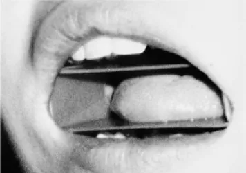
모든 예에서 먼저 기존의 고식적인 방법인 closed-mouth 영 상으로 먼저 CT와 MRI를 촬영하였다. 그리고 closed-mouth 영상과 같은 위치와 절편 두께로서 본원에서 제작한 마우스 피스 (Fig. 1)를 구강 내에 삽입한 후 open-mouth MR과 CT 영상을 얻었다.
마우스 피스는 자원자가 입을 다물어도 변형이 되지 않을 정도로 내구성이 있고 이물감을 최대한 느끼지 않도록 부드 러운 감촉의 재질을 갖춘 의료용 용기에 사용되는 무해 플라 스틱 재료(Boin medica CO., LTD. & Becton Dickinson Korea, Seoul, Korea)를 이용하여 제작하였다. X선 경화 인 공물(beam harding artifact)과 감수성 인공음영(susceptibil- ity artifact)의 여부를 확인하기 위하여 모형 연구(phantom study)와 자원자 1명을 대상으로 시행한 결과 CT에서 Hounsfield 수치가 -80으로 측정되었고 영상에서 공기와 구 분이 되지 않을 정도였고, MRI에서는 T1 강조 영상, T2 강 조 영상 모두 무신호(signal void)로 나타났다. 인공물이나 영 상의 왜곡은 전혀 관찰되지 않았다. 이 마우스피스는 1 mm 두께의 5.5×3.5×2.5 cm 크기의 장방형의 형태로서, 상하 어 금니 사이에 위치하여, 설부가 마우스 피스 내부에 위치하여 구강 표면과 접촉되지 않고 마우스 피스와의 공간이 협소하 여 설부의 가동성(movability)이 없도록 설계하였다. 촬영 전 저자가 직접 삽입하고 closed-mouth영상을 촬영한 후 즉시 자세의 변화 없이 저자가 직접 구강 내에 삽입하고 자원자에 게 자세유지에 대하여 주의를 환기시킴과 함께 open-mouth 와 closed-mouth간의 자세와 위치가 입을 열고 닫은 것 외 에는 동일하게 유지되고 있는 지를 확인하였다. MRI과 CT에 서 각각 지방성 중심선 설중격(fatty midline lingual septum), 중심선 저밀도면(midline low density plane)으로 명명되는 해 부학적으로 설부의 정중을 지나는 지방면(fat plane)의 위치 와 방정중(paramedian)으로 위치하는 한쌍의 이설근 (genioglossus muscle)의 형태가 동일한지를 open-mouth와
closed-mouth의 T1강조 MR 횡단과 관상, CT 횡단 영상에 서 관찰하여 설부의 대칭성 여부를 판정하고 운동 인공물의 여부를 확인하였다. 조직과 접촉하지 않고 공기로 경계 지워 지는 면적의 범위를 설부의 상면, 하면, 측면, 전면에서 각각 비교하여 전체적으로 공기와 접촉하는 면적이 open-mouth와 closed-mouth중 어느 방법에서 넓게 나타나는 지를 판정하 였다. 그리고 입을 열고 닫음에 따라 그 경계와 형태가 변화 하고 이동하는 설부와 구강부, 하악부의 표면부와 내부 골근 육계를 구성하는 총 13개 구조물 (5)의 판별 유무를 확인하 였다 (Table 1). 구조물의 해부학적 경계와 형태가 전체적으 로 잘 보인 경우에는‘완전 식별’, 부분적으로 판별이 가능하 였던 경우는‘부분 식별’, 그리고 주변 조직간의 밀착과 구조 물의 변형이 심하여 판별이 불가능하였던 경우를‘식별 불가 능’으로 구분하여 각각의 점수를 2,1,0점으로 하여 2명의 방 사선과 의사가 협의를 거쳐 평가한 후 자원자별로 13개 분석 항목의 open-mouth와 closed-mouth의 영상별 전체 점수의 합계와 각 부위별 영상간의 평균점수를 비교 분석하여 open- mouth 영상이 어느 정도 우수한지를 조사하였다. 통계학적 방 법은 Student t-test를 이용하였다.
결 과
Open-mouth T1 강조 MR 영상과 CT 영상의 전 예에서 설부의 해부학적 구조의 대칭성이 유지되었고, closed-mouth T1 강도 MR 관상영상과 횡단 영상에서는 각각 3예만이 대 칭적이었다. closed-mouth CT 영상에서는 1예만이 설부의 대칭성 유지가 관찰되었다. 운동 인공물의 발생은 open- mouth T1 강조 MR 영상의 전 예에서 관찰되지 않았으나, closed-mouth 방법을 사용한 14예의 T1 강조 MR 영상에서 는 관상영상에서 7예, 횡단영상에서 4예에서 인공물이 발견되 었다. 설부와의 접촉하는 공기음영의 정도와 면적 또한 open- mouth 방법을 사용한 전 예의 T1 강조 MR 영상과 CT 영상 에서 closed-mouth 방법보다 범위가 넓은것으로 관찰되었다.
자원자별로 13개 분석항목의 점수의 합계를 비교하였을 때 open-mouth 상태에서 촬영한 T1 강조 영상과 CT 영상이 모두 closed-mouth 방법보다 높은 점수를 보였다 (Table 2, 3, 4). 마우스 피스로 인한 구강부 조직간의 인공물 발생은 관 찰되지 않았고 MR 신호강도나 하운스필드 수치 또한 모형연
Fig. 1. Oblique view of open-mouth position and mouth-piece.
Note the protruded tongue within rectangular shaped mouth- piece.
Table 1. Anatomic Structures of Oral Cavity which can be seen on CT and MRI(n=13)
A. Oral Cavity
Dorsum and Base of the tongue, Inferior surface of the tongue, Lingual margin, Gingiva, Buccal surface, Buccal vestibule B. Mouth Floor
Geniohyoid and Mylohyoid muscles, Anterior belly of the di- gastric muscle
C. Bone
Surface of the hard palate D. Soft Tissue and Organ
Surface of the soft palate, Uvula
구와 동일하였다. 각 부위별 영상간의 평균점수를 비교하였을 때, 모든 부위에서 open-mouth T1 강조 MR영상과 CT가 closed-mouth하에서의 영상보다 평균점수가 높거나 같았다 (Table 5). 평균점수를 통계학적으로 비교분석하였을 때 T1 강조 관상면 영상에서는 설배부(dorsum of the tongue), 설 기저부(tongue base), 설하면(inferior surface of the tongue), 치은(gingiva), 협면(buccal surface), 협전정(buccal vestibule), 경구개면(surface of the hard palate), 연구개면 (surface of the soft palate), 구개수(uvula)가 open-mouth
방법에서 식별이 잘되었고 (Table 2) (Fig. 2) 통계적으로도 closed-mouth 방법보다 유의한 우의를 보였다 (p<0.05). 특 히 구개수는 open-mouth하의 T1 강조 관상면 영상에서 잘 관찰되었다 (Fig. 3). T1 강조 횡단면 영상에서는 설배부, 설 연(lingual margin), 협면, 협전정, 치은, 설하면, 두복근의 전 팽대부(anterior belly of the digastric muscle)가 (Table 3) (Fig. 4), 그리고 CT 횡단영상에서 설배부, 설연, 협면에서 open-mouth 영상의 각 구조물의 식별이 훨씬 용이하였으며 (Table 4, Fig. 5) 통계에서도 유의한 결과를 보였다 (p<0.05).
A B
Fig. 2. T1-weighted coronal MR im- ages at oral cavity in open-and closed- mouth techniques.
A. Open-mouth image demonstrates well demarcation of tongue dorsum (white arrow), buccal surface (open ar- row), gingiva (arrowhead), inferior sur- face of the tongue (double arrows), hard palate mucosa (short white ar- row). Note the clear interface between the soft tissue and air-space.
B. Closed-mouth image demonstrates poor demarcation of buccal surface (open arrow), inferior surface of the tongue(double arrows), hard palate mucosa (short white arrow). The con- tour of tongue is slightly distorted and the demarcation of the dorsum is not distinct. Also note the close contact with adjacent structures.
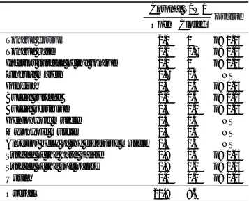
Table 2. Comparative Analysis for Mean Score of Open-mouth and Closed-mouth Coronal T1-Weighted MR Image (T1W1)
Coronal T1W1
Open Closed p value
Tongue dorsum 02.0 0.0 p<0.05
Tongue base 01.1 0.7 p<0.05
Inferior surface of the tongue 02.0 0.0 p<0.05
Lingual margin 01.7 1.4 NS
Gingiva 01.4 0.4 p<0.05
Buccal surface 02.0 0.3 p<0.05
Buccal vestiblue 01.6 1.1 p<0.05
Geniohyoid muscle 01.4 1.4 NS
Mylohyoid muscle 01.4 1.4 NS
Anterior belly of the digastiric muscle 01.4 1.4 NS Surface of the hard palate 01.9 0.3 p<0.05 Surface of the soft palate 01.9 1.1 p<0.05
Uvula 01.0 0.1 p<0.05
Overall 20.8 9.6
Score 2: complete differentiation of anatomic structures Score 1: partial differentiation of anatomic structures Score 0: no differentiation of anatomic structures NS: Not significant
Statistical analysis by Student’s t-test
Table 3. Comparative Analysis for Mean Score of Open-mouth and Closed-mouth Coronal T1-Weighted MR Image (T1W1)
Axial T1W1
Open Closed p value
Tongue dorsum 02.0 0.1 p<0.05
Tongue base 01.0 0.7 NS
Inferior surface of the tongue 02.0 0.3 p<0.05
Lingual margin 01.7 0.3 p<0.05
Gingiva 02.0 0.6 p<0.05
Buccal surface 01.7 0.1 p<0.05
Buccal vestiblue 02.0 0.7 p<0.05
Geniohyoid muscle 01.3 1.0 NS
Mylohyoid muscle 01.3 1.0 NS
Anterior belly of the digastiric muscle 01.3 0.9 p<0.05 Surface of the hard palate 01.0 0.9 NS Surface of the soft palate 00.9 0.7 NS
Uvula 00.0 0.0 NS
Overall 18.2 7.3
Score 2: complete differentiation of anatomic structures Score 1: partial differentiation of anatomic structures Score 0: no differentiation of anatomic structures NS: Not significant
Statistical analysis by Student’s t-test
고 찰
구강은 상부 통기소화기로(upper aerodigestive tract)의 구 부(oral portion)로서 연구개(soft palate), 전구편도궁(ante- rior tonsillar pillars), 유곽유두(circumvallate papillae)에 의 하여 구인두(oropharynx)와 구별되며, 구강의 해부학적 구조 물은 설부, 구저부, 후구치삼각(the retromolar trigone), 경구 개, 구강협부의 점막(buccal mucosa), 치은를 포함한다. 이 부
위에서 대부분 발생하는 편평세포암(squamous cell carcino- ma)과 같은 악성 병변 중에서 (4, 7), 특히 설암의 경우 전 설 절제술(total glossectomy)이 술 후 환자의 삶의 질에 심 각한 영향을 미치기에, 반설절제술(hemiglossectomy)이 가능 한 경계를 결정하는 정중선, 설부의 혈관, 설하신경(hypoglos- sal nerve)과 같은 중요 구조물의 정확한 해부학적 정보를 외 과의들은 최대한 파악하고자 한다 (1, 6, 8). 구강부의 병변 확인은 검사 자의 주의 깊은 시진과 손가락을 이용한 촉진으 로서 대부분 진단에 무리가 없으며, 특히 점막 부의 비정상 소견은 이학적 검사에 의해서만 가능하다 (2, 3). 하지만 일 부의 환자에서는 세밀한 촉진을 견뎌 내지 못하거나, 심한 구 역 반사나 구토가 발생함으로서 정확도가 떨어짐은 물론 검 사를 완결하지 못하는 경우도 있다 (8). 이러한 환자에게 CT 와 MR 영상은 그 진단에 도움이 될 뿐만 아니라, 구인두와 설근부의 저부에 위치하는 종양의 크기와 전이 정도 파악으 로 절제술과 방사선 치료의 계획 수립에 중요하기에 보통 모 든 환자에게 촬영되고 있고 (2, 3, 6), Larsson등 (8)은 가장
A B
Fig. 3. T1-weighted coronal MR im- ages at oropharynx in open-and closed-mouth techniques.
A. Open-mouth image. The oropharyn- geal air-space is now distended and provides clear demarcation of the uvu- la (arrow), tongue base, and tonsillar regions.
B. Closed-mouth image. The oropha- ryngeal air-space is normally col- lapsed.
Table 5. Comparison of Mean Score of the Anatomic Structures (n=3) in Open-mouth and Closed-mouth T1-Weighted MR (T1WI) and CT images
Open>Closed Open=Closed Open<Closed
Axial T1WI 12 1 0
Coronal T1WI 10 3 0
Axial CT 10 3 0
Open>Closed: Open-mouth image is better in tissue demarcation than closed-mouth image.
Open=Closed: Open-mouth image is equal to closed-mouth im- age in tissue demarcation.
Open<Closed: Closed-mouth image is better in tissue demarcation than open-mouth image.
Table 4. Comparative Analysis for Mean Score of Open-mouth and Closed-mouth Axial CT
Axial T1W1
Open Closed p value
Tongue dorsum 02.0 0.7 p<0.05
Tongue base 00.7 0.6 NS
Inferior surface of the tongue 01.3 1.0 NS
Lingual margin 02.0 0.7 p<0.05
Gingiva 00.6 0.4 NS
Buccal surface 02.0 0.3 p<0.05
Buccal vestiblue 00.4 0.1 NS
Geniohyoid muscle 00.6 0.6 NS
Mylohyoid muscle 00.4 0.4 NS
Anterior belly of the digastiric muscle 01.0 0.9 NS Surface of the hard palate 01.0 0.9 NS Surface of the soft palate 00.7 0.6 NS
Uvula 00.0 0.0 NS
Overall 12.4 6.9
Score 2: complete differentiation of anatomic structures Score 1: partial differentiation of anatomic structures Score 0: no differentiation of anatomic structures NS: Not significant
Statistical analysis by Student’s t-test
보기 힘든 부위인 비인두, 설근부, 이상동 부위(pyriform sinus region)의 파악되지 않은 원발성 병변을 찾는 데 도움이 되며 또한 맹생검(blind biopsy)을 위해서는 필히 선행되어야 한다 고 주장하였다. 특히 MR영상은 다면 촬영이 가능하여 두경부 부위의 적절한 평가에 필수적인 관상과 시상면의 영상이 가 능하고 CT보다 연부조직 대조가 우월하기에 종양의 장기 내 또는 장기 외로의 전이여부를 잘 파악할 수 있다고 알려져 있 다 (2, 3). 그러나 기존의 고식적인 영상에서는 구강 내의 설 부가 가지고 있는 특징인 가동성으로 인한 불규칙한 형태와 위치, 그리고 공기 공간의 협소함으로 인한 각 구조물간의 접 촉으로 경계가 흐려지고 정확한 대칭상을 얻을 수 없는 한계
점이 있었다.
저자들은 개선책으로서 입을 벌리고, 설부를 인위적으로 전 방으로 전위시킴과 함께 마우스 피스로서 주위 구조물과의 접 촉을 방지하는 open-mouth 영상을 생각하였다. Open-mouth 영상은 구강 내의 공기 공간의 증가로 인하여 주위 연부 조 직 및 치아와의 대조도가 상승하여 경계면이 명확하여 지고, 또한 설부의 대부분이 주위 조직과 접촉하지 않고 공기와 접 하여 구조물의 변형이 적어지는 장점이 있었다. 예를 들면, MR에서는 구협의 공기 공간의 확장으로 closed mouth에서 잘 보이지 않았던 구개수가 관찰되거나 (Fig. 3), 설부의 상 부와 하부뿐만 아니라 측면의 표면까지 명확해지는 효과가 있
A B
Fig. 4. T1-weighted axial MR images in open-and closed-mouth techniques.
A, B. Open-mouth images show well demarcation of the tongue dorsum (white arrow), lingual margin(short white arrow), buccal surface (open ar- row), and inferior surface of the tongue (double arrows).
C, D. Closed-mouth images. Only a part of the inferior surface of the tongue (double arrows) is seen.
C D
었고 (Fig. 2, 4), CT에서는 설부와 협면의 표면 확인이 용이 하였다 (Fig. 5). 그리고 마우스 피스로 인하여 구강 중앙부 에 설부가 고정되어 운동 인공물이 적어지고 대칭성이 유지 되는 장점도 확인할 수 있었다. 플라스틱 재질을 사용한 마우 스 피스는 MR과 CT촬영에서 마우스 피스 자체가 인공물을 만들지 않았고 아말감(amalgam)이나 의치(denture)와 같은 인공물의 원인과 주위 구조물간의 공기 공간의 확대와 각 구 조물간의 거리의 연장으로 인공물의 최소화가 가능하며 자기 공명의 신호(signal)와 X선의 감쇄(attenuation)에 전혀 영향 을 미치지 않는 잇점도 있을 것이라고 생각한다.
본 연구의 제한점은 첫째, 연구 대상이 많지 않고 둘째, 정
상인 외의 환자를 대상으로 실제 임상에서의 유용성을 시험 한 경험이 적고, 셋째 환자의 마우스 피스로 인한 이물감의 불편 정도를 확인하지 못하였다는 점이다. 즉 각 환자의 연령 과 체구차이로 인한 구강크기의 다양함과 궤양을 동반한 설 암이나 돌출형의 종괴가 구강 내에서 발생한 경우 마우스 피 스의 부적절한 위치와 자극으로 인한 이물감으로 환자의 협 조가 이루어지지 못하여 촬영 자체가 불가능할 수도 있을 것 이다. 이 문제는 연령별로 구강 크기를 감안한 다양한 크기의 마우스 피스를 준비하고, 구강내의 병변의 위치에 따라 장방 형의 마우스 피스의 각진 면이 환자의 병변을 가리고 고통을 가져다 줄 가능성도 있으므로 부드러운 재질과 타원형과 같
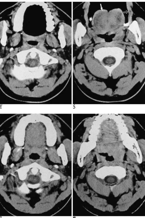
A B
Fig. 5. CT axial scans in open-and closed-mouth techniques.
A, B. Open-mouth images demonstrate tongue dorsum (white arrow), buccal surface (open arrow), lingual margin (short arrow). Note widening of the oropharynx and symmetric tonsillar tissues and the base of the tongue.
C, D. Closed-mouth images: Three structures as described for the open- mouth axial images are not identified.
Also note asymmetry of lingual tonsil- lar tissues and the base of the tongue with obvious narrowing of the oropharynx.
C D
은 여러 종류의 형태 개발과 함께 많은 임상적용의 예를 가 질 수 있다면 충분히 극복할 수 있으리라고 생각한다.
결론적으로 open-mouth 영상은 CT 및 MR에서 경제적이 고 간편하며 공기-연부조직 대조도(air-soft tissue contrast) 의 증가와 설부와 주위 조직간의 분리, 구강 중앙에 설부을 고정시키는 장점으로 구강부 구조물의 평가에 효과적이었다.
참 고 문 헌
1. Stutley J, Cooke J, Parsons C. Normal CT anatomy of the tongue, floor of mouth and oropharynx. Clin Radiol 1989;40:248-253 2. Hoang TA. Hasso AN. Magnetic resonance imaging of the oral cav-
ity and tongue. Top Magn Reson Imaging 1994;6:241-53
3. Kassel EE, Keller MA, Kucharczyk W. MRI of the floor of the
mouth, tongue and orohypopharynx. Radiol Clin North Am 1989;27:331-51
4. Som PM, Oral cavity. In Som PM, Margaret B, Head and Neck imag- ing. 3rd ed. St.Louis: Mosby, 1996:488-544
5. Gray H. The digestive system. In Gross CM, ed. Anatomy of the hu- man body. Philadelphia: Lea & Febiger, 1966,1402-1514
6. Muraki AS, Mancuso AA, Harnsberger HR, Johnson LP, Meads GB. CT of the oropharynx, tongue base, and floor of the mouth:
normal anatomy and range of variations, and applications in stag- ing carcinoma. Radiology 1983;148:725-31
7. Sigal R, Zagdanski AM, Schwaab G, et al. CT and MR imaging of squamous cell carcinoma of the tongue and floor of the mouth.
Radiographics 1996;16:787-810
8. Larsson SG, Mancuso A, Hanafee W. Computed tomography of the tongue and floor of the mouth. Radiology 1982;143:493-500
J Korean Radiol Soc 2001;44:137-144
Address reprint requests to : Chan Ho Kim, M.D., Department of Radiology, College of Medicine, Kosin University 34, Amman-dong, Seo-gu, Busan 602-702, Korea.
Tel. 82-51-240-6341 Fax. 82-51-255-2764 E-mail: kch@medikorea.net
Analysis of Normal Anatomy of Oral Cavity in Open-mouth View with CT and MRI: Comparison with Closed-mouth View1
Chan Ho Kim, M.D., Seong Min Kim, M.D., Bong Jin Cheon, M.D., Jin Do Huh, M.D., Young Duk Joh, M.D.
1Department of Radiology, College of Medicine, Kosin University
Purpose: When MRI and CT of the oral cavity utilize the traditional closed-mouth approach, direct contact be- tween the tongue and surrounding structures may give rise to difficulty in recognizing the anatomy involved and demonstrating the possible presence of pathologic features. we describe a more appropriate scan tech- nique, involving open-mouthed imaging, which may be used to demonstrate the anatomy of the oral cavity in detail.
Materials and Methods: Axial and coronal MR imaging and axial CT scanning were performed in 14 healthy volunteers, using both the closed and open-mouth approach. For the latter, a mouth-piece was put in place pri- or to examination. In all volunteers, open-mouth MR and CT examinations involved the same parameters as the corresponding closed-mouth procedures. The CT and MR images obtained by each method were com- pared, particular attention being paid to the presence and symmetry of motion artifact of the tongue and the extent of air space in the oral cavity. Comparative imaging analysis was based on the recognition of 13 struc- tures around the boundaries of the mouth. For statistical analysis, Student’s t test was used and a p value <
0.05 was considered significant.
Results: Due to symmetry of the tongue, a less severe motion artifact, and increased air space in the oral cavi- ty, the open-mouth method produced excellent images. The axial and coronal MR images thus obtained were superior in terms of demarcation of the inferior surface and dorsum of the tongue, gingiva, buccal surface and buccal vestibule to those obtained with the mouth closed (p<0.05). In addition, axial MR images obtained with the mouth open showed better demarcation of structures at the lingual margin and anterior belly of the digas- tric muscle (p<0.05), while coronal MR images of the base of the tongue, surface of the hard palate, soft palate, and uvula, were also superior (p<0.05). Open-mouth CT provided better images at the lingual margin, dorsum of the tongue and buccal surface than the closed-mouth approach (p<0.05).
Conclusion: Open-mouth MRI and CT are both practical and useful for evaluation of the structures of the oral cavity. The images thus obtained are superior to those acqhired with the month closed.
Index words :Magnetic resonance (MR), technology Computed tomography (CT), technology Mouth