162 Copyright © 2010 Journal of Korean Neurotraumatology Society CASE REPORT
J Kor Neurotraumatol Soc 2010;6:162-164 ISSN 1738-8708
Remote Cerebellar Hemorrhage after Spinal Surgery
Do Keun Kim, MD, Chong Oon Park, MD, PhD,
Seung Hwan Yoon, MD, PhD and Dong Keun Hyun, MD, PhD
Deparment of Neurosurgery, Inha University Hospital, School of Medicine, Inha University, Incheon, Korea
This report details a case of a 56-year-old woman who presented with a herniated lumbar disc at L4-5 with congenital spon- dylolisthesis (grade II) and developed remote cerebellar hemorrhage following an iatrogenic acute reduction in cerebro- spinal fluid (CSF) pressure during spinal interbody fusion. Possible mechanisms are discussed: however, pathological events leading to this complication are unclear. Intracranial hemorrhage (ICH) must be considered in patient presenting with unexplained neurological deterioration not attributable to the spinal surgery, especially when the dura has been opened fol- lowed by significant CSF loss. (J Kor Neurotraumatol Soc 2010;6:162-164)
KEY WORDS: Cerebellar hemorrhage ㆍSpinal surgery ㆍCerebrospinal fluid.
Introduction
Intracranial hemorrhage (ICH) after spinal surgery is extremely rare, but it is very serious clinical problem due to the location of the bleeding. Some authors suggest that re- mote cerebellar hemorrhage (RCH) occurs due to venous infarction, but the pathophysiology and etiology of this con- dition are unknown.5,12,13) The widely assumed pathomech- anism for this type of complication is a decrease in intracra- nial pressure due to loss of cerebrospinal fluid (CSF), which results in caudal shift of brain tissue, thus leading to trac- tion and finally rupture of cortical blood vessel.2,13) This type bleeding pattern includes blood in the sulci of one or both tentorial surface of cerebellum (zebra sign), ICH and ven- tricular hemorrhage.2)
We report a case in which a postoperative RCH occurred after spinal surgery, and discuss possible causative factors.
The related pathomechanism and the diagnostic imaging findings and literature are reviewed and discussed.
Case Report
A 56-year-old woman presented with back pain and both leg pain. She had a history of radiotherapy and chemother- apy for cervix cancer 10 years ago. However, she had no oth- er history of hypertension, trauma, coagulopathy. Magnetic resonance image (MRI) of lumbar spine showed the congeni- tal spondylolisthesis (grade II) with disc herniation (Figure 1). During the operation of the spinal fusion under the prone position, the dura mater was breached accidentally, and gushed out approximately 80 mL of CSF before the dura closed water-tightly. After confirmed no CSF leakage, a Jackson-pratt drain was placed in the surgical bed and con- nected percutaneously to Hemovac suction under positive pressure. When the patient awoke from anesthesia, she was neurologically intact. In the first 24 hours after surgery, the drain drained 700 mL of blood with serosanguinous fluid.
She had mild headache and it was resolved when drainage of the wound was discontinued and by lying flat under the suggestion of intracranial hypotension. Forty three hours af- ter surgery, she complained nausea and vomiting. On neuro- logical examination, she had no new focal neurological sign and improved the leg pain. We thought that such symptoms may be as a complication of the Fentanyl and ZofranTM for postoperative back pain and changed it with nonsteroidal anti-inflammatory drug. Two hours later, she became more disoriented and showed progressive dysarthria. Computed Tomography (CT) scan was done and it demonstrated com- pression of the fourth ventricle by a predominantly right- Received: February 8, 2010 / Revised: March 11, 2010
Accepted: April 8, 2010
Address for correspondence: Chong Oon Park, MD
Deparment of Neurosurgery, Inha University Hospital, School of Medicine, Inha University, 7-206 Sinheung-dong 3-ga, Jung-gu, Incheon 400-711, Korea
Tel: +82-32-890-2370, Fax: +82-32-890-3560 E-mail: nspco@inha.ac.kr
online©MLComm
www.neurotrauma.or.kr 163
Do Keun Kim, et al.
sided ICH with edema, ventricular hemorrhage, and sub- arachnoid hemorrhage (SAH) with mass effect (Figure 2).
Emergency operation was performed for removal of hema- toma and posterior fossa decompression. Ventriculostomy was also performed. On the operation field, there were no specific arterial bleeding sources, but venous oozing be- tween the cerebellar tentorium and upper portion of the cerebellum was noticed. Cerebellar hematoma was orga-
nized, which showed bleeding may be occurred from at least 1 day ago.
Postoperative coma therapy has been performed for 5 days. CT angiography was performed for detecting vascular lesions, which showed no specific findings (Figure 3). Fol- lowed brain MRI showed the fluid collection and low signal change at both hemisphere (right>left) with moderate hy- drocephalus (hydrocephalic index=45%).10) Ventriculoperi- toneal shunt was performed.
At 10 months after surgery, although she can’t walk by herself, she can move by a wheelchair and communicate with us by a simple sentence.
Discussion
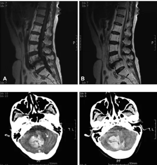
RCH is an infrequent complication resulting from lum- bar spinal procedures. The phenomenon was first de- scribed by Chadduck4) in 1981, after cervical laminectomy in which the dura was opened widely and the dentate liga- ments were sectioned while the patient was in a sitting po- sition. The author proposed that the gradient between the intravascular pressure and the CSF pressure might have in- creased in the sitting position followed by brain displace- FIGURE 1. Sagittal T1-weighted MR
image (A) and T2-weighted MR image (B) show spondylolisthesis (grade II) with herniated disc at L4-5 level. Whole lumbar vertebral body shows the fat mar-
row change on T1- and T2-weighted MRI. A B
FIGURE 2. Axial CT scan obtained 45 hours after lumbar surgery, reveals large intracerebellar hemorrhage (central to right) with a streaky, curvilinear bleed- ing pattern in the cerebellar sulci (arrow).
FIGURE 3. CT angiography shows no specific abnormal vas- cular lesion.
164 J Kor Neurotraumatol Soc 2010;6:162- 164 Remote Cerebellar Hemorrhage after Spinal Surgery
ment and vascular stretching, which would contribute to the development of hemorrhage. Some authors proposed that extensive CSF loss may cause down-ward displacement or
“sag” of cerebellum and stretching of the superior verm- ian veins and their tributaries.1-3,7) Hemorrhage may occur as a consequence of venous tears caused by stretching or ve- nous obstruction because of kinking of draining veins.1,7,13) Most recently, Thomas et al.12) reported a case in which su- pratentorial and infratentorial intraparenchymal hemor- rhage developed after spinal surgery. There are various ex- planations of the pathogenesis of this complication; 1) an excessive loss of CSF because of intraoperative aspiration or drainage with infratentorial venous stretching and rup- ture secondary to upward cerebellar herniation, 2) pre-ex- isting coagulopathies, 3) arterial systemic hypertension, 4) venous obstruction from extreme head rotation, 5) vascu- lar anomalies, 6) anticoagulant therapy, 7) neoplastic an- gioma. But most authors mention excessive CSF loss as the most likely cause of RCH.1-9,11,12)
On our case, the dura was inadvertently opened on the operation and there was CSF leakage through a drainage catheter postoperatively. We thought this case supports the association between low CSF pressure and cerebellar hem- orrhage remote from the operative site. With the abrupt loss of CSF that may occur during and immediately after spinal surgery, collateral venous drainage would not have time to develop. This may cause intracerebellar hemorrhage in patients with insufficient venous collaterals.
Brain CT scan showed the ICH in the upper vermis with SAH (atypical “zebra sign”).2) On the operation field, there were no specific arterial bleeding source, but shown orga- nized ICH with venous oozing between the cerebellar ten- torium and upper portion of the cerebellum. We suspect that this RCH may be originated from venous system by the pres- sure gradient on the operation.
CT scanning is the neuroradiologic investigation with most practical value in patients with cerebellar hemorrhage, but may not be diagnostic in the case of early cerebellar in- farction.
The treatment of acute cerebellar hemorrhage can be operative or conservative. This depends on the size of he- matoma and patient’s condition. Small amount of RCH can be managed medically, but a large amount of acute cerebellar hemorrhage with progressive brain stem dys- function has a mortality that approaches 95% without sur-
gical decompression.1)
Conclusion
In patient with persistent symptoms of headache and nau- sea after spinal surgery in which the dura has been opened, the diagnosis of intracranial hemorrhage should be consid- ered. Such patient should undergo immediate neurological examination and CT investigation under the diagnosis of in- tracranial hemorrhage. Small amount of RCH can be man- aged medically, but monitored with serial imaging. A larger lesion with mass effect must be treated surgically.
REFERENCES
1) Andrews RT, Koci TM. Cerebellar herniation and infarction as a complication of an occult postoperative lumbar dural defect.
AJNR Am J Neuroradiol 16:1312-1315,1995
2) Brockmann MA, Nowak G, Reusche E, Russlies M, Petersen D. Ze- bra sign: cerebellar bleeding pattern characteristic of cerebrospinal fluid loss. Case report. J Neurosurg 102:1159-1162, 2005 3) Cevik B, Kirbas I, Cakir B, Akin K, Teksam M. Remote cerebellar
hemorrhage after lumbar spinal surgery. Eur J Radiol 70:7-9, 2009 4) Chadduck WM. Cerebellar hemorrhage complicating cervical
laminectomy. Neurosurgery 9:185-189, 1981
5) Friedman JA, Ecker RD, Piepgras DG, Duke DA. Cerebellar hem- orrhage after spinal surgery: report of two cases and literature re- view. Neurosurgery 50:1361-1363; discussion 1363-1364, 2002 6) Gelfenbeyn M, Vasil’ev S, Krylov V. Cerebellar haemorrhage after
supratentorial aneurysm surgery with lumbar drainage. Neuro- surg Rev 24:214-219, 2001
7) Konya D, Ozgen S, Pamir MN. Cerebellar hemorrhage after spinal surgery: case report and review of the literature. Eur Spine J 15:95- 99, 2006
8) Mikawa Y, Watanabe R, Hino Y, Ishii R, Hirano K. Cerebellar hem- orrhage complicating cervical durotomy and revision C1-C2 fusion.
Spine (Phila Pa 1976) 19:1169-1171, 1994
9) Morandi X, Riffaud L, Carsin-Nicol B, Guegan Y. Intracerebral hemorrhage complicating cervical “hourglass” schwannoma re- moval. Case report. J Neurosurg 94:150-153, 2001
10) Park CO, Chae KB, Lee SD, Kim Y, Ha YS. The study in frontal ventricular measurement and correlation between cerebroven- tricular index and cephalic index on normal computed tomography.
J Korean Neurosurg Soc 19:608-614, 1990
11) Satake K, Matsuyama Y, Iwata H, Sato K, Kawakami N. Cerebel- lar haemorrhage complicating resection of a cervical intramedul- lary tumour. Spinal Cord 38:504, 2000
12) Thomas G, Jayaram H, Cudlip S, Powell M. Supratentorial and in- fratentorial intraparenchymal hemorrhage secondary to intracra- nial CSF hypotension following spinal surgery. Spine (Phila Pa 1976) 27:E410-E412, 2002
13) Yoshida S, Yonekawa Y, Yamashita K, Ihara I, Morooka Y. Cerebel- lar hemorrhage after supratentorial craniotomy--report of three cases. Neurol Med Chir (Tokyo) 30:738-743, 1990