355
책임저자: 류승완, 대구시 중구 동산동 194번지
700-712, 계명대학교 의과대학 외과학교실 Tel: 053-250-7322, Fax: 053-250-7322
E-mail: [email protected]
접수일:2009년 1월 7일, 게재승인일:2009년 3월 18일 본 논문의 주요내용은 2008년 11월 대한외과학회 추계학술대회 에서 구연 발표되었음.
점막하 위암에서 림프절 전이 예측 인자에 대한 연구
계명대학교 의과대학 외과학교실, 1병리학교실
손영길ㆍ류승완ㆍ김인호ㆍ손수상ㆍ강유나
1Predictive Factors for Lymph Node Metastasis in Submucosal Gastric Cancer
Young Gil Son, M.D., Seung Wan Ryu, M.D., In Ho Kim, M.D., Soo Sang Sohn, M.D., Yu Na Kang, M.D.1 Departments of Surgery and 1Pathology, Keimyung University School of Medicine, Daegu, Korea
Purpose: Lymph node metastasis is an important prognostic factor in patients with early gastric cancer. Therefore,
we analyzed the predictive factors for lymph node metastasis in submucosal gastric cancer and explored the feasibi- lity of minimally invasive surgery.
Methods: The clinicopathological features of 317 patients with submucosal gastric cancer, who underwent radical
gastrectomy with lymph node dissection at Department of Surgery, Keimyung University School of Medicine from January 2003 to December 2007, were examined retrospectively. The lesions were divided into 3 layers according to the depth of submucosal invasion of the cancer cell (SM1, SM2, and SM3). We analyzed the clinicopathological variables regarding lymph node metastasis.
Results: Of the 317 patients, 74 patients (23.3%) had lymph node metastasis. Tumor size, histological type, Lauren
classification, depth of invasion, lymphatic invasion, vascular invasion, and perineural invasion showed a positive correlation with lymph node metastasis by univariate analysis. In multivariate analysis, tumor size (≥4 cm vs <2 cm, P=0.034 and 2∼4 cm vs <2 cm, P=0.043), histological type (P=0.013), and lymphatic invasion (P=0.000) were significantly correlated with lymph node metastasis.
Conclusion: Tumor size, histological type, and lymphatic invasion were independent risk factors for lymph node
metastasis in submucosal gastric cancer. Minimally invasive surgery, such as endoscopic submucosal dissection may be applied to submucosal gastric cancer with a tumor size less than 2 cm, differentiated histological type, and no lymphatic invasion. (J Korean Surg Soc 2009;76:355-359)
Key Words: Submucosal gastric cancer, Lymph node metasatsis, Minimally invasive surgery
중심 단어: 점막하암, 림프절 전이, 최소 침습 수술
서 론
조기위암은 림프절 전이 여부에 관계없이 위의 점막층
및 점막하층에 국한된 위암으로 정의된다.(1) 최근 진단 기 술의 발달과 건강 검진의 증가로 전체 위암 중 조기위암이 차지하는 비율이 1995년에 28.6%, 1999년에 32.8%, 그리고 2004년에 47.4%로 증가하는 추세이다.(2) 조기위암환자에 서 근치적 위절제술 후 5년 생존율은 90% 이상으로 보고되 고 있으며, 림프절 전이가 가장 중요한 예후인자 중의 하나 로 알려져 있다.(3-5) 조기위암에서 림프절 전이의 빈도는 점막암의 경우 5% 내외, 점막하암의 경우 20% 내외로 보고 되어 있다.(5-8) 최근 조기위암의 치료로 수술의 안전성과 기능 보존성을 높이며 수술 후 환자의 삶의 질 향상을 위한
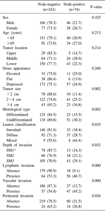
Table 1. Relationship between clinicopathological factors and lymph node metastasis (n=317)
Node-negative (n=243)
Node-positive
(n=74) P-value Sex
Male Female
166 (78.3) 77 (73.3)
46 (21.7) 28 (26.7)
0.325
Age (years) <65 ≥65
151 (79.1) 92 (73.0)
40 (20.9) 34 (27.0)
0.213
Tumor location Upper Middle Lower
29 (85.3) 64 (71.1) 150 (77.7)
5 (14.7) 26 (28.9) 43 (22.3)
0.214
Gross appearance Elevated Flat Depressed
33 (75.0) 38 (86.4) 172 (75.1)
11 (25.0) 6 (13.6) 57 (24.9)
0.260
Tumor size <2 cm 2∼4 cm ≥4 cm
78 (88.6) 122 (74.8) 43 (65.2)
10 (11.4) 41 (25.2) 23 (34.8)
0.002
Histological type Differentiated Undifferentiated
125 (84.5) 118 (69.8)
23 (15.5) 51 (30.2)
0.002
Lauren classification Intestinal Diffuse Mixed
146 (81.6) 92 (71.3) 5 (55.6)
33 (18.4) 37 (28.7) 4 (44.4)
0.035
Depth of invasion SM1*
SM2 SM3
78 (85.7) 60 (76.9) 105 (70.9)
13 (14.3) 18 (23.1) 43 (29.1)
0.032
Lymphatic invasion Absence
Presence
179 (90.9) 64 (53.3)
18 (9.1) 56 (46.7)
0.000
Vascular invasion Absence Presence
186 (87.3) 57 (54.8)
27 (12.7) 47 (45.2)
0.000
Perineural invasion Absence Presence
219 (78.5) 24 (63.2)
60 (21.5) 14 (36.8)
0.036
*SM = submucosa.
여러 최소 침습 수술이 시행되어져 오고 있는데, 이는 림프 절 전이가 없거나 적을 것으로 예상되는 경우 시행되는 내 시경적 점막절제술이나 복강경 수술 등이 대표적이다. 최 근 내시경적 점막하 박리술(endoscopic submucosal dissection, ESD)의 발달로 내시경을 이용한 위암 수술의 적용 범위를 점차 점막하암으로 넓혀가는 가운데, 수술 전 점막하암에 서의 림프절 전이 여부에 대한 평가가 최소 침습 수술의 적용을 위해 필수적이라 하겠다.
본 연구는 근치적 위절제술을 시행 받은 점막하층을 침 범한 조기위암환자에서 수술 전 림프절 전이를 예측할 수 있는 인자를 조사하고 내시경을 이용한 최소 침습 수술의 적용 가능성을 밝히고자 한다.
방 법
2003년 1월부터 2007년 12월까지 계명대학교 의과대학 외과학교실에서 위암으로 진단받고 근치적 위절제술을 시 행한 환자 중 조직검사 상 점막하층을 침범한 317명의 조기 위암 환자를 대상으로 후향적 연구를 하였다.
여러 가지 임상병리학적 인자들(성별, 나이, 종양의 위치, 육안적 소견, 종양의 크기, 조직학적 소견, Lauren 분류, 점 막하층 침윤 깊이, 림프관 침윤, 혈관 침윤, 주위신경 침윤) 과 림프절 전이와의 상관관계에 대하여 비교 분석하였다.
또 점막하층을 침윤한 깊이와 임상병리학적인 지표들과의 상관관계에 대해서도 비교 분석하였다.
점막하층을 동등하게 3등분으로 나누어(상1/3, 중1/3, 하 1/3) 암 세포의 점막하층 침윤 깊이에 따라 상1/3을 SM1, 중1/3을 SM2 그리고 하1/3을 SM3로 한 명의 병리의사에 의 하여 기존의 병리 표본을 재조사하여 분류하였다. 종양의 육안적 소견은 1962년 일본 소화기내시경학회(Japan Gas- troenterological Endoscopy Society)에서 규정한 조기위암의 육안적 분류(macroscopic classification of early gastric cancer) 인 I형 융기형, II형 표면형(IIa형 표면 융기형, IIb형 표면 평탄형, IIc형 표면 함몰형), III형 함몰형 등의 분류에 따랐 으며,(9) 육안적 형태에 따라 융기형(elevated type: I, IIa, IIa
+IIb), 표면형(flat type: IIb, IIb+IIa), 그리고 함몰형(de- pressed type: IIc, IIc+IIa, IIc+IIb, IIc+III, III) 등 세 군으로 분류하였다. 종양의 크기는 장경을 기준으로 하였다. 종양 의 조직학적 소견은 WHO기준에 따랐으며,(10) 분화 정도 에 따라 분화형(유두상 선암, 고분화형 관상 선암, 중분화형 관상 선암)과 미분화형(저분화형 관상 선암, 인환세포암, 점
액성암)으로 재분류하였다.
통계 분석은 SPSS for Window 14.0을 이용하였으며, 각각 변수들은 단변량 분석과 다변량 분석을 통하여 유의성을 평가하였다. 단변량 분석은 Chi-square test, Student's t-test, one-way ANOVA test를 이용하였으며, 다변량 분석은 logis- tic regression analysis를 이용하였다. 모든 분석에서 P값이 0.05 미만일 경우 유의하다고 판정하였다.
Table 2. Multivariate analysis of potential risk factors for lymph node metastasis
Variables Odds ratio 95% CI* P-value
Tumor size <2 cm 2∼4 cm ≥4 cm Histological type Differentiated Undifferentiated Lauren classification Intestinal Diffuse Mixed
Depth of invasion SM1†
SM2 SM3
Lymphatic invasion Absence Presence Vascular invasion Absence Presence Perineural invasion Absence Presence
2.423 2.816
2.903
1.450 2.293
1.115 1.151
14.262
0.640
1.092
1.028∼5.712 1.080∼7.345
1.257∼6.701
0.649∼3.239 0.425∼12.356
0.440∼2.828 0.507∼2.610
4.549∼44.714
0.221∼1.860
0.466∼2.560 0.043 0.034 0.013
0.364 0.334
0.819 0.737 0.000
0.413
0.839
*CI = confidence interval; †SM = submucosa.
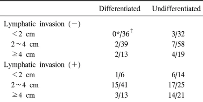
Table 3. Lymph node metastasis in submucosal gastric cancer by tumor size, histological type, and lymphatic invasion
Differentiated Undifferentiated Lymphatic invasion (−)
<2 cm 2∼4 cm ≥4 cm
Lymphatic invasion (+) <2 cm
2∼4 cm ≥4 cm
0*/36† 2/39 2/13
1/6 15/41 3/13
3/32 7/58 4/19
6/14 17/25 14/21
*number of cases with lymph node metastasis; †number of total cases.
결 과
1) 임상병리학적 특성과 림프절 전이와의 상관관계
전체 317명의 환자 중 74명(23.3%)에서 림프절 전이가 관 찰되었다. 성별, 나이, 종양의 위치 및 육안적 소견은 림프 절 전이와 유의한 상관관계는 없었다. 종양의 크기, 조직학 적 소견, Lauren 분류, 점막하층 침윤 깊이, 림프관 침윤, 혈 관 침윤 및 주위신경 침윤은 림프절 전이와 통계학적으로 유의한 상관관계를 보였다(Table 1).
2) 림프절 전이 예측 인자에 대한 다변량 분석
단변량 분석 결과 림프절 전이와의 상관관계에서 통계학 적 유의성을 보인 7가지 인자를 대상으로 다변량 분석을 시행한 결과 종양의 크기(≥4 cm vs <2 cm: odds ratio, 2.816; 95% confidence interval, 1.080∼7.345; P=0.034 및 2∼
4 cm vs <2 cm: odds ratio, 2.423; 95% confidence interval, 1.028∼5.712; P=0.043), 조직학적 소견(odds ratio, 2.903;
95% confidence interval, 1.257∼6.701; P=0.013), 그리고 림프
관 침윤(odds ratio, 14.262; 95% confidence interval, 4.549∼
44.714; P=0.000)이 림프절 전이와 관련된 중요한 위험 인자 였다(Table 2).
3) 점막하암에서 종양의 크기, 조직학적 소견 및 림프관 침윤에 따른 림프절 전이
림프관 침윤이 없고 크기 2 cm 미만의 분화형인 점막하 암에서는 림프절 전이가 관찰되지 않았으나, 미분화형 또 는 림프관 침윤이 있는 점막하암에서는 크기에 관계없이 림프절 전이가 있었다(Table 3).
고 찰
림프절 전이가 조기위암의 가장 중요한 예후인자로 잘 알려져 있다.(7,11) 조기위암의 근치적 수술 후 5년 생존율 은 림프절 전이가 없는 경우 93∼99%, 림프절 전이가 있는 경우 73∼90%로 보고하고 있다.(4,5,12,13) 또한, 점막암과 점막하암의 5년 생존율도 각각 90∼100%, 78∼90%로 보고 되어 있는데 이는 점막암에 비해 점막하암이 림프절 전이 빈도가 높기 때문으로 설명하고 있다. 점막하암에서 림프 절 전이 빈도가 많은 이유로는 점막에 비해 점막하층에 림 프관이 많이 발달되어 있기 때문이며,(14) 많은 연구들에서 림프관 침윤이 림프절 전이의 중요한 위험인자 중 하나로 보고하고 있다.(7,15,16) 따라서, 조기위암의 치료에 있어 림 프절 전이 여부의 예측은 매우 중요한 요소 중 하나이며 본 교실에서는 이미 림프절 전이가 없는 조기위암의 유형 은 육안적 형태가 융기형 혹은 표면형이면서 장경이 20 mm 이하인 점막암, 분화도가 좋은 점막암 및 분화도가 좋으면 서 장경이 10 mm 이하인 융기형 혹은 표면형 점막하암이라
고 보고한 바 있다.(8)
많은 연구에서 점막하암에서의 림프절 전이에 대한 위험 인자들을 보고하였으며 종양의 크기, 점막하층 침윤 깊이, 림프관 침윤, 혈관 침윤, Lauren 분류, 조직학적 소견 등이 림프절 전이의 예측인자로 알려져 있다.(11,16-22) 본 연구 에서도 단변량 분석 결과 종양의 크기, 조직학적 소견, Lauren 분류, 점막하층 침윤 깊이, 림프관 침윤, 혈관 침윤, 주위신경 침윤 등이 림프절 전이와 유의한 상관관계를 보 였으며, 이들 중 다변량 분석 결과 종양의 크기, 조직학적 소견 및 림프관 침윤이 림프절 전이와 관련된 독립적인 위 험인자로 나타나 여러 연구들과 비슷한 결과를 보였다.
현재 점막암에 대해서는 내시경적 치료가 받아들여지고 있으나 점막하암에 대해서는 많은 논란이 있으며 아직까지 는 근치적 위절제술 및 림프절 절제술이 표준 술식으로 인 정되고 있다. 전통적인 내시경적 점막절제술(endoscopic mucosal resection, EMR)의 적응 대상은 점막암에서 20 mm 미만의 분화도가 좋은 융기형 병변과 10 mm 이하의 궤양이 없고 분화도가 좋은 함몰형 병변이다.(23,24) 그러나, 기준 이 너무 제한적이라는 우려와 내시경적 점막하 박리술 (endoscopic submucosal dissection, ESD)의 개발로 내시경적 치료의 적응 대상을 확대하자고 제안되어오고 있다. Gotoda 등(24)은 분화형의 점막암에서는 림프관-혈관 침윤이 없고 궤양이 없다면 크기에 관계없이, 미분화형의 점막암에서는 림프관-혈관 침윤이 없고 궤양이 없는 3 cm 미만의 암까지 대상을 확대하자고 하였으며, 분화형의 점막하암에서는 림 프관-혈관 침윤이 없고 크기가 3 cm 이하, 그리고 침윤 깊이 가 500μm 이하의 SM1 까지도 가능하다고 보고하였다. 반 면 Ishikawa 등(22)과 Nagano 등(25)은 확대된 적응증에 맞 는 침윤 깊이가 500μm 미만의 점막하암에서 림프절 전이 를 보고하면서 일률적인 내시경적 치료의 적용을 우려하였 으며 점막하암에서는 근치적 위절제술 및 림프절 절제술을 시행함이 필요하다고 하였다. 점막하층을 침범한 조기위암 에서 림프절 전이에 대한 또 다른 연구들을 살펴보면 크기 2 cm 이하, 깊이 500μm 이하의 점막하암 경우는 내시경적 절제술 후 추가적인 수술 및 림프절 절제술이 불필요하다 고 보고하고 있다.(17,26,27) 본 연구에서는 점막하암에서 크기가 2 cm 미만의 미분화암인 경우 림프절 전이가 관찰 되었으며, 분화형암일 경우 림프절 전이가 없었다. 따라서 점막하암에서 최소 침습 수술의 적용에는 종양의 조직학적 소견이 중요하다고 생각된다. 또한 림프관 침윤이 있는 점 막하암에서는 크기에 관계없이 림프절 전이가 있어 내시경
적 점막하 박리술 후 림프관 침윤 여부에 대한 병리학적 검사도 시행되어야 할 것이다.
본 연구에서 점막하암에서의 림프절 전이와 관련된 독립 적인 위험인자는 종양의 크기, 조직학적 소견 및 림프관 침 윤으로 나타났다. 위암의 수술 전 검사로 일반적으로 실시 되는 내시경 검사와 내시경적 초음파 검사로 종양의 육안 적 형태, 침윤 깊이 및 종양의 장경을 측정할 수 있다. 그리 고 내시경적 조직 생검에서 조직학적 분화도를 알 수 있으 며, 현재 많이 이용되는 진단적 내시경적 점막하 박리술 후 병리학적 검사를 통해서 림프관 침윤 여부도 진단할 수 있 다. 이들을 종합하면 술 전 조기위암 환자에서의 림프절 전 이를 충분히 예측할 수 있을 것으로 생각된다. 즉 크기 2 cm 미만의 분화형 암에서는 내시경적 절제술의 적응이 될 수 있으나, 병리학적 검사 상 림프관 침윤이 있는 경우 추가 적인 위절제술 및 림프절 절제술이 필요할 것으로 사료된 다. 그러나 본 연구로 점막하암에서의 림프절 전이 가능 여 부를 예측하는 것은 대상 환자군의 수가 작아 한계가 있을 것으로 생각되며, 향후 적절한 전향적인 임상 연구가 필요 할 것으로 사료된다.
결 론
점막하층을 침범한 조기위암에서의 림프절 전이와 관련 된 독립적인 위험인자는 종양의 크기, 조직학적 소견 및 림 프관 침윤으로 나타났다. 크기 2 cm 미만의 분화형 암이면 서 림프관 침윤이 없는 점막하암에서 내시경적 절제술과 같은 최소 침습 수술이 가능할 것으로 사료되며, 미분화형 이나 림프관 침윤 등으로 림프절 전이가 의심되는 경우 근 치적 위절제술 및 림프절 절제술이 적절한 수술 방법이라 생각된다.
REFERENCES
1) Kajitani T. The general rules for the gastric cancer study in surgery and pathology. Part I. Clinical classification. Jpn J Surg 1981;11:127-39.
2) Korean Gastric Cancer Association. 2004 Nationwide gastric cancer report in Korea. J Korean Gastric Cancer Assoc 2007;7:47-54.
3) Kim JP, Hur YS, Yang HK. Lymph node metastasis as a sig- nificant prognostic factor in early gastric cancer: analysis of 1,136 early gastric cancers. Ann Surg Oncol 1995;2:308-13.
4) Hyung WJ, Cheong JH, Kim J, Chen J, Choi SH, Noh SH.
Analyses of prognostic factors and gastric cancer specific sur- vival rate in early gastric cancer patients and its clinical implication. J Korean Surg Soc 2003;65:309-15.
5) Lim KH, Chung HY, Yu W. Prognosis of early gastric cancer:
impact of lymph node metastasis. J Korean Surg Soc 2003;
65:18-22.
6) Sano T, Kobori O, Muto T. Lymph node metastasis from early gastric cancer: endoscopic resection of tumour. Br J Surg 1992;79:241-4.
7) Kitamura K, Yamaguchi T, Taniguchi H, Hagiwara A, Sawai K, Takahashi T. Analysis of lymph node metastasis in early gastric cancer: rationale of limited surgery. J Surg Oncol 1997;
64:42-7.
8) Hwang JY, Lee HJ, Ryu SW, Kim IH, Sohn SS. Preoperative predictive factors of lymph node metastasis in early gastric cancer. J Korean Surg Soc 2005;68:457-63.
9) Murakami T. Pathomorphological Diagnosis: Definition and Gross Classification of Early Gastric Cancer. Gann Monogra- ph on Cancer Research 11. Tokyo: University of Tokyo Press;
1971. p.53-5.
10) Watanabe H, Jass JR, Sobin LH, Ota K. Histological Typing of Oesophageal and Gastric Tumours. WHO International Histological Classification of Tumors. 2nd ed. Berlin:
Springer-Verlag; 1990.
11) Roviello F, Rossi S, Marrelli D, Pedrazzani C, Corso G, Vindigni C, et al. Number of lymph node metastases and its prognostic significance in early gastric cancer: a multicenter Italian study. J Surg Oncol 2006;94:275-80.
12) Seto Y, Nagawa H, Muto T. Impact of lymph node metastasis on survival with early gastric cancer. World J Surg 1997;
21:186-9.
13) Yasuda K, Shiraishi N, Suematsu T, Yamaguchi K, Adachi Y, Kitano S. Rate of detection of lymph node metastasis is correlated with the depth of submucosal invasion in early stage gastric carcinoma. Cancer 1999;85:2119-23.
14) Yamao T, Shirao K, Ono H, Kondo H, Saito D, Yamaguchi H, et al. Risk factors for lymph node metastasis from intra- mucosal gastric carcinoma. Cancer 1996;77:602-6.
15) Lo SS, Wu CW, Chen JH, Li AF, Hsieh MC, Shen KH, et al. Surgical results of early gastric cancer and proposing a treatment strategy. Ann Surg Oncol 2007;14:340-7.
16) Okabayashi T, Kobayashi M, Nishimori I, Sugimoto T, Namikawa T, Onishi S, et al. Clinicopathological features and
medical management of early gastric cancer. Am J Surg 2008;195:229-32.
17) Kurihara N, Kubota T, Otani Y, Ohgami M, Kumai K, Sugiura H, et al. Lymph node metastasis of early gastric cancer with submucosal invasion. Br J Surg 1998;85:835-9.
18) Ko SJ, Suh JH, Park HK, Lee HG, Cho SY, Lee WG, et al.
Predictors of lymph node metastasis in submucosal gastric carcinomas. J Korean Gastric Cancer Assoc 2001;1:155-60.
19) Kunisaki C, Akiyama H, Nomura M, Matsuda G, Otsuka Y, Ono HA, et al. Lymph node status in patients with submucosal gastric cancer. Ann Surg Oncol 2006;13:1364-71.
20) Ohashi S, Okamura S, Urano F, Maeda M. Clinicopathological variables associated with lymph node metastasis in submucosal invasive gastric cancer. Gastric Cancer 2007;10:241-50.
21) An JY, Baik YH, Choi MG, Noh JH, Sohn TS, Kim S.
Predictive factors for lymph node metastasis in early gastric cancer with submucosal invasion: analysis of a single institu- tional experience. Ann Surg 2007;246:749-53.
22) Ishikawa S, Togashi A, Inoue M, Honda S, Nozawa F, Toyama E, et al. Indications for EMR/ESD in cases of early gastric cancer: relationship between histological type, depth of wall invasion, and lymph node metastasis. Gastric Cancer 2007;10:35-8.
23) Tsujitani S, Oka S, Saito H, Kondo A, Ikeguchi M, Maeta M, et al. Less invasive surgery for early gastric cancer based on the low probability of lymph node metastasis. Surgery 1999;
125:148-54.
24) Gotoda T, Yanagisawa A, Sasako M, Ono H, Nakanishi Y, Shimoda T, et al. Incidence of lymph node metastasis from early gastric cancer: estimation with a large number of cases at two large centers. Gastric Cancer 2000;3:219-25.
25) Nagano H, Ohyama S, Fukunaga T, Hiki N, Seto Y, Yamaguchi T, et al. Two rare cases of node-positive differ- entiated gastric cancer despite their infiltration to sm1, their small size, and lack of lymphatic invasion into the submucosal layer. Gastric Cancer 2008;11:53-7.
26) Yamada H, Nihei Z, Yamashita T, Shirota Y, Ichikawa W, Sugihara K. Is lymphadenectomy needed for all submucosal gastric cancers? Eur J Surg 2001;167:199-203.
27) Park DJ, Lee HK, Lee HJ, Lee HS, Kim WH, Yang HK, et al. Lymph node metastasis in early gastric cancer with sub- mucosal invasion: feasibility of minimally invasive surgery.
World J Gastroenterol 2004;10:3549-52.