ISSN 0378-6471 (Print)⋅ISSN 2092-9374 (Online)
http://dx.doi.org/10.3341/jkos.2015.56.7.1065
Original Article
RTVue 빛간섭단층촬영기로 측정한 맥락막 두께와 연관인자에 관한 연구
Analysis of Choroidal Thickness Measured Using RTVue and Associated Factors in Open-Angle Glaucoma
진상욱1⋅최우석1⋅서홍융2⋅노승수3⋅노세현1
Sang Wook Jin, MD1, Woo Seok Choi, MD1, Hong Ryung Seo, MD, PhD2, Seung Soo Rho, MD, PhD3, Sae Heun Rho, MD, PhD1
동아대학교 의과대학 안과학교실1, 왈레스기념 침례병원 안과2, 차의과대학교 의학전문대학원 분당차병원 안과학교실3 Department of Ophthalmology, Dong-A University College of Medicine1, Busan, Korea
Department of Ophthalmology, Wallace Memorial Baptist Hospital2, Busan, Korea
Department of Ophthalmology, CHA Bundang Medical Center, CHA University School of Medicine3, Seongnam, Korea
Purpose: To compare the macular choroidal thickness, ganglion cell complex thickness, peripapillary choroidal thickness and retinal nerve fiber layer thickness among normal, primary open angle glaucoma (POAG) and normal tension glaucoma (NTG) patients using RTVue (Fourier-domain optical coherence tomography; Optovue, Fremont, CA, USA).
Methods: A retrospective analysis of 32 normal controls, 32 POAG and 52 NTG patients was performed. Choroidal thickness, ganglion cell complex thickness and retinal nerve fiber layer thickness were compared among normal controls, POAG and NTG subjects. Additionally, the factors influencing choroidal thickness (age, axial length, spherical equivalent, central corneal thick- ness, mean deviation, nocturnal dip, blood pressure variability) were analyzed.
Results: A total of 32 normal controls, 32 POAG and 52 NTG patients were enrolled in this study. Macular and peripapillary cho- roidal thicknesses were significantly thinner in the NTG patients. In NTG subjects, the significant influencing factors associated with macular and peripapillary choroidal thicknesses were age, axial length, nocturnal dip (diastolic blood pressure), diastolic blood pressure variability and ganglion cell complex thickness. In POAG patients, significant influencing factors associated with macular and peripapillary choroidal thicknesses were age and axial length.
Conclusions: Choroidal thickness was significantly thinner in NTG patients compared with normal controls and POAG patients.
Factors influencing choroidal thickness in NTG patients were age, axial length, nocturnal dip (diastolic blood pressure), diastolic blood pressure variability and ganglion cell complex thickness. In POAG patients, significant factors influencing choroidal thick- ness were age and axial length.
J Korean Ophthalmol Soc 2015;56(7):1065-1074
Key Words: Blood pressure variability, Choroidal thickness, Nocturnal dip, RTVue
■Received: 2014. 11. 28. ■ Revised: 2015. 2. 16.
■Accepted: 2015. 6. 4.
■Address reprint requests to Sae Heun Rho, MD, PhD Department of Ophthalmology, Dong-A University Hospital,
#26 Daesingongwon-ro, Seo-gu, Busan 602-715, Korea Tel: 82-51-240-5227, Fax: 82-51-254-1987
E-mail: shrho@dau.ac.kr
* This study was presented as a narration at the 110th Annual Meeting of the Korean Ophthalmological Society 2013.
* This article has been supported by the Research Fund of the Dong-A University.
ⓒ2015 The Korean Ophthalmological Society
This is an Open Access article distributed under the terms of the Creative Commons Attribution Non-Commercial License (http://creativecommons.org/licenses/by-nc/3.0/) which permits unrestricted non-commercial use, distribution, and reproduction in any medium, provided the original work is properly cited.
녹내장은 망막신경절세포와 그 축삭 돌기의 소멸과 시야 장애가 만성적으로 진행되는 시신경병증이다.1,2 녹내장의 발생 기전으로는 기계적 기전과 허혈성 기전이 있다. 기계 적 기전은 높은 안압에 의해 시신경 유두에 있는 사상판이 후전되고, 이로 인해 사상판의 세공을 통과하는 축삭 돌기 가 기계적인 압력을 받아 축삭원형질 수송의 정체와 망막 신경절세포의 손상과 사멸을 초래한다는 기전이다. 허혈성 기전은 증가된 안압이나 다른 위험인자 등에 의해 시신경
유두의 사상판에 불충분한 혈류 공급이 발생하고 안관류압 과 안혈류 또한 감소하여, 망막신경절세포와 축삭 돌기가 허혈성 손상을 받아 녹내장이 진행한다는 기전이다.3,4 즉 녹내장성 시신경병증은 시신경 유두에 있는 사상판으로 혈 액이 충분히 공급되지 못하는 것과 관련이 있으며, 이곳의 혈류는 유두 주위의 맥락막 혈관의 분지로부터 공급되기 때문에 맥락막을 구성하고 있는 혈관의 밀도나 직경에 이 상이 발생하는 것은 녹내장성 시신경병증과 밀접한 관련이 있다.5-7
과거에는 망막색소상피의 색소와 맥락막 자체가 검안경 검사나 안저 촬영을 방해하기 때문에 해부학적 구조와 혈류 변화를 관찰하기 어려웠으나 최근 진단 기술의 발달로 인해 빛간섭단층촬영(optical coherence tomography, OCT)을 이용하 여 비침습적인 방법으로 맥락막 관찰이 가능하게 되었다.그 중 RTVue (Optovue, Fremont, CA, USA)는 퓨리에 영역 빛간 섭단층촬영(Fourier-domain optical coherence tomography, FD-OCT)으로서 Deep choroidal image technique를 통하여 맥 락막 두께 측정이 가능하다.8,9 맥락막 두께는 맥락막 내의 혈류에 비례하기 때문에 맥락막 두께는 맥락막의 상태를 반영하는 중요한 단위가 될 수 있다.10,11 또한 RTVue를 이 용하여 신경섬유층, 신경절세포층, 내망상층을 포함하고 있 는 신경절세포복합체라고 일컬어지는 황반의 내망막층을 검사할 수 있다. 신경절세포복합체에는 녹내장과 많은 관 련이 있는 망막신경절세포의 축삭 돌기, 세포체, 그리고 수 상돌기가 있으므로 신경절세포복합체 두께를 검사하는 것 은 녹내장의 진단과 진행에 있어 중요한 의미를 가진다.12-15 저자들은 본 연구를 통해 RTVue를 이용하여 황반 부위 의 맥락막과 신경절세포복합체의 두께, 시신경 유두 주위 의 맥락막과 망막신경섬유층의 두께를 구하여 이들 간의 상관 관계 및 맥락막 두께에 영향을 미치는 인자들에 대하 여 알아보고자 하였다.
대상과 방법
2010년 3월부터 2013년 12월까지 본원에서 원발성개방 각녹내장, 정상안압녹내장으로 진단 받고 경과 관찰 중인 환자의 의무 기록을 후향적으로 조사하였다.
원발성개방각녹내장은 골드만 압평안압계로 측정한 안 압이 21 mmHg 이상이며 특징적인 녹내장성 시신경 소견 이 관찰된 환자 중, 자동시야검사에서 녹내장성 시야 결손 이 있고 전방각경 검사에서 개방각인 환자를 대상으로 하 였으며, 정상안압녹내장은 앞에서 언급한 원발성개방각녹 내장의 조건을 만족하고, 적어도 2년 이상 경과 관찰했으 며, 과거력에서 가장 높았던 안압과 매번 내원 시 측정한
안압이 모두 20 mmHg 이하로 측정된 환자를 대상으로 하
였다.16,17 양안 녹내장 환자는 무작위로 편안을 선택하였다.
대조군은 본원 안과에 내원한 환자 중 굴절 이상 이외 다른 안과 질환이 없는 경우를 대상으로 하였다.
전체 대상자 중 녹내장 이외의 시력이나 시야에 영향을 끼칠 수 있는 안과 질환을 이미 진단 받았거나 경과관찰 중 진단 받은 경우, 백내장 수술 등 눈 수술의 병력이 있거나 경과관찰 중 수술 받은 환자, 맥락막과 망막의 질환이 발견 된 경우, 최대교정시력이 0.5 미만인 경우, 녹내장 이외의 시신경 손상을 일으킬 만한 원인 질환이 있는 경우나 당뇨 또는 고혈압이 있는 경우, 고도 근시 등 시신경 유두 주위 위축부가 병적으로 큰 경우, 경과관찰 기간 동안 안압 측정 시간이 제대로 기록되어 있는 않은 환자, 정기적으로 외래 를 방문하지 못한 환자는 대상에서 제외하였다.
골드만압평안압계를 이용한 안압 측정 검사, 현성굴절검 사, 세극등 검사, red free 안저촬영 검사, RTVue를 이용한 빛간섭단층촬영 검사, 전방각경 검사 및 시야검사를 시행 하였다.
환자의 안압은 항녹내장 점안제만으로 치료하였으며 치 료 후 안압을 낮 시간에 한 명의 동일 검사자가 골드만 압 평안압계를 이용하여 3회 측정한 평균치를 사용하였으며 시야 검사는 험프리 자동시야검사(Humphrey field analyzer, Carl-Zeiss Meditec, Dublin, CA, USA)를 이용하여 C24-2 Swedish Interactive Thresholding Algorithm (SITA) standard program으로 측정하였으며, 2년 이상 경과 관찰한 환자를 대상으로 6개월 간격으로 최소 4회 이상 시행하였 으며, false-positive error와 false-negative error가 33% 미만 이고 fixation loss도 20% 미만일 때의 시야 검사를 적절한 것으로 보았다. 24시간 활동 혈압 측정치와의 비교를 위해 24시간 활동 혈압을 측정한 날과 가장 가까운 시기에 검사 한 시야 검사 결과치를 분석에 이용하였다.
빛간섭단층촬영은 숙련된 단일 검사자에 의해 RTVue를 이용하여 시행되었다. 황반하 맥락막 두께 측정은 중심와를 관통하는 선형의 수평 스캔을 이용하여 황반하에서 브루크 막의 고반사선의 외측에서 맥락막-공막의 경계까지 수직선 을 그어 측정하였고 황반에서 이측과 비측으로 750 μm 떨 어진 부위도 같은 방식을 이용하여 측정하였다(Fig. 1).11 유 두 주위 맥락막은 망막신경섬유층을 측정할 때와 동일한, 시신경을 중심으로 한 360°, 3.4 mm 직경의 원형으로 촬영 하였으며, 원형 스캔을 45° 간격으로 나누는 8개 지점에서 브루크막 고반사선의 외측에서 맥락막-공막의 경계까지 수 직선을 그어 측정하였다(Fig. 2).18 측정은 환자의 정보를 알 지 못하는 숙련된 2명의 측정자에 의해 이루어졌으며 두 측정치의 평균치를 사용하였다.
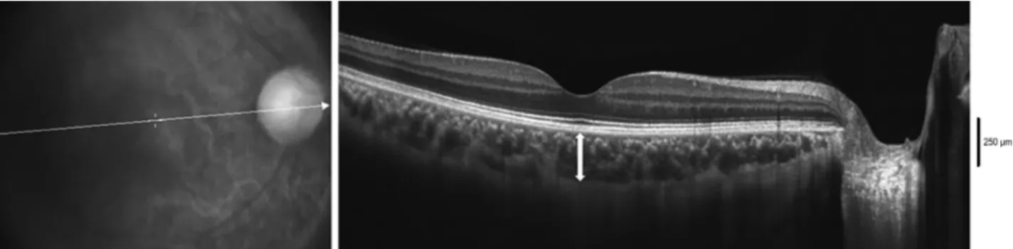
Figure 1. Choroidal thickness of the macula. The image was acquired using the RTVue FD-OCT. The macular thickness is meas-
ured horizontally at 3 points: fovea, 750 μm nasal from the fovea, 750 μm temporal from the fovea (double headed white arrow:measurements line between the retinal pigment epithelium and sclera at the fovea). FD-OCT = Fourier domain-optical coherence tomography.
Figure 2. Choroidal thickness of the peripapillary area. The image was acquired using the RTVue FD-OCT. The peripapillary thick-
ness is measured at 8 points around the optic disc. FD-OCT = Fourier domain-optical coherence tomography.망막신경섬유층 두께는 시신경 유두를 중심으로 360°, 3.4 mm 직경의 원형 스캔을 이용하여 Temporal upper (TU), Superior temporal (ST), Superior nasal (SN), Nasal upper (NU), Nasal lower (NL), Inferior nasal (IN), Inferior temporal (IT), Temporal lower (TL)로 나누어 측정하였다.
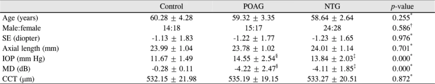
24시간 활동 혈압 측정은 외래에 내원한 개방각 녹내장 환자 중 정상안압녹내장 환자, 원발성개방각녹내장 환자 중 안압은 목표 안압으로 유지되고 있지만 시야 검사상 녹 내장성 시야 손상이 진행되는 환자를 대상으로 전자 혈압 계인 24시간 활동 혈압 측정기(TONOPORT V. GM Medical System, Freiburg, Germany)를 이용하여 비우세완 에서 측정하였다(Fig. 3).
주간혈압(오전 7시-오후 10시)은 30분 간격으로, 야간혈 압(오후 10시-오전 7시)은 1시간 간격으로 측정하였다. 수 축기 혈압(Systolic Blood Pressure, SBP), 이완기 혈압 (Diastolic Blood Pressure, DBP), 심박동수(Heart rate, HR) 를 측정하여 최고치, 최저치, 평균치를 조사하였고, 평균 동 맥압(mean arterial pressure, MAP)과 야간혈압하강(nocturnal dip, ND)은 다음의 식을 이용하여 구하였다.19
MAP = DBP + 1/3 (SBP-DBP)
Nocturnal dip20,21 = (Average of day time blood pressure – Average of night time blood pressure)/Average of day time blood pressure × 100
위의 식을 이용하여 야간 혈압 하강이 10% 미만을 non-dip, 10-15%를 dip, 15% 초과를 over-dip으로 정의하였다.
활동 혈압에서 혈압의 변동성을 반영하는 지표로는 주 간과 야간의 측정 횟수와 평균을 반영한 가중표준편차 (weighted standard deviation, wSD)를 이용하였으며 다음의 식으로 구하였다.22
wSD = (daytime standard deviation [SD] × daytime valid measurement) + (nighttime SD × nighttime valid measurement)/All time valid measurement
통계적 분석은 SPSS® version 22.0 (SPSS Inc., Chicago, IL, USA)을 사용하였으며, 원발성개방각녹내장, 정상안압 녹내장, 정상 대조군 간 맥락막 두께의 차이는 one way ANOVA를 이용하였고 그 중 두 군 간의 두께 차이는 post hoc analysis를 이용하여 통계 분석하였다. 맥락막 두께에 영향을 미치는 안구 인자와 전신 인자는 단변량 회귀 분석,
Figure 3. 24-hour ambulatory blood pressure monitoring (24-hr ABPM). (A) 24-hr ABPM (TONOPORT V. GM Medical System,
Freiburg, Germany). (B) A picture of patient who is undergoing 24-hr ABPM. (C) Printouts from the commercially available 24-hr ABPM. syst. = systolic blood pressure; diast = diastolic blood pressure.다변량 회귀 분석을 이용하여 통계 분석하였다. 또한 두 검 사자 사이의 맥락막 두께 측정치의 신뢰도를 비교하기 위 하여 급내상관계수를 계산하였다. 통계학적 유의 수준은 0.05 미만으로 하였다.
결 과
개방각녹내장 84명, 정상 대조군 32명을 대상으로 하였 으며, 개방각녹내장 중 원발성개방각녹내장은 32명, 정상안 압녹내장은 52명이었다. 평균 나이는 정상 대조군은 60.28
A B
C
Table 1. Patient characteristics in each group
Control POAG NTG p-value
Age (years) 60.28 ± 4.28 59.32 ± 3.35 58.64 ± 2.64 0.255*
Male:female 14:18 15:17 24:28 0.586†
SE (diopter) -1.13 ± 1.83 -1.22 ± 1.77 -1.23 ± 1.65 0.976*
Axial length (mm) 23.99 ± 1.04 23.78 ± 1.02 24.01 ± 1.14 0.701*
IOP (mm Hg) 11.67 ± 1.49 14.55 ± 2.54§ 13.84 ± 2.03‡ 0.000*
MD (dB) -0.28 ± 0.11 -4.22 ± 2.47§ -4.11 ± 1.85‡ 0.000*
CCT (μm) 532.15 ± 21.98 535.19 ± 19.15 533.27 ± 20.51 0.872*
Values are presented as mean ± SD unless otherwise indicated.
POAG = primary open angle glaucoma; NTG = normal tension glaucoma; SE = spherical equivalent; IOP = intraocular pressure; MD = mean deviation; CCT = central corneal thickness.
*Comparison between 3 groups by one-way analysis of variance; †Comparison between 3 groups by chi-square test; ‡Significantly different compared with control by post hoc multiple comparison; §Significantly different compared with control by post hoc multiple comparison.
세, 원발성개방각녹내장은 59.32세, 정상안압녹내장은 58.64 세로 세 군 간 통계적으로 유의한 차이는 없었다(p-value
=0.255). 성별 비교, 구면대응치(spherical equivalent, SE), 안축장(axial length, AL) 모두 세 군 간 통계적으로 유의한 차이는 없었다(성별 비교 p-value=0.586, SE p-value=0.976, AL p-value=0.701). 평균 안압은 정상 대조군 11.67 mmHg, 원발성개방각녹내장 14.55 mmHg, 정상안압녹내장 13.84 mmHg 로 원발성개방각녹내장과 정상안압녹내장에서 정상 대조군 에 비해 높았으나(control vs. primary open angle glaucoma [POAG] p-value=0.000, control vs. normal tension glaucoma [NTG] p-value=0.001), 원발성개방각녹내장과 정상안압녹 내장 간에 통계적으로 유의한 차이는 없었다(p-value=0.636).
평균 mean deviation (MD)은 정상 대조군은 -0.28 dB, 원발 성개방각녹내장에서 -4.22 dB, 정상안압녹내장에서 -4.11 dB로 원발성개방각녹내장과 정상안압녹내장에서 정상 대 조군에 비해 낮았으나(control vs. POAG p-value=0.000, control vs. NTG p-value=0.000), 원발성개방각녹내장과 정상안 압녹내장 간 통계적으로 유의한 차이는 없었다(p-value=0.636).
평균 중심각막두께(central corneal thickness, CCT)는 정상 대조군 532.15 μm, 원발성개방각녹내장 535.19 μm, 정상안 압녹내장 533.27 μm로 세 군 간 통계적으로 유의한 차이는 없었다(p-value=0.872) (Table 1).
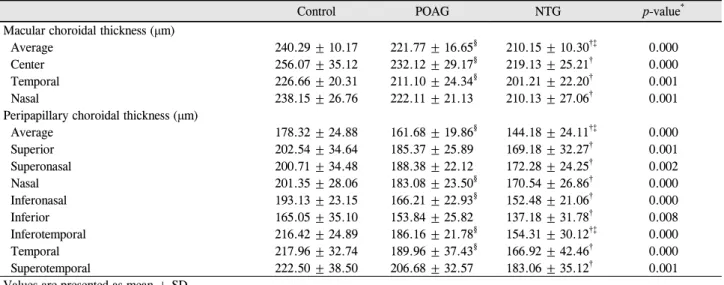
각 군 간 맥락막 두께를 비교한 결과에서는 평균 황반하 맥락막 두께는 정상 대조군 240.29 μm, 원발성개방각녹내 장 221.77 μm, 정상안압녹내장 210.15 μm로 정상 대조군에 비해 원발성개방각녹내장과 정상안압녹내장의 황반하 맥 락막 두께가 더 얇았고(control vs. POAG p-value=0.000, control vs. NTG p-value=0.000), 원발성개방각녹내장에 비 해 정상안압녹내장에서 더 얇았다(p-value=0.006). 황반하 맥락막 두께의 부위별 비교 결과, 중심와 부위에서는 정상 대조군에 비해 원발성개방각녹내장과 정상안압녹내장의 맥
락막 두께가 더 얇았으나(control vs. POAG p-value=0.019, control vs. NTG p-value=0.000), 원발성개방각녹내장과 정상안압녹내장 간 통계적으로 유의한 차이는 없었다 (p-value=0.395). 중심와에서 이측으로 750 μm 떨어진 부위 에서는 정상 대조군에 비해 원발성개방각녹내장과 정상안 압녹내장의 맥락막 두께가 더 얇았으나(control vs. POAG p-value=0.049, control vs. NTG p-value=0.000), 원발성개방 각녹내장과 정상안압녹내장 간 통계적으로 유의한 차이는 없었다(p-value=0.366). 중심와에서 비측으로 750 μm 떨어 진 부위에서는 정상 대조군에 비해 정상안압녹내장의 맥락 막 두께가 더 얇았으나(p-value=0.001) 정상 대조군과 원발 성개방각녹내장 간, 원발성개방각녹내장과 정상안압녹내장 간 통계적으로 유의한 차이는 없었다(control vs. POAG p-value=0.081, POAG vs. NTG p-value=0.289). 평균 유두 주위 맥락막 두께는 정상 대조군 178.32 μm, 원발성개방 각녹내장 161.68 μm, 정상안압녹내장 144.18 μm로 정상 대조군에 비해 원발성개방각녹내장과 정상안압녹내장의 유두 주위 맥락막 두께가 유의하게 더 얇았고(control vs.
POAG p-value=0.039, control vs. NTG p-value=0.000), 원 발성개방각녹내장에 비해 정상안압녹내장에서 얇았다(p-value
=0.027). 유두 주위 맥락막 두께의 부위별 비교에서는 세 군 간 모든 영역에서 통계적으로 유의한 차이가 있었으나, 원발성 개방각녹내장과 정상안압녹내장 간 비교에서는 inferotemporal 부위를 제외한 나머지 영역에서는 통계적으로 유의한 차이 가 없었다(Table 2).
각 군에서 야간혈압하강 정도와 맥락막 두께를 비교한 결과, 원발성개방각녹내장에서는 수축기 혈압과 이완기 혈 압에 따른 non-dip, dip, over-dip 환자 사이에 맥락막 두께 에 통계적으로 유의한 차이는 없었다. 그러나 정상안압녹 내장에서는 수축기 혈압과 이완기 혈압에 따른 non-dip 환 자에서 황반하 및 유두 주위 맥락막 두께가 가장 두꺼웠고
Table 2. Comparison of the macular and peripapillary choroidal thickness between groups
Control POAG NTG p-value*
Macular choroidal thickness (μm)
Average 240.29 ± 10.17 221.77 ± 16.65§ 210.15 ± 10.30†‡ 0.000
Center 256.07 ± 35.12 232.12 ± 29.17§ 219.13 ± 25.21† 0.000
Temporal 226.66 ± 20.31 211.10 ± 24.34§ 201.21 ± 22.20† 0.001
Nasal 238.15 ± 26.76 222.11 ± 21.13 210.13 ± 27.06† 0.001
Peripapillary choroidal thickness (μm)
Average 178.32 ± 24.88 161.68 ± 19.86§ 144.18 ± 24.11†‡ 0.000
Superior 202.54 ± 34.64 185.37 ± 25.89 169.18 ± 32.27† 0.001
Superonasal 200.71 ± 34.48 188.38 ± 22.12 172.28 ± 24.25† 0.002
Nasal 201.35 ± 28.06 183.08 ± 23.50§ 170.54 ± 26.86† 0.000
Inferonasal 193.13 ± 23.15 166.21 ± 22.93§ 152.48 ± 21.06† 0.000
Inferior 165.05 ± 35.10 153.84 ± 25.82 137.18 ± 31.78† 0.008
Inferotemporal 216.42 ± 24.89 186.16 ± 21.78§ 154.31 ± 30.12†‡ 0.000
Temporal 217.96 ± 32.74 189.96 ± 37.43§ 166.92 ± 42.46† 0.000
Superotemporal 222.50 ± 38.50 206.68 ± 32.57 183.06 ± 35.12† 0.001
Values are presented as mean ± SD.
POAG = primary open angle glaucoma; NTG = normal tension glaucoma.
*Comparison between 3 groups by one-way analysis of variance; †Significantly different compared with control by post hoc multiple comparison; ‡Significantly different compared with POAG by post hoc multiple comparison; §Significantly different compared with control by post hoc multiple comparison.
Table 3. Comparison of average macular and peripapillary choroidal thickness among non-dip, dip, and over-dip groups in POAG
Macular choroidal thickness (μm) Peripapillary choroidal thickness (μm)Non-dip (systolic) (n = 13) 221.46 ± 12.59 163.51 ± 12.71
Dip (systolic) (n = 15) 224.60 ± 15.53 165.85 ± 9.56
Over-dip (systolic) (n = 4) 215.75 ± 4.5 163.83 ± 10.79
p-value*** 0.502 0.981
Non-dip (diastolic) (n = 11) 222.73 ± 13.36 162.51 ± 13.44
Dip (diastolic) (n = 17) 223.41 ± 14.92 164.46 ± 9.19†
Over-dip (diastolic) (n = 4) 215.82 ± 13.44 164.75 ± 10.84
p-value*** 0.599 0.889
Values are presented as mean ± SD unless otherwise indicated.
POAG = primary open-angle glaucoma.
*Comparison between 3 groups by one-way analysis of variance; †Significantly different compared with non-dip by post hoc multiple comparison.
Table 4. Comparison of average macular and peripapillary choroidal thickness among non-dip, dip, and over-dip groups in NTG
Macular choroidal thickness (μm) Peripapillary choroidal thickness (μm)Non-dip (systolic) (n = 24) 213.18 ± 11.70 155.50 ± 9.67
Dip (systolic) (n = 23) 206.78 ± 14.65 145.30 ± 17.24
Over-dip (systolic) (n = 5) 202.91 ± 12.59 135.41 ± 9.35†
p-value*** 0.044 0.029
Non-dip (diastolic) (n = 21) 213.62 ± 9.80 156.70 ± 10.91
Dip (diastolic) (n = 26) 207.44 ± 12.17 145.72 ± 16.19
Over-dip (diastolic) (n = 5) 201.57 ± 13.15 148.05 ± 14.95†
p-value*** 0.047 0.018
Values are presented as mean ± SD unless otherwise indicated.
NTG = normal tension glaucoma.
*Comparison between 3 groups by one-way analysis of variance; †Significantly different compared with non-dip by post hoc multiple comparison.
over-dip 환자에서 가장 얇았다(Table 3, 4).
맥락막 두께에 영향을 미칠 수 있는 인자들을 다변량 회 귀 분석을 이용하여 통계 분석한 결과, 원발성개방각녹내장
에서는 나이, 안축장, 평균동맥압만이 통계적으로 유의한 영 향을 미치는 인자였고 정상안압녹내장에서는 황반하 맥락막 두께와 유두 주위 맥락막 두께에 영향을 미치는 인자는 나
Table 5. Association of choroidal thickness with demographic factor, ocular factors and 24-hr ABPM variables by multivariate line-
ar regression analysis in POAGPOAG Macular choroidal thickness Peripapillary choroidal thickness
β ± SE p-value* β ± SE p-value*
Age -2.024 ± 0.620 0.000 -2.674 ± 1.921 0.000
Axial length -0.407 ± 0.895 0.029 -0.912 ± 0.563 0.001
MAP 3.385 ± 1.006 0.003 3.447 ± 1.011 0.003
Values are presented as mean ± SD unless otherwise indicated.
24-hr ABPM = 24-hour ambulatory blood pressure monitoring; POAG = primary open angle glaucoma; SE = spherical equivalent; MAP = mean arterial pressure.
*Multivariate linear regression analysis.
Table 6. Association of choroidal thickness with demographic factor, ocular factors and 24-hr ABPM variables by multivariate
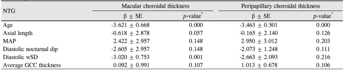
linear regression analysis in NTGNTG Macular choroidal thickness Peripapillary choroidal thickness
β ± SE p-value* β ± SE p-value*
Age -3.621 ± 0.668 0.000 -3.463 ± 0.501 0.000
Axial length -0.618 ± 2.878 0.057 -0.165 ± 2.140 0.126
MAP 2.422 ± 2.957 0.148 2.950 ± 3.012 0.203
Diastolic nocturnal dip -2.605 ± 2.957 0.148 -2.073 ± 1.248 0.111
Diastolic wSD -3.020 ± 0.753 0.001 -2.663 ± 2.093 0.216
Average GCC thickness 0.092 ± 0.991 0.107 1.013 ± 0.678 0.106
Values are presented as mean ± SD unless otherwise indicated.
24-hr ABPM = 24-hour ambulatory blood pressure monitoring; NTG = normal tension glaucoma; SE = spherical equivalent; MAP = mean arterial pressure; wSD = weighted standard deviation; GCC = ganglion cell complex.
*Multivariate linear regression analysis.
Table 7. Comparison of choroidal thickness measurement
value’s reliability which was measured by 2 examinersChoroidal thickness ICC (95% CI, p-value) 0.904 (0.712 to 0.964, p = 0.000) ICC = intra-class correlation coefficients; CI = confidence interval.
이, 안축장, 평균동맥압, 이완기 혈압의 야간혈압하강, 이완 기 혈압의 변동성, 신경절세포복합체 두께였다(Table 5, 6).
두 명의 검사자 간의 급내상관계수는 0.904였다(Table 7).
고 찰
전체 안구 혈류의 95%는 포도막으로 공급되고 그 중 70% 이상이 다시 맥락막으로 공급된다. 맥락막은 신체 조 직 중 단위 무게당 혈류량이 가장 많은 곳 중의 하나로서 외망막층, 망막색소상피와 더불어 시신경의 전사상판에도 혈류를 보내 산소와 영양분을 공급하는 기능을 한다.23-25 녹 내장성 시신경병증은 시신경의 전사상판에 불충분한 혈류 공급으로 인한 망막신경절세포와 축삭 돌기의 허혈성 손상 이 원인이 되어 발생할 수 있으므로 맥락막의 비정상적인 혈 류 공급은 녹내장의 병태생리에 중요한 역할을 할 수 있다.
최근 빛간섭단층촬영 등과 같은 생체 측정 기술의 발달 로 인하여 생체에서 맥락막 두께 측정이 가능해졌고 이를
이용하여 측정된 맥락막 두께와 녹내장 발생의 연관성에 관한 여러 연구가 진행되고 있다.
Hirooka et al26은 50명의 정상인과 52명의 정상안압녹내 장 환자를 대상으로 스펙트럼영역 빛간섭단층촬영을 이용 하여 맥락막 두께를 비교한 결과, 정상인에 비해 정상안압 녹내장에서 하측, 하비측, 하이측의 유두 주위 맥락막 두께 가 얇았다고 하였다. Usui et al27은 정상인 12명과 고도근시 인 정상안압녹내장 환자 8명을 대상으로 high-penetration OCT를 이용하여 맥락막 두께를 비교하였으며 그 결과 정 상인에 비해 고도근시인 정상안압녹내장 환자에서 유두 주 위 맥락막 두께가 얇았다고 하였고, 또한 Park et al28도 48 명의 정상인과 108명의 원발성개방각녹내장과 정상안압녹 내장 환자를 대상으로 enhanced depth imaging-OCT를 이 용해서 맥락막 두께를 비교한 결과, 정상안압녹내장에서 유두 주위 맥락막 두께가 얇았다고 하였다.
그러나 맥락막 두께와 녹내장의 발생과는 연관성이 없었 다는 연구 결과도 있었는데 Ehrlich et al18은 녹내장 의증 환자와 원발성개방각녹내장 환자의 유두 주위 맥락막 두께 를 비교한 연구에서 두 군 간에 맥락막 두께의 차이가 없었 다고 하였고, Li et al29은 원발성개방각녹내장 환자와 정상 인을 대상으로 유두 주위 맥락막 두께를 비교한 결과, 정상 인과 원발성개방각녹내장 환자 사이에는 유두 주위 맥락막 두께 차이가 없었다고 하였다.
RTVue를 이용한 본 연구에서는 정상인에 비해 원발성개 방각녹내장과 정상안압녹내장 환자에서 황반하와 유두 주 위 맥락막 두께가 얇았고, 원발성개방각녹내장에 비해 정 상안압녹내장에서 황반하와 유두 주위 맥락막 두께가 얇았 다. 이는 녹내장 환자에서 정상인에 비해 맥락막 두께가 얇 았다는 이전 연구 결과와 비슷한 결과로 개방각녹내장 환 자들에서 맥락막에 혈류를 공급하는 짧은 뒤섬모체동맥과 맥락막 동맥의 혈류량 감소가 시신경 유두 부위와 황반 부 위의 충분하지 못한 혈류 공급과 자가조절기능의 이상을 초래하여, 황반하와 유두 주위 맥락막 두께가 얇아진 것으 로 생각된다.
맥락막 두께에 영향을 미치는 인자로 고령의 나이와 긴 안축장이 있다. 인간의 사체를 이용한 조직학적 연구에 의 하면 나이가 증가할수록 맥락막 모세혈관의 밀도는 감소하 고, 망막색소상피는 위축되며, 광수용체는 변성되어 맥락막 두께가 감소한다고 하였고,30 빛간섭단층촬영을 이용하여 정상인을 대상으로 맥락막 두께를 조사한 결과, 나이가 증 가할수록 맥락막의 두께는 감소한다고 하였다.31
또한 고도 근시로 인해 안축장이 길어진 눈의 조직학적 연구에 의하면 혈관이 결핍되고 맥락막모세혈관층(chori- ocapillaries)이 얇아져서 맥락막 두께가 감소되어 있었고,32 31명의 고도근시 환자를 대상으로 스펙트럼영역 빛간섭단 층촬영의 enhanced depth image 기법을 이용하여 맥락막 두께를 측정한 결과, 고도 근시에서 맥락막 두께는 매우 얇 았고 나이가 많고 근시 정도가 심할수록 맥락막 두께는 더 얇았다고 하였다.12
본 저자들은 나이와 안축장 이외에도 저혈압, 야간혈압 하강, 그리고 혈압의 변동성 등 맥락막에 공급되는 혈류량 에 연관된 인자들과 맥락막 두께와의 연관성을 알아보았다.
저혈압이나 야간혈압하강에 의해 안관류압이 감소하고, 안혈류량이 부족해지면 시신경이 허혈성 손상을 받아, 녹 내장성 시신경 손상과 시야 결손이 유발되거나 악화될 수 있고,33 Thessaloniki Eye Study34에서는 고혈압을 치료하기 위하여 과도한 항고혈압 약제의 사용은 심한 안관류압의 감소를 유발하여 녹내장의 위험을 증가시킨다고 하였다.
또한 Los Angeles Latino Eye Study35에 따르면 낮은 이완 기 혈압과 높은 수축기 혈압 모두에서 개방각녹내장의 발 생 빈도가 증가되고 특히 이완기 혈압이 낮으면 낮을수록 개방각녹내장의 발생 빈도가 높다고 하였다. 혈압의 조절 과 더불어 혈압의 변동성 또한 추적 장기에 중요한 영향을 미치는데 Parati et al36은 큰 폭의 혈압 변동성이 여러 장기 의 손상에 영향을 미치므로 평균 혈압과 함께 혈압의 변동 성도 같이 조절해야 한다고 하였고, Rothwell et al37은 외래 에서 측정한 큰 폭의 혈압 변동성이 뇌졸중의 중요한 위험
인자라고 하였다.
본 연구에서는 24시간 활동혈압 측정을 이용하여 야간혈 압하강과 혈압의 변동성을 측정하였고 이 측정치가 맥락막 두께에 미치는 영향을 알아본 결과, 원발성개방각녹내장과 는 달리 정상안압녹내장에서는 야간혈압하강과 혈압의 변 동성이 클수록 맥락막 두께가 더 얇았다. 이는 녹내장 환자 에서 혈류량 저하뿐만 아니라 혈압의 변동성 또한 맥락막 두께에 영향을 미친다는 것을 의미한다.
녹내장성시신경손상을 알아보기 위한 방법으로 망막신 경절세포의 50%가 위치하고 있는 황반 두께를 측정하여 망막신경절세포의 소실을 평가해 보고자 하는 여러 연구 결과가 있었다.이들 연구 결과에 의하면 정상인에 비해 녹 내장 환자의 황반 두께가 망막신경절세포와 망막신경섬유 층의 소실로 인해 더 얇았으며, 녹내장군이 대조군에 비해 빛간섭단층촬영으로 측정한 황반 두께가 더 얇았다고 하였
다.38-41 그러나 황반 두께에는 신경절세포층뿐만 아니라 외
망막층 두께도 포함되어 있어서 녹내장성 시신경손상을 정 확히 반영한다고 보기 어려운 단점이 있다.
본 연구는 이런 단점을 보완하기 위하여 RTVue를 이용 하여 황반 부위의 신경절세포복합체 두께만을 측정하였고 이를 맥락막과 망막신경절세포층 두께와 비교한 결과 신경 절세포복합체 두께가 맥락막 두께 및 망막신경절세포층 두 께와 연관성이 있는 것으로 나타났다.
본 연구의 제한점은 다음과 같다. 첫째, 후향적인 연구여 서 맥락막 두께와 맥락막 두께에 영향을 미치는 인자들과 의 인과적 선후관계가 명확하지 않다. 둘째, 연구에 포함된 녹내장 환자가 대부분 초기 녹내장 환자여서 중등도 이상 의 녹내장 환자에 적용하기 어렵다. 셋째, 각 검사 시기가 상이하여 각 검사 결과치 사이의 연관성을 분석한 결과 해 석에 오류가 있을 수 있다. 넷째, 맥락막 두께의 일 중 변동 및 안약이 맥락막 두께에 미치는 영향을 반영하지 못하였 다. 다섯째, 맥락막 두께 측정의 신뢰도를 높이기 위해 두 명의 측정자가 각각 두께를 측정하여 평균치를 이용하였으 나 수동으로 두께를 측정하는 방식으로 인해 정확성에 제 한이 있을 수 있다. 추후 추가적인 연구를 통해 이를 보완 해 나가야 할 것으로 생각된다.
결론적으로 정상인에 비해 원발성개방각녹내장과 정상 안압녹내장에서 RTVue로 측정한 황반하와 유두 주위 맥락 막 두께가 얇았고 원발성개방각녹내장에 비해 정상안압녹 내장에서 황반하와 유두 주위 맥락막 두께가 얇았다. 또한 맥락막 두께에 영향을 미치는 요소는 정상안압녹내장에서 는 나이, 안축장, 평균동맥압, 이완기 혈압의 야간혈압하강 과 이완기 혈압의 변동성, 망막신경절세포층 두께였고, 원 발성개방각녹내장에서는 나이, 안축장, 평균동맥압이었다.
REFERENCES
1) Van Buskirk EM, Cioffi GA. Glaucomatous optic neuropathy. Am J Ophthalmol 1992;113:447-52.
2) Quigley HA, Nickells RW, Kerrigan LA, et al. Retinal ganglion cell death in experimental glaucoma and after axotomy occurs by apoptosis. Invest Ophthalmol Vis Sci 1995;36:774-86.
3) Fechtner RD, Weinreb RN. Mechanisms of optic nerve damage in primary open angle glaucoma. Surv Ophthalmol 1994;39:23-42.
4) Grieshaber MC, Flammer J. Blood flow in glaucoma. Curr Opin Ophthalmol 2005;16:79-83.
5) Hayreh SS. Blood supply of the optic nerve head and its role in op- tic atrophy, glaucoma, and oedema of the optic disc. Br J Ophthalmol 1969;53:721-48.
6) Duijm HF, van den Berg TJ, Greve EL. Choroidal haemodynamics in glaucoma. Br J Ophthalmol 1997;81:735-42.
7) Grunwald JE, Piltz J, Hariprasad SM, DuPont J. Optic nerve and choroidal circulation in glaucoma. Invest Ophthalmol Vis Sci 1998;39:2329-36.
8) Spaide RF, Koizumi H, Pozzoni MC. Enhanced depth imaging spectral-domain optical coherence tomography. Am J Ophthalmol 2008;146:496-500.
9) Branchini L, Regatieri CV, Flores-Moreno I, et al. Reproducibility of choroidal thickness measurements across three spectral domain optical coherence tomography systems. Ophthalmology 2012;119:
119-23.
10) Flammer J, Orgül S, Costa VP, et al. The impact of ocular blood flow in glaucoma. Prog Retin Eye Res 2002;21:359-93.
11) Bayhan HA, Aslan Bayhan S, Can I. Evaluation of the macular choroidal thickness using spectral optical coherence tomography in pseudoexfoliation glaucoma. J Glaucoma 2014 Aug 18. [Epub ahead of print]
12) Fujiwara T, Imamura Y, Margolis R, et al. Enhanced depth imaging optical coherence tomography of the choroid in highly myopic eyes. Am J Ophthalmol 2009;148:445-50.
13) Margolis R, Spaide RF. A pilot study of enhanced depth imaging optical coherence tomography of the choroid in normal eyes. Am J Ophthalmol 2009;147:811-5.
14) Ishikawa H, Stein DM, Wollstein G, et al. Macular segmentation with optical coherence tomography. Invest Ophthalmol Vis Sci 2005;46:2012-7.
15) Tan O, Li G, Lu AT, et al. Mapping of macular substructures with optical coherence tomography for glaucoma diagnosis. Ophthal- mology 2008;115:949-56.
16) Collaborative Normal-Tension Glaucoma Study Group. The effec- tiveness of intraocular pressure reduction in the treatment of nor- mal-tension glaucoma. Am J Ophthalmol 1998;126:498-505.
17) Wolfs RC, Borger PH, Ramrattan RS, et al. Views on open-Angle glaucoma: definitions and prevalences-The Rotterdam Study.
Invest Ophthalmol Vis Sci 2000;41:3309-21.
18) Ehrlich JR, Peterson J, Parlitsis G, et al. Peripapillary choroidal thickness in glaucoma measured with optical coherence tomo- graphy. Exp Eye Res 2011;92:189-94.
19) Wilkins MR, Fitzke FW, Khaw PT. Pointwise linear progression criteria and the detection of visual field change in a glaucoma trial.
Eye (Lond) 2006;20:98-106.
20) Hirotsu C, Ohta E, Hirose N, Shimizu K. Profile analysis of 24-hours measurements of blood pressure. Biometrics 2003;59:
907-15.
21) Choi J, Jeong J, Cho HS, Kook MS. Effect of nocturnal blood pres- sure reduction on circadian fluctuation of mean ocular perfusion pressure: a risk factor for normal tension glaucoma. Invest Ophthalmol Vis Sci 2006;47:831-6.
22) Eguchi K, Hoshide S, Schwartz JE, et al. Visit-to-visit and ambula- tory blood pressure variability as predictors of incident car- diovascular events in patients with hypertension. Am J Hypertens 2012;25:962-8.
23) Alm A, Bill A. Ocular and optic nerve blood flow at normal and in- creased intraocular pressures in monkeys (Macaca irus): a study with radioactively labelled microspheres including flow determi- nations in brain and some other tissues. Exp Eye Res 1973;15:
15-29.
24) Hayreh SS. The blood supply of the optic nerve head and the evalu- ation of it-myth and reality. Prog Retin Eye Res 2001;20:563-93.
25) Parver LM, Auker C, Carpenter DO. Choroidal blood flow as a heat dissipating mechanism in the macula. Am J Ophthalmol 1980;
89:641-6.
26) Hirooka K, Tenkumo K, Fujiwara A, et al. Evaluation of peripapil- lary choroidal thickness in patients with normal-tension glaucoma.
BMC Ophthalmol 2012;12:29.
27) Usui S, Ikuno Y, Miki A, et al. Evaluation of the choroidal thick- ness using high-penetration optical coherence tomography with long wavelength in highly myopic normal-tension glaucoma. Am J Ophthalmol 2012;153:10-6.e1.
28) Park HY, Lee NY, Shin HY, Park CK. Analysis of macular and peri- papillary choroidal thickness in glaucoma patients by enhanced depth imaging optical coherence tomography. J Glaucoma 2014;
23:225-31.
29) Li L, Bian A, Zhou Q, Mao J. Peripapillary choroidal thickness in both eyes of glaucoma patients with unilateral visual field loss. Am J Ophthalmol 2013;156:1277-84.
30) Curcio CA, Saunders PL, Younger PW, Malek G. Peripapillary chorioretinal atrophy: Bruch's membrane changes and photo- receptor loss. Ophthalmology 2000;107:334-43.
31) Margolis R, Spaide RF. A pilot study of enhanced depth imaging optical coherence tomography of the choroid in normal eyes. Am J Ophthalmol 2009;147:811-5.
32) Grossniklaus HE, Green WR. Pathologic findings in pathologic myopia. Retina 1992;12:127-33.
33) Millar-Craig MW, Bishop CN, Raftery EB. Circadian variation of blood-pressure. Lancet 1978;1:795-7.
34) Harris A, Topouzis F, Wilson MR, et al. Association of the optic disc structure with the use of antihypertensive medications: the thessaloniki eye study. J Glaucoma 2013;22:526-31.
35) Jiang X, Varma R, Wu S, et al. Baseline risk factors that predict the development of open-angle glaucoma in a population: the Los Angeles Latino Eye Study. Ophthalmology 2012;119:2245-53.
36) Parati G, Pomidossi G, Ramirez A, et al. Variability of the haemo- dynamic responses to laboratory tests employed in assessment of neural cardiovascular regulation in man. Clin Sci (Lond) 1985;69:
533-40.
37) Rothwell PM, Howard SC, Dolan E, et al. Prognostic significance of visit-to-visit variability, maximum systolic blood pressure, and episodic hypertension. Lancet 2010;375:895-905.
38) Curcio CA, Allen KA. Topography of ganglion cells in human retina. J Comp Neurol 1990;300:5-25.
= 국문초록 =
RTVue 빛간섭단층촬영기로 측정한 맥락막 두께와 연관인자에 관한 연구
목적: 정상안압녹내장(normal tension glaucoma, NTG) 및 원발성개방각녹내장(primary open angle glaucoma, POAG)에서 RTVue (Optovue, Fremont, CA, USA)를 이용하여 측정한 황반하와 유두 주위 맥락막 두께를 비교하고 이에 영향을 미치는 인자들에 대해 알아보고자 하였다.
대상과 방법: 퓨리에 영역 빛간섭단층촬영(Fourier-domain optical coherence tomography, FD-OCT)으로 측정한 황반하와 유두 주 위 맥락막, retinal nerve fiber layer (RNFL), 그리고 ganglion cell complex (GCC) 두께를 비교, 분석하였다. 또한 황반하와 유두 주위 맥락막 두께에 영향을 미칠 수 있는 인자들과의 상관관계를 비교, 분석하였다.
결과: 정상 대조군 32명, POAG 32명, NTG 52명이 포함되었다. 각 군 간 맥락막 두께를 비교한 결과, 황반하와 유두 주위 맥락막 두께는 NTG에서 가장 얇았다. NTG에서는 황반하와 유두 주위 맥락막 두께에 영향을 미치는 인자는 나이, 안축장, 이완기 혈압의 야간혈압하강, 이완기 혈압의 변동성, GCC 두께였고, POAG에서는 나이, 안축장만이 영향을 미치는 인자였다.
결론: 정상인에 비해 POAG와 NTG에서 RTVue로 측정한 황반하와 유두 주위의 맥락막 두께가 더 얇았고 POAG보다 NTG에서 더 얇았다. NTG에서는 나이, 안축장, 큰 폭의 이완기 야간혈압하강과 혈압의 변동성이, POAG에서는 나이, 안축장이 맥락막 두께에 영향 을 미치는 인자였다.
<대한안과학회지 2015;56(7):1065-1074>
39) Zeimer R, Asrani S, Zou S, et al. Quantitative detection of glau- comatous damage at the posterior pole by retinal thickness mapping. A pilot study. Ophthalmology 1998;105:224-31.
40) Leung CK, Chan WM, Yung WH, et al. Comparison of macular and peripapillary measurements for the detection of glaucoma: an
optical coherence tomography study. Ophthalmology 2005;112:
391-400.
41) Kim JW, Rhew JY, Choi KR. Choroidal thickness in primary open-angle glaucoma using spectral-domain optical coherence tomography. J Korean Ophthalmol Soc 2014;55:868-76.