ISSN 0378-6471 (Print)⋅ISSN 2092-9374 (Online)
http://dx.doi.org/10.3341/jkos.2016.57.2.243
Original Article
망막혈관종성증식에서 유리체강내 라니비주맙과 애플리버셉트 주입술의 단기 임상결과 비교
Comparison of Short-Term Clinical Outcomes between Intravitreal Ranibizumab and Aflibercept in Retinal Angiomatous Proliferation
김오제⋅김재휘⋅김종우⋅이태곤⋅조성원⋅이동원⋅한정일⋅김철구
Oh Jae Kim, MD, Jae Hui Kim, MD, Jong Woo Kim, MD, Tae Gon Lee, MD, Sung Won Cho, MD, Dong Won Lee, MD, Jung Il Han, MD, Chul Gu Kim, MD
건양대학교 의과대학 김안과병원 안과학교실
Department of Ophthalmology, Kim’s Eye Hospital, Konyang University College of Medicine, Seoul, Korea
Purpose: To evaluate the 6-month outcomes of intravitreal ranibizumab and aflibercept treatment for patients with retinal an- giomatous proliferation (RAP).
Methods: A retrospective review of medical records of 28 patients (31 eyes) diagnosed with RAP was performed. All patients were initially treated with 3 consecutive intravitreal ranibizumab or aflibercept injections after diagnosis. Additional treatment was performed when exudation recurred. The best-corrected visual acuity (BCVA) and central foveal thickness were measured be- fore the first injection and 3 and 6 months after the first injection. The values measured before the treatment were compared with those after treatment.
Results: Sixteen eyes were treated with ranibizumab and 15 eyes with aflibercept. The logarithm of minimal angle of resolution (log MAR) values of BCVA before the first injection and 3 and 6 months after the first injection were 0.78 ± 0.50, 0.47 ± 0.30 and 0.59 ± 0.41 in the ranibizumab group and 0.96 ± 0.52, 0.83 ± 0.52 and 0.74 ± 0.56 in the aflibercept group, respectively. Central foveal thickness was 315.75 ± 115.44, 188.38 ± 57.33 and 218.50 ± 96.49 μm in the ranibizumab group and 249.00 ± 74.88, 143.73 ± 32.73 and 196.73 ± 94.08 μm in the aflibercept group, respectively. BCVA was significantly improved and central foveal thickness was significantly reduced at 6 months (p < 0.05) compared to measurements before the first injection in both groups.
However, BCVA improvement and central foveal thickness were not significantly different between the 2 groups.
Conclusions: Both intravitreal ranibizumab and aflibercept treatments were beneficial for both normalizing macular thickness and improving visual acuity in patients with RAP. The efficacy of the 2 drugs was not noticeably different.
J Korean Ophthalmol Soc 2016;57(2):243-247
Keywords: Aflibercept, Clinical outcomes, Ranibizumab, Retinal angiomatous proliferation
■Received: 2015. 9. 10. ■ Revised: 2015. 10. 7.
■Accepted: 2015. 12. 12.
■Address reprint requests to Chul Gu Kim, MD
Department of Ophthalmology, Kim’s Eye Hospital, #136 Yeongsin-ro, Yeongdeungpo-gu, Seoul 07301, Korea Tel: 82-2-2671-7665, Fax: 82-2-2671-6359 E-mail: [email protected]
ⓒ2016 The Korean Ophthalmological Society
This is an Open Access article distributed under the terms of the Creative Commons Attribution Non-Commercial License (http://creativecommons.org/licenses/by-nc/3.0/) which permits unrestricted non-commercial use, distribution, and reproduction in any medium, provided the original work is properly cited.
망막혈관종성증식은 망막-망막 혹은 망막-맥락막 문합을 특징으로 하는 특이한 형태의 삼출성 나이관련황반변성1으 로 전체 삼출성 나이관련황반변성의 약 5-20% 정도를 차지 하는 것으로 알려져 있다.2-4
과거 광역학치료 혹은 유리체강내 트리암시놀론 주입술 등의 방법을 이용한 경우 망막혈관종성증식의 치료 결과는 만족스럽지 못한 경우가 많았으나,5-7 유리체강내 항혈관내 피성장인자 치료가 도입되면서 망막혈관종성증식에 대한
보다 효과적인 치료가 이루어지게 되었다. 기존의 보고에 따르면, 유리체강내 라니비주맙 주입술로 치료한 경우 병 변의 축소 및 장기적인 시력호전과 중심황반두께의 감소를 얻을 수 있었다.8-10
유리체강내 애플리버셉트 주입술은 보다 최근 도입된 삼 출성 나이관련황반변성 치료 방법이다.11 삼출성 나이관련 황반변성 진단 후 1개월 간격으로 3회 주사 후 2개월 간격 으로 애플리버셉트를 주사하는 방법은 매달 라니비주맙을 주사하는 방법과 비슷한 효과를 보였다.11 애플리버셉트 주 입술은 라니비주맙 주입술에 반응하지 않는 일부 삼출성 나이관련황반변성에서 좋은 효과를 보이는 것으로 알려져
있으며,12-14 특히 결절맥락막혈관병증의 치료에 보다 우수
한 효과를 보이는 것으로 나타났다.15 최근 몇몇 연구들은 애플리버셉트 주입술을 이용하여 망막혈관종성증식을 치 료한 결과를 보고하고 있으나,16,17 연구에 포함된 안의 수가 적어 아직까지 망막혈관종성증식 치료에 있어서 애플리버 셉트 주입술의 효과는 완전히 알려져 있지 않다. 또한 망막 혈관종성증식에서 라니비주맙과 애플리버셉트 주입술의 임상 결과를 직접 비교한 연구는 없었다.
본 연구에서는 망막혈관종성증식 환자를 대상으로 유리 체강내 라니비주맙 주입술과 애플리버셉트 주입술의 6개월 임상 결과를 알아보고, 두 약제의 효과를 서로 비교하고자 한다.
대상과 방법
2014년 5월부터 2015년 1월까지 망막혈관종성증식으로 진단 받고 유리체강내 항혈관내피성장인자 주입술을 시행 받은 6개월 이상 경과관찰된 환자를 대상으로 후향적 의무 기록 분석을 시행하였다. 처음 진단된 경우만을 연구에 포 함시켰으며, 이전에 삼출성 나이관련황반변성으로 진단 후 치료 받은 병력이 있는 경우에는 제외하였다. 6개월간의 경 과관찰 기간 동안 유리체강내 라니비주맙 혹은 애플리버셉 트 단일요법으로 치료 받은 경우만을 연구에 포함시켰으며, 경과관찰 기간 동안 약제를 교체 투여하였거나 광역학치료 혹은 베바시주맙 주입술 등 삼출성 나이관련황반변성에 대 한 다른 치료를 시행 받은 경우는 연구에서 제외하였다.
삼출성 나이관련황반변성이 진행하는 경우 망막-맥락막 문합이 나타날 수 있는 것으로 알려져 있는데,18 이와 같은 경우를 제외하기 위해 황반중심에 지도모양 위축이나 원반 형 반흔이 관찰되는 경우 또는 증상 발생 후 6개월 이상 경 과한 경우 연구에서 제외하였다. 증식당뇨망막병증, 망막혈 관폐쇄가 관찰된 경우, 황반원공이나 망막앞막 등 황반 미 세구조와 시기능에 영향을 미칠 수 있는 기타 유리체-망막
질환이 동반된 경우에는 연구에서 제외하였다.
환자를 대상으로 최대교정시력을 측정하고, 세극등을 이용 한 안저검사, 형광안저혈관조영 및 인도사이아닌그린혈관조 영(HRA-2; Heidelberg Engineering, Dossenheim, Germany) 을 시행하였으며, 파장영역 빛간섭단층촬영(Spectral OCT/SLO®; OTI Ophthalmic Technologies Inc., Miami, FL, USA)을 시 행하였다.
유리체강내 주입술은 외래 수술실에서 시행되었고, 시술 전 0.5% proparacaine (Alcaine®, Alcon, Fort Worth, TX, USA)을 점안한 뒤 1.25% 혹은 5% povidone iodine을 시술 할 눈에 점안하고 속눈썹을 포함하여 눈 주위를 닦았다. 개 검기를 끼우고 생리식염수로 충분히 세척한 뒤 주사기의 바 늘 끝이 눈꺼풀 가장자리나 속눈썹에 닿지 않도록 주의하면 서 각막 윤부에서 3.0 mm 혹은 3.5 mm 하측 사분면 혹은 상측 사분면에 30게이지 일회용 바늘을 이용하여 0.05 mL의 라니비주맙(Lucentis®, Genentech Inc., South San Francisco, CA, USA) 혹은 애플리버셉트(Eylea®, Regeneron, New York, NY, USA and Bayer HealthCare, Berlin, Germany)를 주사하였다. 모든 환자들은 초기 치료로 1개월 간격으로 3회 의 유리체강내 주입술을 시행 받았다. 마지막 주사 후 1개 월(처음 진단 후 3개월)에 형광안저혈관조영술과 빛간섭단 층촬영을 시행하였으며, 치료자의 판단에 따라 인도사이아 닌그린혈관조영을 추가로 시행하였다. 이후 환자 상태에 따라 1-2개월 간격으로 추적관찰을 진행하였으며, 빛간섭 단층촬영상 이전 주사 치료에도 불구하고 망막하액 혹은 망막내액이 완전히 소실되지 않았거나 경과관찰 중 망막하 액 혹은 망막내액이 재발하면서 시력이 저하되는 경우 추 가 주사를 시행하였다. 시력의 저하를 유발하지 않는 소량 의 망막하액 혹은 망막내액이 재발하는 경우에는 추가 주 사 없이 관찰하였다.
중심망막두께는 빛간섭단층촬영 결과를 이용하여 중심 와 위치에서 내경계막으로부터 망막색소상피까지의 거리 를 수직으로 측정하였다. 진단 당시, 진단 후 3개월(3번째 주사 후 1개월) 및 진단 후 6개월의 시력과 중심망막두께를 측정하였다. 라니비주맙 단일요법으로 치료 받은 안을 라 니비주맙 주입군, 애플리버셉트 단일요법으로 치료 받은 안을 애플리버셉트 주입군으로 분류하였다. 양 군 내에서 진단 당시의 시력을 진단 후 6개월의 시력과 비교하였으며, 6개월 동안의 시력 변화 정도를 양 군 사이에 서로 비교하 였다. 추가적으로 첫 3회의 주입술 후 추가 주입술을 시행 받은 빈도를 양 군 간에 서로 비교하였다.
통계 분석에는 SPSS 프로그램(SPSS ver. 12.0 for Windows;
SPSS Inc., Chicago, IL, USA)을 이용하였다. Wilcoxon sign- ed ranks test를 이용하여 서로 다른 두 시점에 측정된 값을
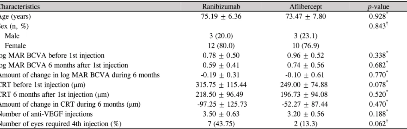
Table 1. Comparisons of characteristics of 31 included eyes (28 patients) with retinal angiomatous proliferation
Characteristics Ranibizumab Aflibercept p-value
Age (years) 75.19 ± 6.36 73.47 ± 7.80 0.928*
Sex (n, %) 0.843†
Male 3 (20.0) 3 (23.1)
Female 12 (80.0) 10 (76.9)
log MAR BCVA before 1st injection 0.78 ± 0.50 0.96 ± 0.52 0.338*
log MAR BCVA 6 months after 1st injection 0.59 ± 0.41 0.74 ± 0.56 0.682*
Amount of change in log MAR BCVA during 6 months -0.19 ± 0.31 -0.10 ± 0.61 0.770*
CRT before 1st injection (μm) 315.75 ± 115.44 249.00 ± 74.88 0.078*
CRT 6 months after 1st injection (μm) 218.50 ± 96.49 196.73 ± 94.08 0.520*
Amount of change in CRT during 6 months (μm) -97.25 ± 125.73 -52.27 ± 87.44 0.470*
Number of anti-VEGF injections 3.50 ± 0.63 3.20 ± 0.56 0.188*
Number of eyes required 4th injection (%) 7 (43.75) 2 (13.3) 0.062†
Values are presented as mean ± SD unless otherwise indicated.
SD = standard deviation; logMAR = logarithm of minimal angle of resolution; BCVA = best-corrected visual acuity; CRT = central retinal thickness; VEGF = vascular endothelial growth factor.
*p-value by Mann-Whitney U-test; †p-value by Chi-square test.
Figure 1. Six-months changes in BCVA (log MAR) and central foveal thickness in eyes with retinal angiomatous proliferation that
were treated with ranibizumab (solid line) or aflibercept (dotted line) treatment, according to the follow-up period. (A) BCVA (log MAR). (B) Central foveal thickness. BCVA = best-corrected visual acuity; log MAR = logarithm of minimal angle of resolution.비교하였으며, 서로 다른 두 군 간의 차이는 Mann-Whitney U-test를 이용하였다. 추가주사가 필요했던 안의 빈도는 카 이제곱검정을 이용하여 비교하였다. 0.05 미만의 p값을 통 계적으로 유의한 값으로 정의하였다.
결 과
전체 31안(28명)을 대상으로 분석을 시행하였고, 남자는 6명(21.4%), 여자는 22명(78.6%)이었으며, 평균 연령은 74.35 ± 7.03세였다(Table 1).
라니비주맙 주입군은 16안이었으며, 애플리버셉트 주입 군은 15안이었다. 라니비주맙 주입군의 경우 6개월간의 추 적관찰 기간 동안 평균 3.50 ± 0.63회의 유리체강내 항혈관 내피성장인자 치료를 시행하였다. 진단 당시 및 진단 후 3 개월, 6개월에 측정된 평균 최대교정시력(logMAR)은 각각 0.78 ± 0.50, 0.47 ± 0.30, 0.59 ± 0.41이었으며(Fig. 1A), 같
은 시기에 측정된 평균 중심망막두께는 각각 315.75 ± 115.44, 188.38 ± 57.33, 218.50 ± 96.49 μm였다(Fig. 1B).
진단 후 6개월에 측정된 최대교정시력은 진단 당시와 비교 하여 유의하게 호전되었으며(p=0.027), 중심망막두께 역시 유의하게 감소한 결과를 보였다(p=0.015). 첫 3회의 주사치 료 후 추가 주사가 필요했던 경우는 7안(43.75%)이었다. 애 플리버셉트 주입군의 경우 6개월간의 추적관찰 기간 동안 평균 3.20 ± 0.56회의 유리체강내 항혈관내피성장인자 치 료를 시행하였다. 진단 당시 및 진단 후 3개월, 6개월에 측 정된 평균 최대교정시력(logMAR)은 각각 0.96 ± 0.52, 0.83 ± 0.52, 0.74 ± 0.56이었으며(Fig. 1A), 같은 시기에 측 정된 평균 중심망막두께는 각각 249.00 ± 74.88, 143.73 ± 32.73, 196.73 ± 94.08 μm였다(Fig. 1B). 진단 후 6개월에 측정된 최대교정시력은 진단 당시와 비교하여 유의하게 호 전되었으며(p=0.034), 중심망막두께 역시 유의하게 감소한 결과를 보였다(p=0.020). 첫 3회의 주사치료 후 추가 주사
A B
가 필요했던 경우는 2안(13.33%)이었다.
라니비주맙 주입군과 애플리버셉트 주입군을 서로 비교 하였을 때, 나이(p=0.928), 성별(p=0.843), 6개월간의 경과 관찰 기간 동안의 시력 변화 정도(p=0.770), 중심망막두께 의 감소 정도(p=0.470), 주사 횟수(p=0.188), 첫 3회의 주입 술 후 추가 주입술이 필요했던 안의 비율(p=0.062)은 서로 차이가 없었다(Table 1).
고 찰
망막혈관종성증식을 유리체강내 항혈관내피성장인자 단 일요법으로 치료한 최근 연구 결과들은 시력 및 해부학적 인 호전이 장기간 유지될 수 있음을 보여주었다. 라니비주 맙을 이용한 치료에서, Shin and Yu19는 망막혈관종성증식 을 진단 받은 31안을 대상으로 24개월 동안 7.7 ± 3.4회의 라니비주맙 치료를 시행하여 유의한 시력호전을 얻었다고 보고하였으며, Cho et al8은 망막혈관종성증식을 새롭게 진 단 받은 38안을 대상으로 36개월 동안 평균 9.61 ± 3.1회의 유리체강내 항혈관내피성장인자 치료를 하여 18개월까지 유의한 시력호전을 얻었다고 보고하였고, Gharbiya et al9은 망막혈관종성증식을 새롭게 진단 받은 21안(베바시주맙 9 안, 라니비주맙 7안, 베바시주맙에서 경과관찰 중 라니비주 맙으로 교체투여한 5안)을 대상으로 24개월 동안 평균 8.8
± 4.4회의 라니비주맙 혹은 베바시주맙 치료를 하여 24개 월까지 유의한 시력호전을 얻었다고 보고하였다. Kim et al10은 망막혈관종성증식을 새롭게 진단 받은 33안을 대상 으로 12개월 동안 평균 4.2 ± 1.7회의 라니비주맙 단일치료 를 하여 6개월까지 유의한 시력호전을 얻었으며 12개월까 지 비교적 안정적인 시력을 유지하였다고 발표하였다.
애플리버셉트를 이용한 치료 결과 역시 최근 보고되고 있는데, Tsaousis et al17은 12안을 대상으로 한 달 간격으로 3회의 에플리버셉트 치료 후 1달 뒤 최대교정시력의 호전 및 중심망막두께의 감소를 관찰한 단기 연구결과를 보고하 였다. Oishi et al16은 10명의 망막혈관종성증식 환자를 대 상으로 12개월 동안 한 달 간격으로 3회의 애플리버셉트 주사 후 두 달 간격으로 추가 주사를 시행한 결과 최대교정 시력의 호전 및 중심망막두께의 감소가 나타났음을 보고하 였다.
본 연구에서는 6개월의 경과관찰 기간 동안 라니비주맙 주입군과 애플리버셉트 주입군 모두에서 시력이 유의하게 호전되었으며 중심망막두께가 유의하게 감소한 결과를 보 였는데, 이는 두 약제 모두 망막혈관종성증식의 치료에 효 과적이라는 점을 시사한다. 하지만 라니비주맙 주입군과 애플리버셉트 주입군 사이에 시력의 호전 및 중심망막두께
의 감소에는 유의한 차이가 없어 망막혈관종성증식의 치료 에서 두 약제 사이 효과의 차이는 발견할 수 없었다.
애플리버셉트의 장점은 약제의 효과가 라니비주맙에 비 해 보다 오래 지속된다는 점이다.11,20 이와 같은 이유로 인 해 환자가 병원을 방문하는 횟수를 줄일 수 있다는 점은 애 플리버셉트의 상대적인 장점으로 알려져 있다.21 본 연구에 서 3회의 주입술 후 추가적인 주입술이 필요했던 경우가 애플리버셉트 주입군에서 더 적은 빈도로 나타났지만 라니 비주맙 주입군과의 비교에서 통계적으로 유의한 차이는 없 었으며, 이러한 경향이 애플리버셉트 약제의 효과 때문이 라고 보기는 어려울 것으로 생각된다.
본 연구의 제한점은 다음과 같다. 본 연구는 후향적 연구 이며, 단기 추적관찰하였다. 라니비주맙과 애플리버셉트 약 제를 선택하는 데 있어서 명확한 기준은 없었으며, 치료자 의 개인적인 선호와 판단에 따라 약제를 선택하였다.
요약하면, 본 연구에서는 망막혈관종성증식을 대상으로 6개월 추적관찰한 결과 유리체강내 라니비주맙 주입술과 애플리버셉트 주입술 모두 시력을 개선하고 중심망막두께 를 줄이는 데에 만족할 만한 효과를 보였다. 6개월 동안의 시력 변화 정도와 중심망막두께 감소 정도는 두 약제 사이 에 통계적으로 유의한 차이가 없었다. 이와 같은 연구의 결 과는 망막혈관종성증식을 치료하는 데 있어서 두 약제 모 두 좋은 효과를 기대할 수 있으며, 특정 약제가 더 우위에 있지 않다는 점을 시사한다. 그러나 본 연구가 소수의 안을 대상으로 한 단기 추적관찰 연구라는 점을 고려하였을 때, 보다 정확한 평가를 위해서는 보다 많은 안을 대상으로 장 기간 추적관찰한 추가 연구가 필요할 것으로 판단된다.
REFERENCES
1) Yannuzzi LA, Negrão S, Iida T, et al. Retinal angiomatous pro- liferation in age-related macular degeneration. Retina 2001;21:
416-34.
2) Liu Y, Wen F, Huang S, et al. Subtype lesions of neovascular age-related macular degeneration in Chinese patients. Graefes Arch Clin Exp Ophthalmol 2007;245:1441-5.
3) Maruko I, Iida T, Saito M, et al. Clinical characteristics of exuda- tive age-related macular degeneration in Japanese patients. Am J Ophthalmol 2007;144:15-22.
4) Massacesi AL, Sacchi L, Bergamini F, Bottoni F. The prevalence of retinal angiomatous proliferation in age-related macular degener- ation with occult choroidal neovascularization. Graefes Arch Clin Exp Ophthalmol 2008;246:89-92.
5) Reche-Frutos J, Calvo-Gonzalez C, Donate-Lopez J, et al. Retinal angiomatous proliferation reactivation 6 months after high-dose intravitreal acetonide triamcinolone and photodynamic therapy.
Eur J Ophthalmol 2007;17:979-82.
6) Saito M, Shiragami C, Shiraga F, et al. Comparison of intravitreal triamcinolone acetonide with photodynamic therapy and intra-
= 국문초록 =
망막혈관종성증식에서 유리체강내 라니비주맙과 애플리버셉트 주입술의 단기 임상결과 비교
목적: 망막혈관종성증식에서 유리체강내 라니비주맙과 애플리버셉트 주입술을 이용하여 치료한 6개월의 임상 결과를 보고하고자 한다.
대상과 방법: 망막혈관종성증식으로 진단 후 유리체강내 라니비주맙 혹은 애플리버셉트 주입술로 치료 받은 31안(28명)을 대상으로 후향적 의무기록 분석을 시행하였다. 진단 후 1개월 간격으로 3회의 주사 후, 이후에는 재발하는 경우 추가 치료를 시행하였다. 치료 전 및 첫 번째 주사 후 3, 6개월에 최대교정시력 및 중심망막두께를 측정하여 비교하였다.
결과: 라니비주맙군은 16안, 애플리버셉트군은 15안이었다. 치료 전 및 치료 후 3개월, 6개월에 측정된 평균 최대교정시력(logMAR)은 라니비주맙군에서 0.78 ± 0.50, 0.47 ± 0.30, 0.59 ± 0.41, 애플리버셉트군에서 0.96 ± 0.52, 0.83 ± 0.52, 0.74 ± 0.56이고, 평균 중심망막두께는 라니비주맙군에서 315.75 ± 115.44, 188.38 ± 57.33, 218.50 ± 96.49 μm, 애플리버셉트군에서 249.00 ± 74.88, 143.73 ± 32.73, 196.73 ± 94.08 μm였다. 두 군 모두에서 진단 후 6개월에 측정된 최대교정시력은 진단 당시와 비교하여 유의하게 호전되었으며(p<0.05), 중심망막두께 역시 유의하게 감소하였다(p<0.05). 두 군 사이에 시력(p=0.770) 및 중심망막두께 (p=0.470)의 감소 정도는 유의한 차이가 없었다.
결론: 유리체강내 라니비주맙 혹은 애플리버셉트 주입술은 망막혈관종성증식 환자에서 시력을 호전시키고 황반두께를 정상화시키는 데 효과적인 방법으로 생각된다.
<대한안과학회지 2016;57(2):243-247>
vitreal bevacizumab with photodynamic therapy for retinal an- giomatous proliferation. Am J Ophthalmol. 2010;149:472-81.e1.
7) Krebs I, Krepler K, Stolba U, et al. Retinal angiomatous pro- liferation: combined therapy of intravitreal triamcinolone aceto- nide and PDT versus PDT alone. Graefes Arch Clin Exp Ophthalmol 2008;246:237-43.
8) Cho HJ, Lee TG, Han SY, et al. Long-term visual outcome and prognostic factors of Intravitreal anti-vascular endothelial growth factor treatment for retinal angiomatous proliferation. Graefes Arch Clin Exp Ophthalmol 2015 Apr 1. [Epub ahead of print]
9) Gharbiya M, Parisi F, Cruciani F, et al. Intravitreal anti-vascular endothelial growth factor for retinal angiomatous proliferation in treatment-naive eyes: long-term functional and anatomical results using a modified PrONTO-style regimen. Retina 2014;34:298-305.
10) Kim DB, Kim JH, Jeong SH, et al. Twelve-month outcomes of in- travitreal anti-vascular endothelial growth factor therapy for retinal angiomatous proliferation. J Korean Ophthalmol Soc 2013;54:
1700-7.
11) Heier JS, Brown DM, Chong V, et al. Intravitreal aflibercept (VEGF trap-eye) in wet age-related macular degeneration.
Ophthalmology 2012;119:2537-48.
12) Bakall B, Folk JC, Boldt HC, et al. Aflibercept therapy for exuda- tive age-related macular degeneration resistant to bevacizumab and ranibizumab. Am J Ophthalmol 2013;156:15-22.e1.
13) Cho H, Shah CP, Weber M, Heier JS. Aflibercept for exudative AMD with persistent fluid on ranibizumab and/or bevacizumab. Br
J Ophthalmol 2013;97:1032-5.
14) Yonekawa Y, Andreoli C, Miller JB, et al. Conversion to afli- bercept for chronic refractory or recurrent neovascular age-related macular degeneration. Am J Ophthalmol 2013;156:29-35.e2.
15) Kawashima Y, Oishi A, Tsujikawa A, et al. Effects of aflibercept for ranibizumab-resistant neovascular age-related macular degen- eration and polypoidal choroidal vasculopathy. Graefes Arch Clin Exp Ophthalmol 2015;253:1471-7.
16) Oishi A, Tsujikawa A, Yamashiro K, et al. One-year result of afli- bercept treatment on age-related macular degeneration and pre- dictive factors for visual outcome. Am J Ophthalmol 2015;159:
853-60.e1.
17) Tsaousis KT, Konidaris VE, Banerjee S, Empeslidis T. Intravitreal aflibercept treatment of retinal angiomatous proliferation: a pilot study and short-term efficacy. Graefes Arch Clin Exp Ophthalmol 2015;253:663-5.
18) Riusala AM, Immonen IJ. Predictors of structural findings in old disciform lesions. Am J Ophthalmol 2004;138:245-53.
19) Shin JY, Yu HG. Optical coherence tomography-based ranibizu- mab monotherapy for retinal angiomatous proliferation in Korean patients. Retina 2014;34:2359-66.
20) Stewart MW, Rosenfeld PJ. Predicted biological activity of intra- vitreal VEGF Trap. Br J Ophthalmol 2008;92:667-8.
21) Browning DJ, Kaiser PK, Rosenfeld PJ, Stewart MW. Aflibercept for age-related macular degeneration: a game-changer or quiet ad- dition? Am J Ophthalmol 2012;154:222-6.