pISSN: 0378-6471 eISSN: 2092-9374
http://dx.doi.org/10.3341/jkos.2012.53.9.1352
= 증례보고 =
중심장액맥락망막병증에 레이저광응고술 후 생긴 맥락막신생혈관의 유리체강내 베바시주맙 주입술
정종현1⋅문 건2⋅김광수1⋅김유철1
계명대학교 의과대학 동산의료원 안과학교실1, 성균관대학교 의과대학 삼성창원병원 안과학교실2
목적: 중심장액맥락망막병증에 대한 직접 레이저광응고술 후 속발성 맥락막 신생혈관이 발생한 환자에서 유리체강내 베바시주맙 주입 술로 치료한 1예를 경험하였기에 이를 보고하고자 한다.
증례요약: 8개월 전부터 발생한 좌안의 변시증으로 내원한 44세 남자환자를 대상으로 최대교정시력, 안저검사, 형광안저혈관조영검사 및 빛간섭단층촬영검사를 시행하여 중심장액맥락막병증으로 진단하였고, 한달 후, 형광안저혈관조영상에서 보이는 누출점에 레이저광 응고술을 시행하였다. 레이저광응고술 8주 후 시력이 0.4로 저하되고, 형광안저혈관조영검사 및 빛간섭단층촬영검사에서 망막하출혈 을 동반한 맥락막신생혈관으로 진단되어 유리체강내 베바시주맙 주입술을 시행하였다. 주입술 4주 후 황반부종과 망막하출혈이 감소 하고 시력이 1.0으로 호전되어 잘 유지되었으나, 2년 뒤 중심장액맥락막병증이 재발되었고, 3개월 후 자연 치유되었다.
결론: 유리체강내 베바시주맙 주입술은 중심장액맥락망막병증에 대한 직접 레이저광응고술 후 발생한 속발성 맥락막신생혈관의 치료 에 있어 안전하고 효과적인 방법이었으며, 한 번의 시술로 장기간 동안 맥락막신생혈관의 재발 없이 유지될 수 있었다.
<대한안과학회지 2012;53(9):1352-1356>
■ 접 수 일: 2012년 2월 17일 ■ 심사통과일: 2012년 3월 27일
■ 게재허가일: 2012년 7월 21일
■ 책 임 저 자: 김 유 철
대구광역시 중구 달성로 56 계명대학교 동산의료원 안과
Tel: 053-250-8026, Fax: 053-250-7705 E-mail: [email protected]
자연치유되지 않는 중심장액맥락망막병증에서 누출 부 위의 레이저광응고술은 좋은 치료로 인정받아 널리 행해져 왔다.1,2 그러나 레이저치료 후 1-5%에서 맥락막신생혈관 이 생길 수 있으며 그 발생 경과 및 치료에 대해서 알려진 바는 드물다.3-5과거 레이저광응고술 후 발생한 맥락막신 생혈관의 치료로 추가적인 아르곤 레이저광응고술이나 수 술적인 치료가 행해졌으나 현재는 나이관련황반변성과 연 관된 맥락막신생혈관에서 좋은 효과를 보이고 있는 광역학 치료나 유리체강내 항혈관내피성장인자항체(anti-vascular endothelial growth factor antibody) 주입술이 좋은 대안으 로 제시되고 있다.5-8
저자들은 최근 중심장액맥락망막병증에서 누출점에 대한 직접 레이저광응고술 후 속발성 맥락막 신생혈관이 발생한 환자에서 유리체강내 베바시주맙(Bevacizumab, AvastinⓇ; Genentech Inc., San Francisco, CA, USA) 주입술로 시 력 향상과 해부학적 호전을 이루었다가 중심장액맥락망막 병증이 재발된 드문 예를 경험하였기에 이를 보고하고자 한다.
증례보고
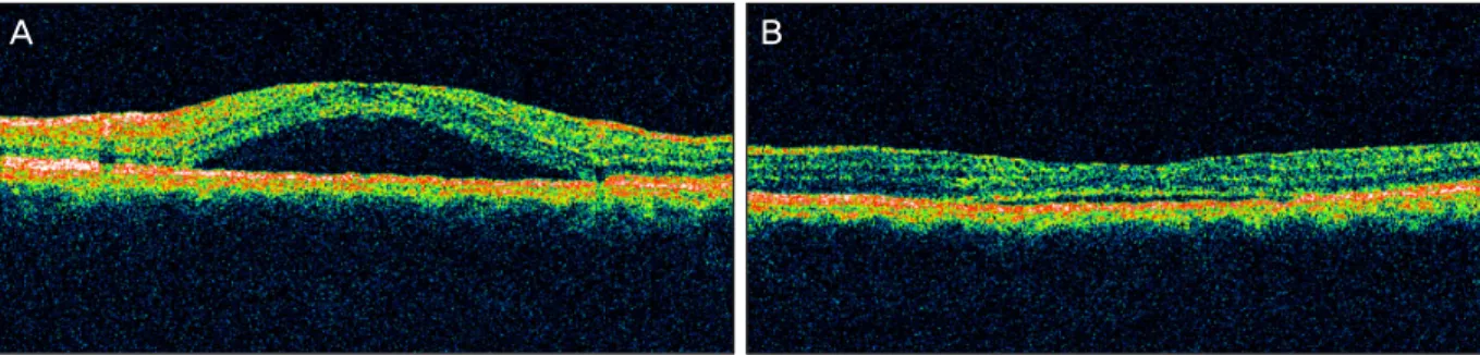
44세의 남자환자가 8개월 전부터 시작된 좌안의 변시증 을 주소로 내원하였다. 특별한 과거력이나 전신증상은 없었 고, 내원 시 실시한 갑상선기능검사, 부신수질호르몬검사는 정상이었다. 우안은 2년 전부터 만성 중심성장액성맥락망 막병증으로 진단을 받았으며, 최대교정시력은 우안이 0.06, 좌안이 1.0, 굴절검사에서 우안이 -0.50디옵터, 좌안은 정 시였다. 안통, 충혈 또는 눈부심 등의 다른 안과적 증상은 없었으며 세극등현미경 및 안압 검사에서 정상소견을 보였 다. 안저검사상 우안은 황반부의 망막색소상피 위축 소견이 보였고(Fig. 1A), 좌안은 황반부에 동심원상으로 장액성망 막박리 소견이 발견되었다(Fig. 1B). 형광안저혈관조영검 사에서 우안은 황반부에 창문비침(window defect)과 망막 색소상피위축역(atrophic tract)이 관찰되었고(Fig. 1C), 좌 안의 중심와로부터 상이측 영역에 굴뚝 연기 모양(smoke stack appearance)의 형광누출이 관찰되었으며(Fig. 1D), 빛간섭단층촬영검사(stratus OCT, Carl Zeiss Meditec Inc., Dublin, CA, USA)에서 장액성망막박리 소견이 관찰 되었다(Fig. 2A). 좌안을 중심장액맥락막병증으로 진단하 고, 누출 부위에 광속직경 50-100 μm, 광속시간 50 msec, 화상을 확인할 수 있을 정도의 강도로 레이저광응고술을 시행하였다. 레이저광응고술 4주 후 망막하액이 대부분 흡 수되었고 변시증이 호전을 보였다(Fig. 2B). 레이저광응고
Figure 1. Initial fundus photographs and fluorescein angiographs. The right eye shows retinal pigment epithelium atrophy at
the macula (A). The left eye shows elevated serous lesion involving the macula (B). FAG of the right eye shows atrophic tract and window defect around the macular area in the late phase (C). FAG of the left eye shows a smokestack appearance around the macular area in the late phase (D). FAG = fluorescein angiograph.Figure 2. The baseline optical coherence tomography (OCT) of the left eye shows a neurosensory retinal detachment in the
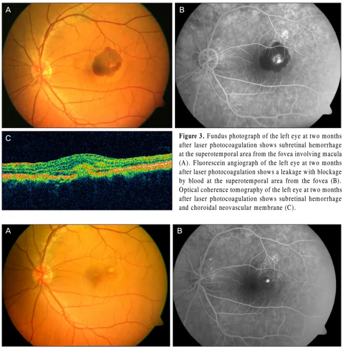
macular region (A), OCT of the left eye at four weeks after focal laser photocoagulation shows resolution of neurosensory reti- nal detachment (B).술8주후좌안 최대교정시력이 0.5로 저하되었고, 좌안 안 저검사 상 레이저치료를 받은 부위를 중심으로 황반하 출 혈 소견이 관찰되었다(Fig. 3A). 형광안저혈관조영검사에 서 좌안의 중심와로부터 상이측에초기 형광염색(stain)과 후기 형광누출(leak)이 관찰되었고(Fig. 3B), 빛간섭단층
촬영검사에서 망막하 맥락막신생혈관막이 관찰되었다(Fig.
3C). 좌안을 속발성 맥락막신생혈관으로 진단하고 유리체 강내 베바시주맙(0.05 ml; 1.25 mg) 주입술을 시행하였다.
주입술 5주 후 좌안 최대교정시력 0.9로 호전되었고, 안저 검사상 망막하 출혈이 감소되었으며(Fig. 4A), 형광안저혈
A B
C D
A B
Figure 3. Fundus photograph of the left eye at two months
after laser photocoagulation shows subretinal hemorrhage at the superotemporal area from the fovea involving macula (A). Fluorescein angiograph of the left eye at two months after laser photocoagulation shows a leakage with blockage by blood at the superotemporal area from the fovea (B).Optical coherence tomography of the left eye at two months after laser photocoagulation shows subretinal hemorrhage and choroidal neovascular membrane (C).
Figure 4. Fundus photograph of the left eye at five weeks after intravitreal bevasizumab injection shows decreased subretinal
hemorrhage at the superotemporal area from the fovea (A). Fluorescein angiograph of the left eye at five weeks after intra- vitreal bevasizumab injection shows stained lesion without leakage at the superotemporal area from the fovea (B).Figure 5. OCT of the left eye at two years after intravitreal bevasizumab injection shows a neurosensory retinal detachment
in the macular region (A). OCT of the left eye at four months after a neurosensory retinal detachment shows resolution of neurosensory retinal detachment (B). OCT = optical coherence tomography.관조영검사에서형광누출없이염색만 되었다(Fig. 4B). 이 후 안저소견과 시력에서 변화 없이 지내다가, 베바시주맙
주입술 2년 후 다시 빛간섭단층촬영검사(spectral OCT/SLO;
OTI, Ophthalmic Technology, Toronto, Ontario,Canada)에
A B
C
A B
A B
서 좌안에 장액성망막박리 소견이 관찰되었으나 4개월 후 자연 치유되었다(Fig. 5).
고 찰
중심장액맥락망막병증는 거의 90%에서는 20/25 이상의 시력으로 회복되지만 5%에서는 반복되는 재발이나 만성화 가 되어 심한 시력장애를 남긴다.9그런 이유로 첫 내원 시 에는 스트레스나 스테로이드 계열의 약물 등의 병의 유발 인자에 대해 설명을 한 후, 증상 3개월까지는 경과관찰하고 호전이 없는 경우는 치료를 해서 4개월째 치료가 되는 것을 목표로 하고 있다. 그러나 환자가 빠른 시력 회복을 원하는 경우에는 일찍 치료를 시작할 수도 있다. 레이저광응고술과 광역학치료(photodynamic therapy, PDT)가 현재 가장 널 리 사용되는 대표적인 치료의 방법이며 광역학치료는 최근 좋은 결과가 많이 보고되고 있다.10그러나 중심장액맥락망 막병증에서 광역학치료는 국내에서 아직 적응증을 인정받 지 못하고 환자 또한 이틀간 실내에서 머물면서 강한 빛으 로부터 격리되어야하는 불편함이 있다. 여기에 비해 레이저 광응고술은 편의성과 경제성을 함께 갖추고 있기는 하나 장액성망막박리 주변에 레이저광응고술을 하는 간접 레이 저광응고술(indirect photocoagulation)은 효과가 없다는 보고가 많고 누출 부위에 레이저를 하는 직접 레이저광응 고술(direct photocoagulation)은 누출점이 중심소와에 접 해있는 경우는 시행하기가 힘들고 레이저 후 속발성 맥락 막신생혈관이 발생할 위험이 있다.11 속발성 맥락막신생혈 관은 중심장액맥락망막병증으로 직접 레이저광응고술을 받 은 환자의 1-5%에서 보고되고 있으며 레이저로 부르크막이 국소적으로 찢어져서 밑에 있는 맥락막 혈관이 그 틈으로 자 라는 것으로 설명되고 작은 크기로 강하게 레이저를 할 경우 부르크막이 잘 찢어져서 잘 생기는 것으로 알려졌다.7
이 환자의 경우 반대편 눈은 현재는 비활동성인 만성 중 심장액맥락망막병증을 보이고 노인성 황반변성 같은 다른 망막질환을 의심할 안저소견은 없어 레이저 반흔에 의한 이차적인 맥락막신생혈관으로 생각한다. 최근 Pikkel and Rumelt7도 중심장액맥락망막병증에서 레이저 치료를 한 후 생긴 맥락막신생혈관에서 베바시주맙주사를 하고 나서 좋 은 결과를 보고하였으며 노인성 황반변성에 의한 맥락막신 생혈관보다 예후가 양호해서 3번 연속으로 주사할 필요는 없다고 하였는데 본 증례도 기존의 보고와 마찬가지로 단 한번의 주사로 맥락막신생혈관이 좋아졌고 맥락막신생혈관 의 재발은 없었다. 그러나 기존의 보고와 달리 후에 다시 중심장액맥락망막병증이 재발하였는데 병변이 중심장액맥 락망막병증에서 맥락막신생혈관이 발생했다가 다시 중심장
액맥락망막병증이 재발된 특이한 경우로 중심장액맥락망막 병증과 맥락막신생혈관의 연관성을 연구할 만한 좋은 사례 로 생각되나 중심장액맥락망막병증 재발 당시 형광안저촬 영을 촬영하지 않아 맥락막신생혈관이 있었던 부분이 다시 중심장액맥락망막병증의 누출부분이었는지 아니면 다른 부 분이 누출이 되었는지 알 수 없는 것은 아쉬운 점이다.
이차적 맥락막신생혈관을 다시 레이저 광응고술로 치료 하는 것은 중심암점발생의 위험이 있고 현재 일부 중심와 밖 병변(extrafoveal lesion)을 제외하고는 맥락막신생혈관 에서는 거의 일차치료로 사용하고 있지 않으며 광역학치료 는 시력보존에는 효과적이나 시력 개선이 없어, 항혈관내피 성장인자(anti-vascular endothelial growth factor) 치료 가 나이관련황반변성을 비롯한 다른 맥락막신생혈관에서 일차적인 치료법으로 인정받고 있다.7그러나 이번 증례에 서 속발성 맥락막신생혈관을 치료 후 다시 중심장액맥락망 막병증이 재발할 수 있다는 점을 볼 때 광역학치료가 맥락 막신생혈관의 치료 외에도 중심장액맥락망막병증의 예방효 과도 있을 수 있다고 생각한다면 일반적인 다른 맥락막신 생혈관의 경우에서와는 달리 베바시주맙보다 나은 장점도 있을 수 있다고 생각할 수 있다. 이는 더 많은 증례를 통하 여 연구되어야 할 것으로 생각한다.
이 증례는 직접 레이저광응고술로 중심장액맥락망막병 증을 치료를 한 후에 생긴 속발성 맥락막신생혈관에서 베 바시주맙은 장기간 맥락막신생혈관의 재발이 없는 효과적 인 치료 효과를 보여 이를 보고하는 바이다.
참고문헌
1) Clais CM, Ober DM, Ciardella AP, Yannuzzi LA. Central serous chorioretinopathy. In: Ryan SJ, ed. Retina, 4th ed. Vol. 2.
Philadelphia: Elsevier-Mosby, 2006;1136-61.
2) Burumcek E, Mudun A, Karacorlu S, Arslan MO. Laser photo- coagulation for persistent central serous retinopathy: results of long-term follow-up. Ophthalmology 1997;104:616-22.
3) Gass JD. Photocoagulation treatment of idiopathic central serous choroidopathy. Trans Sect Ophthalmol Am Acad Ophthalmol Otolaryngol 1977;83(3 Pt 1):456-67.
4) Matsunaga H, Nangoh K, Uyama M, et al. [Occurrence of choroi- dal neovascularization following photocoagulation treatment for central serous retinopathy]. Nihon Ganka Gakkai Zasshi 1995;
99:460-8.
5) Ha TW, Ham DI, Kang SW. Management of choroidal neo- vascularization following laser photocoagulation for central serous chorioretinopathy. Korean J Ophthalmol 2002;16:88-92.
6) Chan WM, Li KK, Liu DT, et al. Photodynamic therapy with verte- porfin in laser-induced choroidal neovascularization. Am J Ophthalmol 2003;136:565-7.
7) Pikkel J, Rumelt S. Intravitreal bevacizumab for choroidal neo- vascularization secondary to laser photocoagulation for central se-
=ABSTRACT=
Intravitreal Bevacizumab Injection for Choroidal Neovascularization Secondary to Laser Photocoagulation for Central Serous
Chorioretinopathy
Jong Hyun Jung, MD1, Kun Moon, MD2, Kwang Soo Kim, MD1, Yu Cheol Kim, MD1
Department of Ophthalmology, Keimyung University Dongsan Medical Center, Keimyung University School of Medicine1, Daegu, Korea Department of Ophthalmology, Samsung Changwon Hospital, Sungkyunkwan University School of Medicine2, Changwon, Korea
Purpose: To report a case of intravitreal bevacizumab injection for choroidal neovascularization following direct laser pho- tocoagulation for central serous chorioretinopathy.
Case summary: A 44-year-old male patient with an 8-month history of metamorphopsia in his left eye visited our clinic and was diagnosed with central serous chorioretinopathy after performing refraction, fundus examination, fluorescein angiography (FAG) and optical coherence tomography (OCT). After 1 month, laser photocoagulation of the leaking point observed on the FAG was performed. After 8 weeks following laser photocoagulation, visual acuity was reduced to 0.4, subretinal hemorrhage accompanied by choroidal neovascularization was observed on FAG and OCT, and an intravitreal bevacizumab injection was administered. After 4 weeks following the injection, macular edema and subretinal hemorrhage decreased, visual acuity increased to 1.0 and was maintained properly. However, after 2 years, the central serous chorioretinopathy recurred and after 3 months, healed spontaneously.
Conclusions: Intravitreal bevacizumab injection is a safe and effective treatment for secondary choroidal neovascularization occurring after direct laser photocoagulation for central serous chorioretinopathy. In addition, a single treatment can main- tain the patient’s status with no recurrence of choroidal neovascularization over a long-term period.
J Korean Ophthalmol Soc 2012;53(9):1352-1356
Key Words: Bevacizumab, Central serous chorioretinopathy, Choroidal neovascularization
Address reprint requests to Yu Cheol Kim, MD
Department of Ophthalmology, Keimyung University Dongsan Medical Center
#56 Dalseong-ro, Jung-gu, Daegu 700-712, Korea
Tel: 82-53-250-8026, Fax: 82-53-250-7705, E-mail: [email protected] rous chorioretinopathy. Eur J Ophthalmol 2012;22:488-91.
8) Cakir M, Cekiç O, Yilmaz OF. Photodynamic therapy for iatro- genic CNV due to laser photocoagulation in central serous chorioretinopathy. Ophthalmic Surg Lasers Imaging 2009;40:
405-8.
9) Gilbert CM, Owens SL, Smith PD, Fine SL. Long-term follow-up of central serous chorioretinopathy. Br J Ophthalmol 1984;68:815-20.
10) Chung SE, Kang JH, Kang SW. Chronic central serous chorioretin- opathy: photodynamic therapy. J Korean Ophthalmol Soc 2007;
48:279-84.
11) Oh J, Kwon OW, Kim MH, et al. Photodynamic therapy for chronic central serous chorioretinopathy: multicenter study of 65 cases. J Korean Ophthalmol Soc 2009;50:390-8.