INTRODUCTION
Rhinitis and asthma usually occur together. There are increas- ing evidences that allergic rhinitis (AR) may influence the clinical course of asthma. AR patients without symptoms of asthma such as episodic cough, dyspnea, and wheezing often have bronchial hyperresponsiveness (BHR) to nonspecific bronchoconstrictors such as methacholine or histamine (1, 2).
Methacholine responsiveness in the asthmatic range among the patients with rhinitis is associated with variable airflow obstruction and subclinical asthma (3).
Putative mechanisms linking rhinitis to asthma are explained by direct and indirect effects (4). The direct effects are naso- bronchial reflex, postnasal drip of inflammatory cells and/or mediators from the nose into the lower airways, and absorp- tion of inflammatory cells and/or mediators from the nose into the systemic circulation, and ultimately, the lung. The indirect effects are nasal obstruction causing reduction in fil- tration, humidification, and warming function of the nose (5).
Rhinitis patients showed a lower degree of bronchial sensitivi- ty to allergen than asthmatics, but responded to allergen inhala- tion with changes in airway inflammation and in maximal response plateau, very similar to asthma subjects. These data support the hypothesis that both allergic asthma and AR belong to the same population, and that the differences in symptoms depend on a quantitatively different response to
environmental allergen inhalation (5). Eosinophilic inflam- mation may be present in subjects with AR and BHR even when there are no symptoms of asthma (6).
To investigate the mechanism of connecting upper and lower airway inflammation, we conducted methacholine challenge test and nasal eosinophils on nasal smear to patients with AR.
MATERIALS AND METHODS Study design
Patients were recruited between 1998 and 2000 from Seon- am University Hospital. The first visit included history and diagnostic testing procedures to verify inclusion and exclusion criteria and a questionnaire for symptoms and medications was given and physical examination was performed. After then, nasal smear for eosinophils, skin prick tests (SPT), spirometry, and methacholine provocation test were done.
Subjects
The study was performed as a prospective controlled clinical trial. A total of 35 patients (13 perennial AR with exacerbation and 22 seasonal AR) was included. AR was defined as a posi- tive answer to the question, “Do you have any symptoms such
An-Soo Jang
Department of Internal Medicine, Cheju National University College of Medicine, Jeju, Korea
Address for correspondence An Soo Jang, M.D.
Division of Allergy and Pulmonology, Department of Internal Medicine, Cheju National University College of Medicine, 154, Samdo-2-dong, Jeju 690-716, Korea
Tel : +82-64-754-1100, Fax : +82-64-757-8276 E-mail : [email protected]
761 J Korean Med Sci 2002; 17: 761-4
ISSN 1011-8934
Copyright � The Korean Academy of Medical Sciences
Nasal Eosinophilic Inflammation Contributes to Bronchial Hyperresponsiveness in Patients with Allergic Rhinitis
There are increasing evidences that allergic rhinitis (AR) may influence the clinical course of asthma. We conducted methacholine challenge test and nasal eosinophils on nasal smear to patients with allergic rhinitis in order to investigate the mecha- nism of connecting upper and lower airway inflammation in 35 patients with AR during exacerbation. The methacholine concentration causing a 20% fall in FEV1 (PC20) was used as thresholds of bronchial hyperresponsiveness (BHR). Thresholds of 25 mg/dL or less were assumed to indicate BHR. All patients had normal pul- monary function. Significant differences in BHR were detected in the comparison of patients with cough or postnasal drip and without cough or postnasal drip. There were significant differences of PC20 between patients with cough or postnasal drip and those without cough or postnasal drip (3.41±3.59 mg/mL vs 10.2±1.2 mg/mL, p=0.001). The levels of total IgE were higher in patients with seasonal AR than in patients with perennial AR with exacerbation (472.5±132.5 IU/L vs.
389.0±70.9 IU/L, p<0.05). Nasal eosinophils were closely related to log PC20
(r=-0.65, p<0.01). These findings demonstrated that nasal eosinophilic inflam- mation might contribute to BHR in patients with AR.
Key Words : Rhinitis, Allergic; Eosinophils; Bronchial Diseases; Methacholine Chloride; Asthma
Received : 4 July 2002 Accepted : 14 August 2002
762 A.-S. Jang
as sneezing, itching, coryza, and nasal obstruction?” and a positive skin prick test (Allergopharma, Germany) response to 1 or more of 55 inhalant allergens. The type of rhinitis, sea- sonal or perennial was in agreement with the kind of sensitizing allergen, seasonal or perennial. Seasonal AR patients were currently exposed and symptomatic. All patients did not take anti-allergic therapy during the study period. Exclusion criteria were the history of bronchial asthma, the existence of any nasal disease other than AR, pregnancy, any acute or inflammatory disease, anti-allergic therapy such as antihistamines or topical steroids at study entry, presence of parasitic infections, hyper- eosinophilia, respiratory infection for 4 weeks prior to the study. All subjects were informed and gave their consent before starting the study. The ethics committee of Seonam Univer- sity Hospital approved the study protocol.
Recording of symptoms
The patients received a questionnaire to document their symptoms of AR (e.g., rhinorrhea, itching, sneezing, nasal obstruction). The severity of symptoms was scored individually on an arbitrary scale from 0 to 3 (0=free of symptoms, 1=mild, 2=moderate, 3=severe symptoms). The total symptom scores were calculated by adding up all scores.
Nasal swabs for eosinophils
A nasal secretion sample was taken from both nasal cavities by wiping the surface of the inferior turbinate with a cotton- tipped applicator. The sample was smeared over a standard glass slide, fixed, stained, and immediately examined to count the eosinophils. The proportion of eosinophils was expressed as a percentage of the total non-squamous cell count.
Sinus radiography
Waters’view, Candler’s view, and skull lateral view were taken.
Spirometry
Spirometry was performed according to American Thoracic Society standards (7) using SensorMedics 2200 spirometer (Cardiopulmonary Care CompanyTM, Yorba Linda, California).
The representative values for forced vital capacity (FVC) and forced expiratory volume in one second (FEV1) were selected according to International Thoracic Society criteria (8), and the reference values were taken from the reports by Choi et al.
(9) and by Kim et al. (10).
Bronchial hyperresponsiveness
Methacholine challenge tests were carried out by a modified method described by Chai et al. (11), and were performed in
the pollen season. Concentrations of 0.075, 0.15, 0.31, 0.62, 1.25, 2.5, 5, 10, and 25 mg/mL methacholine were prepared by dilution with buffered saline. A Micro-dosimeter (S&M Instrument Co, Doylestown, PA) was used to deliver the aerosol generated by a DeVilbiss 646 nebulizer (Sunrise Medical HHG., Inc., Pittsburgh, PA, U.S.A.). Subjects inhaled 5 breaths of increasing concentrations of methacholine until FEV1fell by more than 20% of its basal value or it reached the highest concentration level. The largest value of triplicate FEV1at 30, 90 or 180 sec after each inhalation was adopted for analysis. If PC20was less than 25 mg/mL, a subject was considered to have BHR to methacholine.
Allegy skin prick tests
Allergy skin prick tests were performed using 55 common allergen extracts (Allergopharma Co, Germany). None of the subjects had received antihistamines orally for 3 days preceding the study. A positive control of histamine (1 mg/mL) along with a negative diluent control was included in all tests. After 15 min, the mean diameter of a wheal formed by the allergen was compared with that formed by histamine. If the former was same or larger than the latter (A/H ratio ≥1.0), the reac- tion was deemed to be positive. Atopy means one or more positive allergy prick tests.
Blood sampling
Venous blood was collected into the tubes containing 5.0 mL ethylenediaminetetraacetic acid (K3 Vacutainer BD, Ruther- fold, N.J.) simultaneously with nasal smear, differential white blood cell count was obtained using of a Coulter STKS instru- ment (Coulter Corp., Hialeah, Fla.). The total serum IgE was measured by enzyme immunoassay.
Statistical analysis
All data were analyzed using the SPSS version 7.5 for Win- dows. Data are expressed as mean±SEM. Comparison of con- tinuous variables was performed using chi-square test, Fisher’s exact test, and Mann-Whitney U test. Spearman’s correlations were used to assess relationships between variables. A p-value of
<0.05 was considered significant.
RESULTS
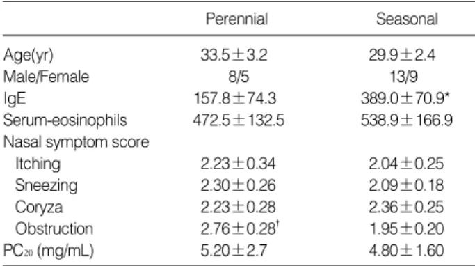
The characteristics of 35 patients (13 perennial AR with exacerbation and 22 seasonal AR) enrolled to the study are given in Table 1. Seasonal AR patients were positive in alder, birch, hazel, rye, timothy, mugwort, ragweed allergens.
Symptom scores
The mean average scores of sneezing, itching, coryza, and
Nasal Eosinophilic Inflammation and Bronchial Hyperresponsiveness 763
nasal obstruction were 2.17, 2.11, 2.31, and 2.25, respectively (2.17±1.03 vs. 2.11±1.21 vs 2.31±1.10 vs 2.25±1.03).
Nasal obstruction scores were higher in patients with perennial AR with exacerbation than in patients with seasonal AR (2.76
±0.28 vs 1.95±0.20, p<0.05).
Nasal eosinophils, IgE, blood eosinophils
The level of total IgE was higher in patients with seasonal AR than in patients with perennial AR with exacerbation (472.5±132.5 IU/L vs 389.0±70.9 IU/L, p<0.05). Nasal eosinophils were 31.7±5.1% (0-95%). There were no differ- ences of nasal eosinophils between perennial and seasonal AR (26.3±6.5% vs 34.8±7.3%). PC20was lower in patients with >10% of nasal eosinophils than in patients with <10%
of nasal eosinophils (4.06±1.36 vs 8.02±3.73). However, nasal eosinophils were closely related to log PC20(Fig. 1, r=
-0.65, p<0.01). There were no correlations between symp- tom scores and nasal eosinophils.
Bronchial hyperresponsiveness
Twenty-two patients had BHR. No significant differences in BHR were detected in the comparison of patients with and without cough, and of patients with and without postnasal drip. Significant differences in BHR were detected in the com- parison of patients with cough or postnasal drip, and without
cough or postnasal drip (Table 2, p<0.01). There were signif- icant differences of PC20in patients between with cough or postnasal drip, and without cough or postnasal drip (3.41± 3.59 mg/mL vs 10.2±1.2 mg/mL, p=0.001).
DISCUSSION
The present study showed that nasal eosinophils were corre- lated with BHR, suggesting that upper airway eosinophilic inflammation contributes to BHR.
There is a link between AR and asthma. Asthma and rhinitis can be associated with both an IgE-mediated allergic reaction and an inflammatory pattern. Twenty eight to fifty percent of asthmatic patients has AR, compared to 10-20% in the general population. Many patients with AR who have no perceived asthma symptoms have BHR to natural stimuli, such as exercise or to bronchial challenge with chemical stimuli, such as his- tamine and methacholine, especially during AR exacerbation (3). Simons (12) suggested that the new term “allergic rhino- bronchitis” accurately describes chronic allergic inflammation throughout the airways of patients with concurrent AR and asthma. He recommended, the key to management of both disorders lies in addressing the common immunopathologic mechanisms and in preventing and relieving chronic allergic inflammation, not only with appropriate pharmacologic treat- ment, but also by recommending allergen avoidance and in selected patients, specific immunotherapy. In this study nasal eosinophils were related to BHR, suggesting that the common immunopathologic mechanisms of upper and lower airway inflammation may occur.
AR is characterized by the temporal relationship of symp- toms to allergen exposure such as dust mites, pollens, animal dander, and mold spores. Symptom scores may provide a com- prehensive picture of airway disease for quality assurance or
Cough or Postnasal drip Yes 17 2 No 5 11 Table 2. Relationship of bronchial hyperresponsiveness and cough or postnasal drip
Bronchial hyperresponsiveness
Yes No
Sensitivity=89.4%. Specificity=68.7%. Overall accuracy=80.0%.
Log PC20(mg/mL)
Nasal eosinophils (%) 1.5
1
0.5 0 -0.5
-1
-1.5
Fig. 1. Correlations between nasal eosinophils and bronchial hyperresponsiveness. PC20values expressed as milligrams per milliter of methacholine.
r=-0.65 p<0.01
0 10 20 30 40 50 60 70 90 100
Age(yr) 33.5±3.2 29.9±2.4
Male/Female 8/5 13/9
IgE 157.8±74.3 389.0±70.9*
Serum-eosinophils 472.5±132.5 538.9±166.9 Nasal symptom score
Itching 2.23±0.34 2.04±0.25
Sneezing 2.30±0.26 2.09±0.18
Coryza 2.23±0.28 2.36±0.25
Obstruction 2.76±0.28� 1.95±0.20 PC20(mg/mL) 5.20±2.7 4.80±1.60
Perennial Seasonal
*p<0.05 compared with perennial allergic rhinitis with exacerbation, �p<
0.05 compared with seasonal allergic rhinitis.
PC20were defined as the methacholine concentration causing a 20%
fall in FEV1.
Table 1. Characteristics of patients with allergic rhinitis
764 A.-S. Jang
research purpose (13). Bousquet et al. (14) demonstrated that quality of life was more affected by rhinoconjuctivitis than by asthma symptoms. Nasal obstruction scores were higher in patients with perennial AR with exacerbation than in patients with seasonal AR, indicating that airway passage be more affect- ed in perennial AR.
The underlying pathologic processes are similar in the upper and lower airways. Patients with grass pollen allergy develop a moderate hyposmia during 3 weeks of natural grass pollen exposure. This is better correlated with inflammatory mediators such as eosinophilic cationic protein in the nasal secretion level than with nasal air flow measured by active anterior rhino- manometry (15). Immune effector cells responsible for allergic reactions in both the lung and the nose include, most promi- nently, mast cell, T lymphocytes, and eosinophils (16-18).
Eosinophils are characteristics for acute and chronic inflam- matory changes observed in bronchial asthma and AR, and have also been implicated in many aspects of tissue damage that occurs at sites of chronic inflammation. In this study there are no differences of nasal eosinophils between perennial and seasonal AR.
AR patients who were hyperresponsive to methacholine were at significantly greater risk of developing asthma than those with normal bronchial challenge (19). Upper airway inflam- matory processes occurring totally or primarily in the upper airway may participate in the pathogenesis of BHR and asthma (20). Perennial rhinitis is much more important than seasonal rhinitis as a risk factor for developing nonspecific BHR (21). In contrary to that study, we had no differences of BHR between seasonal and perennial AR in this study. Further follow up studies are needed to clarify risk factor for developing bronchial asthma. Rhinitis subjects with nonspecific hyperresponsiveness develop asthma more frequently than those without (3, 19).
Inflammatory cells are present, not only in the airways of pa- tients with asthma but also, in airways of patients with seasonal AR, even outside natural exposure.
In summary, the present study shows that there is a rela- tionship of nasal eosinophils and BHR, suggesting that nasal eosinophils may play a role in the development of BHR in rhinitis patients.
REFERENCES
1. Townley RG, Ryo UY, Kolotkin BM, Kang B. Bronchial sensitivity to methacholine in current and former asthmatic and allergic rhini- tis patients and control subjects. J Allergy Clin Immunol 1975; 56:
429-42.
2. Madonini E, Briatico-Vangosa G, Pappacoda A, Maccagni G, Cardani A, Saporiti F. Seasonal increase of bronchial reactivity in allergic rhinitis. J Allergy Clin Immunol 1987; 79: 358-63.
3. Ramsdale EH, Morris MM, Roberts RS, Hargreave FE. Asymptomatic bronchial hyperresponsiveness in rhinitis. J Allergy Clin Immunol 1985;
75: 573-7.
4. Corren J. The impact of allergic rhinitis on bronchial asthma. J Allergy Clin Immunol 1998; 101: S352-6.
5. Alvarez MJ, Olaguibel JM, Garcia BE, Tabar AI, Urbiola E. Compar- ison of allergen-induced changes in bronchial hyperresponsiveness and airway inflammation between mildly allergic asthma patients and allergic rhinitis patients. Allergy 2000; 55: 531-9.
6. Gutierrez V, Prieto L, Torres V, Morales C, Gonzalez E. Peak flow variability and sputum eosinophilia in allergic rhinitis. Ann Allergy Asthma Immunol 1998; 81: 143-50.
7. American Thoracic Society. Standardization of spirometry-1987 update.
Am Rev Respir Dis 1987; 136: 1285-98.
8. Morris AH, Kanner RE, Crapo RO, Gerdner RM. Clinical Pulmonary Function Testing: A manual of uniform Laboratory Procedures. 2nd ed, Salt Lake City, Intermountain Thoracic Society: 1984.
9. Choi IS, Kim JM, Park JO, Park KO. Normal standards of the maxi- mal expiratory flow-volume curve for healthy nonsmoking adults.
Korean J Intern Med 1984; 27: 192-200.
10. Kim JM, Jeong ET, Jeong WJ, Park JO, Choi IS, Park KO. Study in the normal predicted standards of spirometry for the healthy non- smoking Korean adults. Tubercul Respir Dis 1984; 31: 1-9.
11. Chai H, Farr RS, Froehlich LA, Mathison DA, Mclean JA, Rosen- thal RR, Sheffer AL, Spector SL, Townley RG. Standardization of bronchial inhalation challenge procedures. J Allergy Clin Immunol 1975; 56: 323-7.
12. Simons FE. Allergic rhinobronchitis: The asthma-allergic rhinitis link. J Allergy Clin Immunol 1999; 104: 534-40.
13. Wasserfallen JB, Gold K, Schulman KA, Baraniuk JN. Development and validation of a rhinoconjunctivitis and asthma symptom score for use as an outcome measure in clinical trials. J Allergy Clin Immunol 1997; 100: 16-22.
14. Bousquet J, Bullinger M, Fayol C, Marquis P, Valentin B, Burtin B.
Assessment of quality of life in patients with perennial allergic rhinitis with the French version of the SF-36 Health Status Questionnaire. J Allergy Clin Immunol 1994; 94: 182-8.
15. Klimek L, Eggers G. Olfactory dysfunction in allergic rhinitis is relat- ed to nasal eosinophilic inflammation. J Allergy Clin Immunol 1997;
100: 158-64.
16. Church MK, Levi-Schaffer F. The human mast cells. J Allergy Clin Immunol 1997; 99: 155-60.
17. Borish L, Rosenwasser LJ. Update on cytokines. J Allergy Clin Immunol 1996; 97: 719-34.
18. Barnes PJ. Cytokines as mediators of chronic asthma. Am J Respir Crit Care Med 1994; 150: S42-9.
19. Braman SS, Barrows AA, De Cotiis BA, Settipane GA, Corrao WY.
Airway hyperresponsiveness in allergic rhinitis. A risk factor for asth- ma. Chest 1987; 91: 671-74.
20. Eggleston PA. Upper airway inflammatory diseases and bronchial hyperresponsiveness. J Allergy Clin Immunol 1988; 81: 1036-41.
21. Verdiani P, Di Carlo S, Baronti A. Different prevalence and degree of nonspecific bronchial hyperreactivity between seasonal and perennial rhinitis. J Allergy Clin Immunol 1990; 86: 576-82.