Effects of Repetitive Transcranial Magnetic Stimulation on
Behavioral Recovery during Early Stage of Traumatic Brain Injury in Rats
Repetitive transcranial magnetic stimulation (rTMS) is a promising technique that modulates neural networks. However, there were few studies evaluating the effects of rTMS in traumatic brain injury (TBI). Herein, we assessed the effectiveness of rTMS on behavioral recovery and metabolic changes using brain magnetic resonance spectroscopy (MRS) in a rat model of TBI. We also evaluated the safety of rTMS by measuring brain swelling with brain magnetic resonance imaging (MRI). Twenty male Sprague-Dawley rats underwent lateral fluid percussion and were randomly assigned to the sham (n = 10) or the rTMS (n = 10) group. rTMS was applied on the fourth day after TBI and consisted of 10 daily sessions for 2 weeks with 10 Hz frequency (total pulses = 3,000). Although the rTMS group showed an anti-apoptotic effect around the peri-lesional area, functional
improvements were not significantly different between the two groups. Additionally, rTMS did not modulate brain metabolites in MRS, nor was there any change of brain lesion or edema after magnetic stimulation. These data suggest that rTMS did not have beneficial effects on motor recovery during early stages of TBI, although an anti-apoptosis was observed in the peri-lesional area.
Keywords: Transcranial Magnetic Stimulation; Brain Injury; Apoptosis; Recovery; Bcl-2;
BAX; TBI; rTMS Kyung Jae Yoon,1,3 Yong-Taek Lee,1,3
Pil-Wook Chung,2 Yun Kyung Lee,4 Dae Yul Kim,5 and Min Ho Chun5
1Department of Physical Medicine & Rehabilitation,
2Department of Neurology, Kangbuk Samsung Hospital, Sungkyunkwan University School of Medicine, Seoul; 3Medical Research Institute, Regenerative & Neuroscience Laboratory, Kangbuk Samsung Hospital, Seoul; 4Department of Pathology, Samsung Medical Center, Sungkyunkwan University School of Medicine, Seoul; 5Department of Rehabilitation Medicine, Asan Medical Center, University of Ulsan College of Medicine, Seoul, Korea
Received: 13 March 2015 Accepted: 10 July 2015 Address for Correspondence:
Min Ho Chun, MD
Department of Rehabilitation Medicine, Asan Medical Center University of Ulsan College of Medicine, 88 Olympic-ro 43-gil, Songpa-gu, Seoul 05505, Korea
Tel: +82.2-3010-3796, Fax: +82.2-3010-6964 E-mail: mhchun@amc.seoul.kr
Funding: This work was supported by Samsung Biomedical Research Institute grant (2012, #SMX1132351).
http://dx.doi.org/10.3346/jkms.2015.30.10.1496 • J Korean Med Sci 2015; 30: 1496-1502
INTRODUCTION
Traumatic brain injury (TBI) is a major cause of death and per- sistent disability, and functional recovery often requires exten- sive rehabilitation (1). Although direct mechanical insult is the main mechanism behind TBI, additional programmed cell death or apoptosis may lead to neuronal and glial cell loss (2, 3).
Previous trials demonstrated that anti-apoptotic effects im- proved histological and functional outcomes after TBI (4, 5). In addition, neuronal plasticity has a major role in functional re- covery after TBI (6, 7).
Repetitive transcranial magnetic stimulation (rTMS) is a non-invasive intervention that modulates neural connectivity.
Rapid magnetic field changes in the stimulation coil induce electric currents in cortical areas, which are thought to revers- ibly modulate neuronal circuits by depolarizing cortical neu- rons (8-10). Previous clinical trials showed that rTMS has bene- ficial effects on functional recovery after stroke (11-13). One of the proposed therapeutic mechanisms of rTMS is anti-apopto-
sis, putatively by increasing B-cell lymphoma 2 (Bcl-2) expres- sion (14, 15).
Till now, there are few trials which have examined the effects of rTMS on motor recovery after TBI. The purposes of this study were to investigate the effect of rTMS on behavioral recovery during the early stage of TBI in a rat model.
MATERIALS AND METHODS
Adult male Sprague-Dawley rats (n = 20, 240-280 g) were housed in laboratory cages in a controlled environment (22.0-24.0°C) and were maintained under a 12/12 hr light/dark cycle with free access to food and water.
Rats underwent lateral fluid percussion for induction of TBI as described previously (16). Briefly, rats were anesthetized with an intraperitoneal injection of 1.0% ketamine (30 mg/kg, Ketara; YuhanYanghang, Seoul, Korea). A heating pad was used to maintain the body temperature at 37 ± 0.5°C throughout the procedure. The skull was cleaned with a topical depilatory
agent and betadine solution, and a 3-mm craniectomy was per- formed 1.5 mm to the right of the midline bregma over the pri- mary motor cortex without penetrating the dura (Fig. 1A) (17).
A female Luer-Lock fitting was secured over the craniectomy site with ethyl methacrylate. A saline-filled injury device (Model 211 B4, Kistler Instrumental Corp., Amherst, NY, USA) was at- tached to the rat via a Leur-Lock. A single moderate severity pulse (3.5-4.0 atm pressure) was delivered by rapidly injecting a bolus of saline into the craniectomy site.
Twenty TBI rats were randomly assigned to either the rTMS (n = 10) group or the sham (n = 10) group. The rTMS group re- ceived 10 sessions of stimulation over a 2-week period begin- ning on the fourth day after TBI. Each daily session consisted of 15 trains of 2 sec at a rate of 10 Hz with a 1-sec inter-train inter- val (a total of 3,000 impulses; Fig. 1B). The stimulation coil was
set tangentially above the stimulation site. For sham stimula- tion, the TBI rats were stimulated with a sham coil that produced the same noise as the real magnetic coil.
Rotarod and beam balance tests were performed on post-in- jury days 4 (before rTMS), 18 (after ten sessions of rTMS), and 25 by a blinded investigator. Brain magnetic resonance imaging (MRI) and magnetic resonance spectroscopy (MRS) were per- formed on post-injury days 4, 18, and 25. There were no fatali- ties in either group during the experiment. Rats were sacrificed after the last evaluation, and the brains were collected for west- ern blot and immunohistochemical analyses.
A Magstim rapid2 magnetic stimulator (Magstim Co., Whit- land, Defeld, UK) with a figure-of-eight coil (12-mm inner di- ameter, 20-mm outer diameter, 3.5-T peak magnetic field) was used in conscious rats, as described previously (15). Biphasic stimulation with a clockwise current was applied using a 400-µs pulse. Motor evoked potentials (MEPs) in the biceps femoris muscle of the weak hind limb were measured using a Sapphire Premier device (Medelec, Old Woking, Surrey, UK), by a previ- ous described method (18). Briefly, the rats were stimulated at the site at which the stimulus of a slightly suprathreshold inten- sity produced the largest peak-to-peak MEP amplitude in the biceps femoris.
The effects of rTMS on cortical modulation also depend on the stimulation intensity. Previous trials in which 80% to 130%
of the resting motor threshold (RMT) was applied showed ther- apeutic benefits in stroke management (19). To maximize the safety of rTMS for treatment of TBI, the stimulation intensity used in this study was 80% of the RMT, the lowest possible ther- apeutic intensity. Additionally, our previous study showed that 80% of the RMT intensity had beneficial effects on motor recov- ery in a stroke rat model (15). RMT was defined as the lowest stimulator output at which the peak-to-peak MEP amplitude was greater than 5% of its maximal amplitude in at least five of the ten trials.
Rotarod and beam balance tests were performed to assess motor coordination and balance ability. Rats were put on the rotarod at an initial speed of 4 rpm, and the speed was increas- ed to 40 rpm over the course of four minutes. For the beam bal- ance test, rats were placed in the center of a wooden rectangu- lar beam (60 cm in length and 1.5 cm in diameter), which was set horizontally and suspended 40 cm above a foam pad. In both tests, the time to fall was measured. Each test was administered three times by a blinded researcher, and the results were averaged.
Rats were placed on a 4.7-T Bruker Biospec Imager (Bruker Medical Systems, Karlsruhe, Germany) and anesthetized with an intraperitoneal injection of 1.0% ketamine. Intact hemi- spheric volume (VIH), lesioned hemispheric volume (VLH), and lesion volume were measured using Paravision 3.0 software (Bruker Biospec, Ettlingen, Germany) by a blinded rater. Tissue- processing artifacts were minimized by calculating the edema A
B Fig. 1. Schematic location of the 3-mm craniectomy, which was performed at 1.5 mm to the right of the midline bregma over the primary motor cortex (A). Stimulation method of rTMS (B). Rats underwent ten daily sessions for two weeks. Each daily session consisted of 15 trains of 2 sec at a rate of 10 Hz, 80% intensity of the RMT with a 1-sec inter-train interval (3,000 impulses total). B, bregma; rTMS, repetitive transcranial magnetic stimulation; RMT, resting motor threshold.
2 sec 10 Hz 80% of RMT
1 sec Train
1
Train 2
Train 13 Day
1 Day
2 Day
9 Day
10
Train 14
Train 15 rTMS
ratio as follows: edema ratio = VLH/VIH × 100 (20). The voxel of interest was selected over the peri-lesional site (3 × 3 × 3 μL) of the subcortical area on a T2-weighted image (T2WI). All raw spectroscopic data were processed using the Bruker’s XWIN- NMR software installed in the MRI system. The metabolites were identified by the chemical shifts as observed in the MRS.
The values in parts per million were as follows: choline (Cho), 3.2 ppm; creatine (Cr), 3.03 ppm; and N-acetyl-aspartate (NAA), 2.0 ppm. Cho and NAA peak areas were quantitated as relative ratios to Cr, which is constant in the brain tissue and used as a standard (21).
At post-injury day 25, rats were anesthetized with intraperi- toneal injection of 1.0% ketamine and perfused transcardially with 50 mL of phosphate-buffered saline (PBS) and another 50 mL of 4% paraformaldehyde in 0.1 M/L PBS. The brains were removed, fixed in 10% neutral-buffered formalin, dehydrated in a graded ethanol series, and embedded in paraffin for im- munohistochemistry, and they were obtained from the peri-le- sional area. Each block was sectioned at a 6 μm-thickness and mounted on poly-L-lysine coated slides. These slides under- went a serial process of deparaffinization with xylene, dehydra- tion with ethanol, washing with saline, and microwave in citrate buffer for 10 min. Hydrogen peroxide was used to block endog- enous peroxidase activity for 30 min to reduce the non-specific immunoreactivity. Primary antibodies against B-cell lympho- ma 2 (Bcl-2, 1/100, Abcam, Cambridge, MA, USA) and Bcl-2 associated X protein (BAX, 1/100, Abcam) were employed. Bcl- 2 and BAX are anti-apoptotic proteins, as previously described (22). Anti-mouse, HRP/DAB (Labvision, Fremont, CA, USA) was used as the secondary antibody. Immunostained sections were digitized using a 400 × objective (Leica DFC 290, Leica, Heerbrugg, Germany) in conjunction with the Leica Applica-
tion suite (version 3.3.0, Leica). Five views obtained from the peri-lesional area were evaluated by a blinded pathologist, and the stained cells were counted and averaged.
Brains were removed at post-injury day 25 and frozen at -20°C for western blots. The tissues from the peri-lesional area in brains were selected for the blots. Proteins were extracted with a series of procedures including mixing with the protein extraction so- lution, homogenization by vortexing for 5 min, and harvesting the supernatants after centrifugation for 10 min at 15,000 rpm.
Total protein amounts were assayed with the Bradford’s meth- od. Proteins were resolved on a SDS-PAGE gel, and blotting was performed for 90 min. Primary antibodies were applied onto the nitrocellulose membrane for 2 hr. After washing, secondary antibodies were added to the membrane for 1 hr. Primary anti- bodies against B-cell lymphoma 2 (Bcl-2, 1/100, Abcam) and Bcl-2 associated X protein (BAX, 1/1,000, Abcam) were applied for protein analyses. An antibody for glyceraldehyde-3-phos- phate dehydrogenase (GAPDH) was used as a loading control.
Relative band density was assessed with Image J software.
Statistical analysis was performed with PASW software pack- age, version 18.0. Student’s t-test was applied to evaluate the differences in behavioral tests, edema ratio, and metabolites between the two groups on post-injury day 4. Repeated-mea- sures ANOVA was used to analyze behavioral tests, lesion vol- ume, edema ratio, and relative ratios of metabolites. Immunos- taining and western blot data were analyzed using the Student’s t-test. P values less than 0.05 were considered significant.
Ethics statement
All protocols for this study were approved by institutional ani- mal care and use committee at medical research institute of Kangbuk Samsung Hospital, Seoul.
Sham rTMS
Seconds
D4 D18 D25
Post-op Rotarod 200
150
100
50
0
A
Seconds
D4 D18 D25
Beam balance
Post-op 50
40 30 20 10
0
B Fig. 2. Results of Rotarod (A) and beam balance tests (B). There are no significant differences between the two groups (sham, n = 10 rats; rTMS, n = 10 rats). D4, 4 days after injury; D18, 18 days after injury; D25, 25 days after injury.
rTMS Sham
Ratio
D4 D18 D25 NAA/Cr
Post-op 2.0
1.5 1.0 0.5 0 rTMS
Sham
Ratio
D4 D18 D25 Cho/Cr 2.0
1.5 1.0 0.5 0
Post-op
A B C
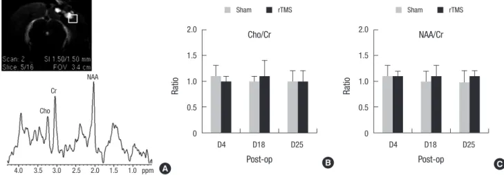
Fig. 3. Results of metabolites with MRS. T2WI showed the location of MRS voxel (white squre, 3 × 3 × 3 μL) in the peri-lesional site (A). There are no significant differences in Cho/Cr (B) or NAA/Cr (C) ratios between the two groups (sham, n = 10 rats; rTMS, n = 10 rats). Cho, choline; Cr, creatine; NAA, N-acetyl-aspartate; D4, 4 days after injury; D18, 18 days after injury; D25, 25 days after injury.
Cho Cr
NAA
4.0 3.5 3.0 2.5 2.0 1.5 1.0 ppm
rTMS Sham
Volume (μL)
D4 D18 D25
Lesion volume 1.5
1.0
0.5
0
Post-op
rTMS Sham
Ratio (%)
D4 D18 D25
Edema ratio 2.0
1.5 1.0 0.5 0
Post-op
B
C Fig. 4. Results of brain swelling with MRI (A). There is no significant difference in lesion volume or brain edema between the two groups (B & C). rTMS, repetitive transcranial magnetic stimulation; D4, 4 days after injury; D18, 18 days after injury; D25, 25 days after injury.
rTMS Sham
D4
D18
D25
A
RESULTS Behavioral tests
Baseline rotarod and beam balance data on post-injury day 4 did not show significant differences between the rTMS and sham groups. There were no significant group × time interac-
tions for the rotarod and beam balance tests among two groups (Fig. 2).
There were no significant differences in baseline MRS data for Cho/Cr and NAA/Cr ratios between the two groups. Group × time interactions for Cho/Cr and NAA/Cr ratios were not signifi- cant among two groups (Fig. 3).
Bcl-2
Sham rTMS
BAX
rTMS Sham
No. of stained cells
* *
Bcl-2 BAX
100 80 60 40 20
A 0 B
Fig. 5. Results of immunohistochemical staining with Bcl-2 and BAX (A). A significantly more number of Bcl-2 stained cells and a lesser number of BAX stained cells in the rTMS group compared with the sham group around the peri-lesional area (B). sham, n=10 rats; rTMS, n=10 rats. *P < 0.05, Student’s t-test. Bcl-2, B-cell lymphoma 2; BAX, Bcl-2 associated X protein; rTMS, repetitive transcranial magnetic stimulation.
B
Relative density (%)
Bcl-2 BAX
* *
150 120 90 60 30 0
rTMS Sham
Sham rTMS
Bcl-2
BAX
GAPDH
A
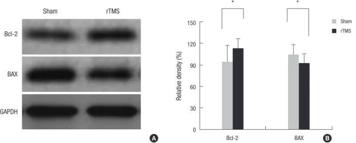
Fig. 6. Results of western blot showed Bcl-2 and BAX bands (A). A significant increase in Bcl-2 expression and a decrease in BAX expression is observed in the rTMS group compared with the sham group (B). sham, n = 10 rats; rTMS, n = 10 rats. *P < 0.05, Student’s t-test. Bcl-2, B-cell lymphoma 2; BAX, Bcl-2 associated X protein; GAPDH, glyc- eraldehyde-3-phosphate dehydrogenase; rTMS, repetitive transcranial magnetic stimulation.
Brain MRI
Baseline data for brain swelling on post-injury day 4 did not show significant differences between the two groups. There was no significant group × time interaction for lesion volume or brain swelling between the groups (Fig. 4).
Immunohistochemistry
Immunohistochemical analyses of Bcl-2 and Bax staining showed that the rTMS group had a significantly greater number
of Bcl-2 stained cells and a lesser number of BAX stained cells around the peri-lesional area compared with the sham group (P < 0.05; Student’s t-test; Fig. 5).
Western blot
The densitometric analyses of western blots showed that the rTMS group had a significantly higher Bcl-2 expression and a lower BAX expression compared with the sham group (P < 0.05;
Student’s t-test; Fig. 6).
DISCUSSION
This is the first report to investigate whether application of rTMS for TBI has a therapeutic effect on motor recovery or not. The major finding is that rTMS did not improve motor function dur- ing the early stage of TBI, although an anti-apoptotic effect was observed in the peri-lesional area without aggravation of brain injury.
Previous experiments showed that rTMS has an anti-apopto- sis in the peri-ischemic areas in models of acute and subacute ischemic stroke (14, 15). The peri-ischemic region, known as
‘ischemic penumbra’, is functionally silent due to impaired blood supply, but it sustains minimal metabolic activities (23). There- fore, most of the apoptosis is observed in the ischemic penum- bra (24-26). In this study, the rTMS group demonstrated signifi- cantly enhanced expression of Bcl-2 and reduced expression of BAX, which are known the anti-apoptotic and pro-apoptotic proteins, respectively (Fig. 5) (22, 27, 28). Apoptosis triggered by TBI leads to progressive neuronal loss (2, 3). However, the ob- served anti-apoptosis did not induce motor recovery in rat TBI model, which could indicate that the mechanical damages caus- ing necrosis of tissue, cell swelling, and plasma membrane rup- ture were more significant than apoptosis in early stage of TBI (29). These could be more serious when the intensity of trauma is severe. Therefore, anti-apoptosis induced by rTMS might be unable to suppress direct cellular injury during the early stage of moderate TBI. The present results are in agreement with those of previous reports, in which Bcl-2 over-expression did not elicit functional improvement in a TBI model (30-32).
Additionally, the damaged brain might be more vulnerable to excitation during the early injury period. Previous studies showed that early exercise aggravated brain injury in an animal model of stroke (33, 34). Considering the previous trials which reported that TBI patients require a longer recovery time than stroke patients (35, 36), it can be argued that the application of rTMS to the damaged brain during the early period of injury counteracted the beneficial effects of anti-apoptosis induced by rTMS after TBI.
Cho and NAA measured by 1H MR spectroscopy are the structural component of cellular membrane and a predecessor of brain lipids, respectively (37). In states of neuronal damage, including TBI, the Cho level is expected to increase with a de- crease of the NAA level. In this study, Cho/Cr and NAA/Cr ratio measured by MRS were not significantly different among the groups (Fig. 3). Additionally, MRI findings showed that lesion volume and brain edema was not improved nor aggravated af- ter rTMS (Fig. 4). These results meant that rTMS was neutral ef- fect on neuronal recovery or deterioration in early stage of mod- erate TBI.
There are some limitations to this study. First of all, we did not include a healthy group. The inclusion of these groups
could have explained the therapeutic effects of rTMS compared to those in the normal control group, and it could also have helped to clarify the effect of TBI on motor function. Addition- ally, adverse events such as seizures or tremors were not mea- sured during the experimental period. Therefore, we could not report any other adverse events associated with rTMS, except for lesion volume, brain edema and metabolic changes.
In conclusions, rTMS did not have beneficial effects on mo- tor recovery during early stages of moderate TBI, although an anti-apoptosis was observed in the peri-lesional area. Although behavioral data did not support the claim that rTMS is benefi- cial for treatment of TBI, further studies should be performed in different periods or severities of TBI, using diverse rTMS pro- tocols and more sensitive behavioral tests to evaluate the po- tential therapeutic effects of rTMS.
DISCLOSURE
There was no potential conflict of interest in the experiment.
AUTHOR CONTRIBUTION
Chun MH designed the study and Yoon KJ performed the ex- periment. Lee YT provided the idea of experiment and Lee YK analyzed the data of immunohistochemistry. Chung PW ana- lyzed data. Yoon KJ, Kim DY, and Chun MH wrote the manu- script. All authors contributed writing the article.
ORCID
Kyung Jae Yoon http://orcid.org/0000-0002-2765-4309 Min Ho Chun http://orcid.org/0000-0001-8666-7225
REFERENCES
1. Ragnarsson KT. Results of the NIH consensus conference on “rehabilita- tion of persons with traumatic brain injury”. Restor Neurol Neurosci 2002;
20: 103-8.
2. Raghupathi R, Graham DI, McIntosh TK. Apoptosis after traumatic brain injury. J Neurotrauma 2000; 17: 927-38.
3. Miñambres E, Ballesteros MA, Mayorga M, Marin MJ, Muñoz P, Figols J, López-Hoyos M. Cerebral apoptosis in severe traumatic brain injury pa- tients: an in vitro, in vivo, and postmortem study. J Neurotrauma 2008;
25: 581-91.
4. Knoblach SM, Alroy DA, Nikolaeva M, Cernak I, Stoica BA, Faden AI.
Caspase inhibitor z-DEVD-fmk attenuates calpain and necrotic cell death in vitro and after traumatic brain injury. J Cereb Blood Flow Metab 2004;
24: 1119-32.
5. Soustiel JF, Palzur E, Nevo O, Thaler I, Vlodavsky E. Neuroprotective an- ti-apoptosis effect of estrogens in traumatic brain injury. J Neurotrauma 2005; 22: 345-52.
6. Monnerie H, Tang-Schomer MD, Iwata A, Smith DH, Kim HA, Le Roux
PD. Dendritic alterations after dynamic axonal stretch injury in vitro.
Exp Neurol 2010; 224: 415-23.
7. Hu B, Liu C, Bramlett H, Sick TJ, Alonso OF, Chen S, Dietrich WD.
Changes in trkB-ERK1/2-CREB/Elk-1 pathways in hippocampal mossy fiber organization after traumatic brain injury. J Cereb Blood Flow Metab 2004; 24: 934-43.
8. Chouinard PA, Van Der Werf YD, Leonard G, Paus T. Modulating neu- ral networks with transcranial magnetic stimulation applied over the dorsal premotor and primary motor cortices. J Neurophysiol 2003; 90:
1071-83.
9. Siebner HR, Peller M, Willoch F, Minoshima S, Boecker H, Auer C, Drzezga A, Conrad B, Bartenstein P. Lasting cortical activation after re- petitive TMS of the motor cortex: a glucose metabolic study. Neurology 2000; 54: 956-63.
10. Strafella AP, Paus T. Cerebral blood-flow changes induced by paired- pulse transcranial magnetic stimulation of the primary motor cortex. J Neurophysiol 2001; 85: 2624-9.
11. Khedr EM, Ahmed MA, Fathy N, Rothwell JC. Therapeutic trial of repet- itive transcranial magnetic stimulation after acute ischemic stroke. Neu- rology 2005; 65: 466-8.
12. Mansur CG, Fregni F, Boggio PS, Riberto M, Gallucci-Neto J, Santos CM, Wagner T, Rigonatti SP, Marcolin MA, Pascual-Leone A. A sham stimu- lation-controlled trial of rTMS of the unaffected hemisphere in stroke pa- tients. Neurology 2005; 64: 1802-4.
13. Takeuchi N, Chuma T, Matsuo Y, Watanabe I, Ikoma K. Repetitive tran- scranial magnetic stimulation of contralesional primary motor cortex improves hand function after stroke. Stroke 2005; 36: 2681-6.
14. Gao F, Wang S, Guo Y, Wang J, Lou M, Wu J, Ding M, Tian M, Zhang H.
Protective effects of repetitive transcranial magnetic stimulation in a rat model of transient cerebral ischaemia: a microPET study. Eur J Nucl Med Mol Imaging 2010; 37: 954-61.
15. Yoon KJ, Lee YT, Han TR. Mechanism of functional recovery after repeti- tive transcranial magnetic stimulation (rTMS) in the subacute cerebral ischemic rat model: neural plasticity or anti-apoptosis? Exp Brain Res 2011; 214: 549-56.
16. McIntosh TK, Vink R, Noble L, Yamakami I, Fernyak S, Soares H, Faden AL. Traumatic brain injury in the rat: characterization of a lateral fluid- percussion model. Neuroscience 1989; 28: 233-44.
17. Fonoff ET, Pereira JF Jr, Camargo LV, Dale CS, Pagano RL, Ballester G, Teixeira MJ. Functional mapping of the motor cortex of the rat using transdural electrical stimulation. Behav Brain Res 2009; 202: 138-41.
18. Ogiue-Ikeda M, Kawato S, Ueno S. The effect of repetitive transcranial magnetic stimulation on long-term potentiation in rat hippocampus de- pends on stimulus intensity. Brain Res 2003; 993: 222-6.
19. Fitzgerald PB, Brown TL, Daskalakis ZJ, Chen R, Kulkarni J. Intensity- dependent effects of 1 Hz rTMS on human corticospinal excitability. Clin Neurophysiol 2002; 113: 1136-41.
20. Kline RA, Negendank WG, McCoy LE, Lester M, Berguer R. MRI quan- titation of edema in focal cerebral ischemia in cats: correlation with cy- tochrome aa3 oxidation state. Magn Reson Med 1990; 13: 319-23.
21. Arnold DL, Matthews PM. Practical aspects of clinical applications of
MRS in the brain. In: Charles CH, Young IR. eds. MR spectroscopy: clini- cal applications and techniques. London: Martin Dunitz, 1996, p139-59.
22. Antonsson B. Bax and other pro-apoptotic Bcl-2 family “killer-proteins”
and their victim the mitochondrion. Cell Tissue Res 2001; 306: 347-61.
23. Ginsberg MD, Zhao W, Alonso OF, Loor-Estades JY, Dietrich WD, Busto R. Uncoupling of local cerebral glucose metabolism and blood flow after acute fluid-percussion injury in rats. Am J Physiol 1997; 272: H2859-68.
24. Charriaut-Marlangue C, Margaill I, Represa A, Popovici T, Plotkine M, Ben-Ari Y. Apoptosis and necrosis after reversible focal ischemia: an in situ DNA fragmentation analysis. J Cereb Blood Flow Metab 1996; 16:
186-94.
25. Clemens JA, Stephenson DT, Smalstig EB, Dixon EP, Little SP. Global ischemia activates nuclear factor-kappa B in forebrain neurons of rats.
Stroke 1997; 28: 1073-80; discussion 80-1.
26. Vexler ZS, Roberts TP, Bollen AW, Derugin N, Arieff AI. Transient cere- bral ischemia. Association of apoptosis induction with hypoperfusion. J Clin Invest 1997; 99: 1453-9.
27. Clark RS, Chen J, Watkins SC, Kochanek PM, Chen M, Stetler RA, Loef- fert JE, Graham SH. Apoptosis-suppressor gene bcl-2 expression after traumatic brain injury in rats. J Neurosci 1997; 17: 9172-82.
28. Reed JC. Proapoptotic multidomain Bcl-2/Bax-family proteins: mecha- nisms, physiological roles, and therapeutic opportunities. Cell Death Differ 2006; 13: 1378-86.
29. Verma A. Opportunities for neuroprotection in traumatic brain injury. J Head Trauma Rehabil 2000; 15: 1149-61.
30. Raghupathi R, Fernandez SC, Murai H, Trusko SP, Scott RW, Nishioka WK, McIntosh TK. BCL-2 overexpression attenuates cortical cell loss af- ter traumatic brain injury in transgenic mice. J Cereb Blood Flow Metab 1998; 18: 1259-69.
31. Nakamura M, Raghupathi R, Merry DE, Scherbel U, Saatman KE, Mc- Intosh TK. Overexpression of Bcl-2 is neuroprotective after experimental brain injury in transgenic mice. J Comp Neurol 1999; 412: 681-92.
32. Tehranian R, Rose ME, Vagni V, Griffith RP, Wu S, Maits S, Zhang X, Clark RS, Dixon CE, Kochanek PM, et al. Transgenic mice that overex- press the anti-apoptotic Bcl-2 protein have improved histological out- come but unchanged behavioral outcome after traumatic brain injury.
Brain Res 2006; 1101: 126-35.
33. Humm JL, Kozlowski DA, Bland ST, James DC, Schallert T. Use-depen- dent exaggeration of brain injury: is glutamate involved? Exp Neurol 1999; 157: 349-58.
34. Risedal A, Zeng J, Johansson BB. Early training may exacerbate brain damage after focal brain ischemia in the rat. J Cereb Blood Flow Metab 1999; 19: 997-1003.
35. Katz DI, Alexander MP, Klein RB. Recovery of arm function in patients with paresis after traumatic brain injury. Arch Phys Med Rehabil 1998;
79: 488-93.
36. Jang SH. Review of motor recovery in patients with traumatic brain in- jury. NeuroRehabilitation 2009; 24: 349-53.
37. Kornienko VN, Pronin IN. Diagnostic neuroradiology. Berlin: Springer- Verlag, 2009, p18-9.