황금추출물의 지방장벽 생성을 통한 염증완화 효과
안상현
1⋅김기봉
2,3,*1세명대학교 한의과대학 해부학교실, 2부산대학교 한의학전문대학원 한방소아과학교실
3부산대학교한방병원 한방소아과
Received: December 21, 2020 ∙ Revised: February 11, 2021 ∙ Accepted: February 22, 2021
*Corresponding Author: Kibong Kim
Department of Korean Pediatrics, Pusan National University Korean Medicine Hospital, Geumo-ro 20, Mulgeum-eup, Yangsan-si, Gyeongsangnam-do, 50612, Republic of Korea
Tel: +82-55-360-5952, Fax: +82-55-360-5952, E-mail: [email protected]
ⓒ The Association of Pediatrics of Korean Medicine. All rights reserved. This is an open-access article distributed under the tenus of the Creative Commons Attribution Non-Commercial License (http://creativecommons.org/licenses/by-nc/3.0/), which permits unrestricted non-commercial use, distribution, and reproduction in any medium, provided the original work is properly cited.
Abstract
Effect of Anti-Inflammation through Creation of Skin Fat Barrier on Scutellaria baicalensis extract
Ahn Sang Hyun1⋅Kim Ki Bong2,3,*
1Dept. of anatomy, college of Korean Medicine, Semyung University
2School of Korean Medicine, Pusan National University
3Dept. of Korean Pediatrics, Korean Medicine Hospital, Pusan National University
Objectives
The purpose of this study is to confirm the effect of alleviating inflammation through creating skin fat barrier from Scutellaria baicalensis extract.
Methods
Four-week-old Balb/C mice were divided into four groups: control group (Ctrl), lipid barrier eliminated group (LBEG), dexamethasone (DM) treated group after lipid barrier elimination (DMTG), and Scutellaria baicalensis (SB) treated group after lipid barrier elimination (SBTG). Scutellaria baicalensis extract were administered for 5 days after removal of the fat barrier. Changes in skin condition, improvement of the fat barrier, and relief of inflammation were observed in each group.
Results
Compared to LBEG and DMTG, pathological skin damage and tissue changes were less in SBTG, and transepidermal water loss (TEWL) and pH were also significantly reduced. Filaggrin was also significantly increased in SBTG. KLK7, PAR-2, and TSLP in SBTG also showed significant reduction compared to the LBEG and DMTG.
Conclusions
Scutellaria baicalensis extract restores skin barrier and relieves inflammation through the creation of skin fat barrier.
This means that the Scutellaria baicalensis extract can regulate Th2 differentiation through the creation of the epithelial fat barrier.
Key words: Scutellaria baicalensis, Skin Fat Barrier, Filaggrin, Inflammation
ISSN 2287-9463(Online), https://doi.org/10.7778/jpkm.2021.35.1.40
Ⅰ. Introduction
피부는 주변 환경과 인체를 분리하는 동시에 인체 를 보호하는 1차 장벽의 역할을 담당한다1). 피부는 표 피와 진피로 나누어지고, 이 중 표피는 각질층 (stratum corneum, SC), 과립층 (granular layer), 가시층 (spinous layer), 기저층 (basal layer)으로 이루어져 있으며, 병원 균, 독소, 건조제와 같은 다양한 외부 유해 요인으로부 터 신체를 보호한다2). 표피의 장벽 기능은 주로 표피의 가장 바깥 층인 SC에서 작동하며, SC는 각질세포와 다 른 세포 사이에 층상 구조를 형성하는 표피 지질의 연 속적인 층을 형성한다. 각질세포는 각질을 생성하는 표피세포로서 표피의 95%를 차지하며, 각질유리과립 (keratohyalin granule)과 층판소체 (lamella body; LB)를 생성한다. 필라그린 (Filaggrin, FLG)은 SC의 세포 사이 성분으로 작용하는 대표적인 각질유리과립이다3). 이러 한 피부 장벽이 손상되면 경피 수분 손실 (transepidermal water loss, TEWL)이 증가하게 되고, 가려움과 관련된 신경 섬유의 활성화, 외부 항원의 침투 및 피부 염증 등을 유발하게 된다4). 따라서 피부 장벽 회복은 피부 질환의 악화를 예방하는 가장 기본적인 치료법이라 할 수 있다.
아토피피부염 (Atopic dermatitis, AD)은 피부 장벽 기능이 감소되어 나타나는 대표적인 만성 염증성 피부 질환이다. 일반적으로 AD에서 FLG의 발현 감소가 두 드러지게 관찰되며, 이러한 FLG의 발현 감소는 Th2 분 화로 인한 염증 반응에 의해 발생하게 된다5,6). 따라서 AD는 건조한 피부, 가려움증, TEWL 증가, 피부 pH 증가 등이 특징적으로 나타난다. FLG 유전자 이상이 있는 소아의 경우 알레르기 행진 (allergic march)을 따 라 더 빨리 진행된다. 따라서 FLG의 회복은 알레르기 질환 치료에서 가장 중요한 부분이라 할 수 있다7).
본 연구에 사용된 황금 (Scutellaria baicalensis, 黃芩)은 임상현장에서 많이 사용되는 약재로서, 청열조습 (淸 熱燥濕), 청열해독 (淸熱解毒)하여 폐열해수 (肺熱咳 嗽), 습열사리(濕熱瀉利), 열림(熱淋), 붕루(崩漏) 같은 내과적 질환과 옹종(癰腫), 정창 (疔瘡) 등과 같은 외과 적 질환에 모두 다용되며, 습진, 건선, 피부염 등에 응 용될 수 있다8,9). 또한 황금추출물에 대한 최근 연구들
10-12)을 통해 황금추출물의 피부에서의 항염증 효과와
표피 회복 효과에 대한 가능성을 확인하였다.
본 연구는 황금추출물 투여 후 피부상태 변화를 확
인하기 위해 TEWL, pH를 관찰하였고, 지방장벽 구조 단백질 변화를 확인하기 위해 피부각질층 내 FLG를 관 찰하였으며, 세린 프로테아제 (serine protease, SP)의 활 성화를 확인하기 위해 각질층내 Kallikrein-related pepti- dase (KLK) 7, Protease activated receptor (PAR)-2, Thymic stromal lymphopoietin (TSLP)를 관찰하였다. 이 연구를 통해 황금추출물의 피부 장벽 생성을 통한 염증완화 효과를 확인하였기에 보고하는 바이다.
Ⅱ. Materials and Methods
1. 실험동물
자바이오 (대한민국)에서 분양 받은 태령 4주 Balb /C계 수컷 생쥐를 무균사육장치내에서 2주일 동안 적 응시킨 후 체중 20 ±1.5g 된 생쥐를 선별하여 사용하였 다. 대조군 (Ctrl), 지방장벽 제거군 (lipid barrier elimi- nated group, LBEG), 지방장벽 제거후 dexamethasone 처 리군 (dexamethasone (DM) treated group after lipid bar- rier elimination, DMTG), 지방장벽 제거후 황금추출물 투여군 (Scutellaria baicalensis (SB) treated group after lipid barrier elimination, SBTG)으로 나누었다. 각 군에 7마 리씩 배정하였다. 동물실험은 세명대학교 동물실험윤 리위원회의 승인 (IACUC No. smecae-20-06-02) 후 실 시되었고, 실험실 동물의 관리와 사용에 대해서는 NIH 가이드라인에 따라 시행되었다.
2. 황금추출물의 제조
황금 200 g을 증류수 2000 ㎖에 넣고 3시간 동안 전탕한 후 여과하였다. 그 여액을 rotary evaporator를 이용하여 50 ㎖으로 감압, 농축한 후 동결 건조하여 추 출물 54.6 g (수득률 27.3%) 획득하였다.
3. 지방장벽 제거와 약물 투여
지방장벽 제거는 생쥐 등쪽 부위 피부를 면도한 다 음 Tape (3M, USA)를 이용하여 각질층의 desqumation 을 제거하였다. 10% sodium dodeecyl sulfate (SDS :Sigma, USA) 500 ㎕를 도포한 후 면봉을 이용하여 20 회 문질러서 각질층의 lipid lamella를 제거하였다. 그런 다음 황금추출물 73 ㎎/㎏량을 생리식염수에 녹인 후 0.2 ㎖씩 SBTG에 5일 동안 경구투여하였다. 대조약물
로 사용된 dexamathasone (Sigma-Aldrich, USA)는 1 ㎎/
㎏량으로 5일 동안 DMTG에 0.2 ㎖씩 경구투여하였다.
4. TEWL과 pH 측정
지방장벽 제거 후 TEWL은 Vapometer (Delfin Technologies, Finland)로 측정하였다. 피부 pH 변화는 SKIN-O-MET (COSMOMED, Germany)으로 측정하였다.
5. 조직표본
피부는 vascular rinse와 10% 중성 포르말린용액 (neutral buffered formalin, NBF)으로 심장관류고정을 실시하였다. 얻어진 등쪽 피부를 10% NBF에 실온에 서 24시간 동안 고정한 후 통상적인 방법으로 paraffin 에 포매하고 5 ㎛ 두께로 연속절편을 만들었다.
6. 조직화학
상피조직내 염증으로 기인된 조직 변화를 관찰하기 위해 Phloxine-tartrazine 염색법을 실시하였다. Mayer's hematoxylin에 5분간 핵 염색한 후 phloxine 용액에 30 분간 반응시켰다. 그런 다음 tartrazine 용액에서 분별 후 관찰하였다.
7. 면역조직화학
상피내 염증관련 미세환경의 면역조학학적 변화를 조사하기 위해 항 filaggrin, Kallikrein (KLK) 7, pro- tease-activated receptor (PAR)-2, thymic stromal lympho- poietin (TSLP) 등의 항체를 이용한 면역조직화학적 염 색을 실시하였다. 우선 피부절편을 proteinase K (20 ㎍ /㎖, Dako, USA)에 5분 동안 proteolysis 과정을 거친 후 1% fetal bovine serum (Sigma, USA)이 포함된 10% nor- mal goat serum (Vector Lab, USA)에서 1시간 동안 blocking 반응시켰다. 1차 항체인 mouse anti-filaggrin (1:100, Santa Cruz Biotec, USA), mouse anti-KLK7 (1:100, Santa Cruz Biotec), mouse anti-PAR-2 (1:100, Santa Cruz Biotec), mouse anti-TSLP (1:100, Abcam, USA)에 4 ℃ humidified chamber에서 72시간 동안 반응 시켰다. 2차 항체인 biotinylated goat anti-mouse IgG (1:100, Abcam)에 실온에서 24시간 link 하였고, avidin biotin complex kit (Vector Lab)에 1시간 동안 실온에서 반응시켰다. 0.05% 3,3'-diaminobenzidine과 0.01% HCl 이 포함된 0.05 M tris-HCl 완충용액 (pH 7.4)에서 발색
시킨 후, hematoxylin으로 대조염색하였다.
8. 영상분석
면역조직화학의 결과는 image Pro Plus (Media cy- bernetics, USA)를 이용한 영상분석을 통해 수치화 (means ± standard error) 되었다. 각 군의 피부표본 10개 를 임의로 선정한 후 x400 배율에서 촬영한 다음 pos- itive pixels (intensity 80 ~100)/20,000,000 pixels로 영상 분석하였다.
9. 통계
통계는 SPSS software (SPSS 25, SPSS Inc., USA)를 사 용하였으며, one-way ANOVA 시행을 통해 유의성 (p<0.05)을 검증하고, 사후 검증은 Tukey HSD를 실시 하였다.
Ⅲ. Results
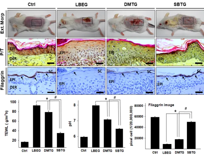
1. 피부손상 회복
LBEG과 DMTG에서 홍반 (erythema), 출혈 (hemor- rhage), 딱지 (scarring), 미란 (erosion)이 관찰되었다. 이 에 반해 SBTG에서는 딱지와 미란이 없었으며, 피부 손 상 역시 적었다 (Fig. 1.). LBEG과 DMTG에서는 상피 세포과형성, 가시층에서의 세포사이공간의 확장, 기저 층에 림프구 침윤증가와 바닥면 붕괴가 관찰되었다.
반면에 SBTG의 상피에서는 LBEG와 DMTG에 비해 이 런 손상이 적었다 (Fig. 1).
각질층에서의 Filaggrin의 변화는 지방장벽제거 후 LBEG, DMTG, SBTG 모두에서 감소하였다. Ctrl (58,964
± 1,175/20,000,000 pixel)에 비해 LBEG는 85% 감소 (9,051 ± 378/20,000,000 pixel)하였으며, DMTG는 70% 감소 (17,778 ± 297/20,000,000 pixel)하였고, SBTG는 15% 감소 (50,191± 990/20,000,000 pixel)하였 다. SBTG의 filaggrin 양성반응은 LBEG에 비해 455%
많았으며, DMTG에 비해 182% 많았다 (Fig. 1.).
TEWL는 LBEG (92.9 ± 2.1 g/m2h), DMTG (78.8 ± 2.5 g/m2h), SBTG (34.7± 2.1 g/m2h), Ctrl (16.7 ± 0.4 g/m2h) 순이었다 (Fig. 1.)
pH는 LBEG (7.97 ± 0.07), DMTG (7.08 ± 0.04), SBTG (6.48 ± 0.04), Ctrl (5.97 ± 0.04) 순이었다 (Fig. 1.).
2. 염증 조절
KLK7 양성반응은 각질층과 과립층에서 강한 양성 반응을 보였는데, LBEG, DMTG, SBTG 모두에서 증가 하였다. Ctrl (6,588 ± 256/20,000,000 pixel)에 비해 LBEG 는 1120% 증가 (80,343 ± 1,251/20,000,000 pixel)하였으 며, DMTG는 734% 증가 (54,926 ± 1,251/20,000,000 pix- el)하였고, SBTG는 366% 증가 (30,809 ± 810/20,000,000 pixel)하였다. SBTG의 KLK7 양성반응은 LBEG에 비해 62% 적었으며, DMTG에 비해 44% 적었다 (Fig. 2.).
PAR2 양성반응은 피부 상피 모든 층에서 강한 양성 반응을 보였으며, LBEG, DMTG, SBTG 모두에서 증가 하였다. Ctrl (6,338 ± 185/20,000,000 pixel)에 비해 LBEG 는 1,221% 증가 (83,750 ± 995/20,000,000 pixel)하였으
며, DMTG는 517% 증가 (39,078 ± 928/20,000,000 pixel) 하였고, SBTG는 243% 증가 (21,760 ± 689/20,000,000 pixel)하였다. SBTG의 PAR2 양성반응은 LBEG에 비해 74%, DMTG에 비해 44% 적었다 (Fig. 2.).
TSLP 양성반응은 피부 상피 각질층과 과립층에서 강한 양성반응을 보였으며, LBEG, DMTG, SBTG 모두 에서 증가하였다. Ctrl (67,658 ± 184/20,000,000 pixel)에 비해 LBEG는 797% 증가 (68,718 ± 1,206/20,000,000 pixel)하였으며, DMTG는 481% 증가 (44,475 ± 1,041/
20,000,000 pixel)하였고, SBTG는 304% 증가 (30,919
± 1,173/20,000,000 pixel)하였다. SBTG의 TSLP 양성반 응은 LBEG에 비해 55%, DMTG에 비해 30% 적었다 (Fig. 2.).
Figure 1. The alleviation effects on symptoms of lipid barrier elimination by SB.
SB relieved the lipid barrier destruction -induced skin lesions (erythema, hemorrhage, scarring, erosion, and excoriation) in SBTG (phloxine-tartrazine).
The expression of filaggrin (arrow indicates light brown particle) was significantly increased in SBTG as compared with LBEG and DMTG, the data of filaggrin image analysis showed the same results (Filaggrin immunohistochemistry), The TEWL and pH is decreased in SBTG, but increased in LBEG and DMTG. Abbreviations. SB, Scutellaria baicalensis Ctrl, normal; LBEG, lipid barrier eliminated group; DMTG, dexamethasone (DM) treated group after lipid barrier elimination; SBGT, Scutellaria baicalensis treated group after lipid barrier elimination; TEWL, Transepidermal water loss; P/T, phloxine-tartrazine; EPI, epithelium; Der, Dermis; SC, stratum corneum; Square, lipid barrier elimination induced Skin lesion; Bar size, 50 ㎛; *, p < 0.05 compared with LBEG; #, p < 0.05 compared with DMTG.
Ⅳ. Discussion
황금은 성미가 苦寒하고 청열조습 (淸熱燥濕), 사화 해독 (瀉火解毒), 지혈, 안태 (安胎)하는 효능이 있어 폐 열해수 (肺熱咳嗽), 서온흉민 (暑溫胸悶), 습열비만 (濕 熱痞滿), 열림 (熱淋), 사리 (瀉痢), 황달 (黃疸), 토혈 (吐血), 육혈 (衄血), 붕루 (崩漏), 옹종창독 (癰腫瘡毒), 태동불안 (胎動不安) 등에 사용되어 왔다8). 황금은 대 표적인 항염증 약물로써, 임상 현장에서도 피부질환에 많이 사용되고 있으며, 현재 다양한 연구들이 진행되 고 있다. 전 등은 접촉피부염에서 황금 열수추출물을 경구 투여한 후 ear edema, 염증세포 침윤 등을 관찰하 여 피부 내 항염증 효과를 보고하였다13). 또한 김 등은 황금추출물이 접촉피부염과 아토피피부염 유발 흰쥐 에서 항산화기능과 표피 회복 기능이 있음을 보고하였
다11), 이러한 연구 결과는 황금은 항산화 효과 및 항염 증 효과가 있어 표피 회복를 시켜주며 아토피피부염 같은 피부질환에 치료 효과가 있음을 의미한다.
피부에서 SC는 주로 인체의 방어막 역할을 하며, SC 의 지질 장벽은 장벽 내에서 중요한 역할을 한다. 지질 장벽은 피부를 통한 과도한 수분 손실을 방지하고 박 테리아 및 유해 물질의 침입을 방지한다14). 일반적으로 이러한 지질 장벽은 "벽돌과 박격포"이론으로 설명된 다. 벽돌은 각질 세포를 의미하고, 모르타르는 지질 매 트릭스, 각질 형성체 및 단단한 접합을 의미한다15). 최 근들어 이러한 피부장벽의 역할의 중요성이 높아지면 서 피부질환과 지질 장벽과의 연관성에 대한 연구가 증가하고 있다4,14-16).
이러한 피부 지방 장벽에서 FLG는 각질 세포막을 형성할 때 각질 섬유를 응집하여 단단하고 평평한 각 질 세포 구조를 형성하여 지질 장벽에서 벽돌 역할을 Figure 2. The regulation of inflammation by SB.
The expression of KLK7, PAR-2, and TSLP (arrow indicates light brown particle) was significantly decreased in SBTG as compared with LBEG and DMTG (Immubohistochemistry; Bar size, 50 ㎛). The data of image analysis for positive reaction showed the same results. Abbreviations.
SS, stratum spinosum; Other Abbreviations same as Fig. 1.
담당하게 된다. 따라서 FLG는 피부질환의 염증, 가려 움, 건조 등을 치료함에 있어 중요한 인자라고 할 수 있다. 피부 수분을 유지하는 기능을 하는 여러 요소 중 아미노산 (amino acid)과 피롤리딘카르복실산 (pyrrolidine carboxylic acid, PCA)은 FLG의 분해로 만들어진다. 또 한 FLG의 분해 물질 중 trans-urocanoic acid (trans-UCA) 는 표피의 pH를 4.5~5.5로 유지시키고 피부는 산도를 유지하여 피부 장벽을 유지하고 회복력을 나타내며 감 염으로부터 보호한다. 따라서 FLG의 결핍은 각질 세포 의 형성을 약화시키고 각질 세포 간의 접착력을 감소 시키며 TEWL을 증가시켜 전체적인 피부 장벽 기능을 감소시키게 된다. 이러한 피부 장벽 기능의 감소는 외 부 알레르겐의 체내 침투를 용이하게 하여 감작 및 알 레르기 반응을 증가시키게 된다. 또한 세포 골격의 위 축으로 인해 trans-UCA의 감소로 각질층의 pH가 증가 하고 아미노산과 PCA의 감소는 피부 건조를 유발하게 된다17).
본 연구에서 SBTG가 LBEG와 DMTG에 비해 FLG 회복 효과가 우수함을 확인하였다. 이는 황금추출물이 FLG를 회복시켜 지방 장벽의 물리적 지지를 형성함을 의미한다.
SC의 pH가 증가함에 따라 LEKTI 1의 활성이 감소 하여 KLK7과 같은 SP의 활성이 증가하고 각질 세포 탈락을 유도한다18). SP의 활성화는 PAR-2의 활성을 증 가시키는데, PAR-2는 G-단백질 결합 수용체 (G-pro- tein-coupled receptor)로서 피부의 염증 매개체 역할을 하며, 가려움증과 피부 장벽 항상성에 관여한다19). PAR-2의 활성은 층상체의 분비를 감소시키고, 지방 장 벽을 약화시키며, TSLP를 과발현하여 NF-κB를 매개하 여 Th2 분화 매개체와 알레르기성 염증 반응을 유도한 다20). SC의 pH를 증가시켜 일련의 과정을 통해 염증 반응과 Th2 분화가 활성화됨을 알 수 있다.
TSLP 과발현은 IL-4와 같은 사이토카인 (cytokine)의 분비를 증가시킨다. IL-4가 분비되면 B세포의 발현을 증가시키고, Th2 분화 활성을 유도하여 CD40L을 발현 한다. CD40L은 B세포의 CD40에 결합하여 B세포를 형 질세포로 분화하여 IgE를 생성하게 된다21). IgE는 비만 세포 표면의 FcεR에 결합하여 비만세포의 활성화를 유
도하고22,23), FcεR의 활성은 NF-κB의 활성을 유도한다.
NF-κB는 염증성 사이토카인과 케모카인의 생성을 증 가시켜 알레르기성 염증 반응에서 중요한 역할을 하는
인자이다24,25). 이러한 일련의 과정에서 FLG 결핍은 염
증 반응과 Th2 분화에 관여하여 아토피피부염과 같은
피부질환을 유발하게 된다.
본 연구에서는 LBEG와 DMTG에 비해 SBTG에서 TEWL과 pH가 현저히 감소하였으며, LBEG와 DMTG 에 비해 염증반응 역시 유의하게 감소하였다. 이러한 결과는 황금추출물이 염증반응과 Th2 분화를 조절하 여 지질 장벽의 지속적인 손상을 방지할 수 있는 가능 성을 보여준다.
황금추출물은 FLG 결함으로 인한 일련의 과정을 완 화시키고, 피부의 지방장벽을 회복시킴으로써 염증반 응을 감소시킬 수 있음을 확인하였다. 나아가 황금추 출물이 FLG을 회복시켜 AD와 같은 염증성 피부질환 을 효과적으로 예방할 수 있는 선제적 치료법이 될 수 도 있을 것이다. 다만 본 연구는 동물실험에 제한되어 있으며, 임상적인 유효성을 예단하기는 힘들다. 향후 황금추출물의 임상적 유효성과 안전성 연구를 통하여 인체에서의 FLG 회복 및 염증 완화 효과를 확인하고, 실제 임상현장에서 피부 질환 치료에 적용될 수 있기 를 바란다.
Ⅴ. Conclusion
황금추출물의 피부지방장벽의 형성 및 기능회복을 통한 염증 완화 효과를 확인하기 위해 본 연구는 태령 4주의 지방장벽을 제거한 4개 군의 Balb/C계 수컷 생쥐 의 피부조직 차이, 피부 면역조직학적 차이를 통해 다 음과 같은 결과를 얻었다.
1. 병리적 피부 손상지표와 손상으로 인한 조직 변 화는 SBTG가 LBEG와 DMTG에 비해 적었으며, TEWL과 pH는 LBEG와 DMTG에 비해 SBTG에 서 유의한 감소 (p<0.05)를 보였다.
2. 각질층내 filaggrin 양성반응이 LBEG와 DMTG에 비해 SBTG에서 유의한 증가 (p<0.05)를 보였다.
3. Th2 skewed condition 유도 변화과정에서 나타나 는 KLK7, PAR-2, TSLP가 LBEG와 DMTG에 비 해 SBTG에서 유의한 감소 (p<0.05)를 보였다.
Ⅵ. Acknowledgement
이 논문은 2020학년도 세명대학교 교내학술연구비 지원에 의해 수행된 연구임.
Ⅶ. References
1. Elias PM, Wakefield JS. Skin barrier function. Curr Allergy Asthma Rep. 2008;8(4):299-305.
2. Matsui T, Amagai M. Dissecting the formation, structure and barrier function of the stratum corneum. Int Immuno.
2015;27(6):269-280.
3. Egawa G, Kabashima K. Barrier dysfunction in the skin allergy. Allegology International. 2018;67(1):3-11.
4. Yosipovitch G, Misery L, Proksch E, Metz M, Ständer S, Schmelz M. Skin Barrier Damage and Itch: Review of Mechanisms, Topical Management and Future Directions. Acta Dermato-Venereologica. 2019;99(13):
1201-9.
5. Takei K, Mitoma C, Hashimoto-Hachiya A, Takahara M, Tsuji G, Nakahara T, Furue M. Galactomyces fermen- tation filtrate prevents T helper2-mediated reduction of filaggrin in an aryl hydrocarbon receptor-dependent manner. Clin Exp Dermatol. 2015;40(7):786-93.
6. Takei K, Mitoma C, Hashimoto-Hachiya A, Uchi H, Takahara M, Tsuji G, Kido Nakahara M, Nakahara T, Furue M. Antioxidant soybean tar Glyteer rescues T-helper-mediated downregulation of filaggrin ex- pression via aryl hydrocarbon receptor. J Dermatol.
2015;42(2):171-180.
7. Tenn MW, Ellis AK. The clinical relevance of filaggrin mutations: Effect on allergic disease. Ann Allergy Asthma Immunol. 2016 Nov;117(5):483-489.
8. The textbook compilation committee of Korean herbology.
Bonchohak. Seoul. Younglimsa. 2010:218-20.
9. Park WS. Effect of Scutellariae Radix Water Extract on Hydrogen Peroxide Production in RAW 264.7 mouse macrophages. Kor. J. Herbology. 2011;26(1):53-8.
10. Yang BD, Kim SR, Choi CH, Jeoung HW, Kim HW.
Effects of Scutellaria baicalensis Extract on Skin Lesion of Contact Dermatitis induced by DNFB in Mice. J
Physiol & Pathol Korean Med. 2017;31(1):59-64.
11. Kim YH, Park YS. Effect of Scutellaria baicalensis Water Extract on Antioxidative Activity and Epidermal Thickness in DNCB-induced Allergic Contact Dermatitis Animal Model. Korean Soc Food Sci Nutr. 2006;35(5):543-8.
12. Kim BA, Kim MS, Kang BM, Byeon SH, Park IH, Park JH, Jung JW, Ahn EM, Jung HA, Jang JH, Bae W, Lee HY, Choi PN, Park CI. Inhibitory Studies of Hwangryunhaedok-tang on Development of Atopic Dermatitis in NC/Nga Mice. Kor. J. Herbology. 2008;
23(2):59-65.
13. Jun JH, Kang YH. The Effects of Scutellaria baicalensis GEORGI on Allergic contact dermatitis induced by DNCB in Mice. D.J.I.O.M. 1998;7(1):119-33.
14. Smeden J, Bouwstra JA. Stratum corneum lipids: their role for the skin barrier function in healthy subjects and atopic dermatitis patients. Curr Probl Dermatol.
2016;49:8-26.
15. Kubo A, Nagao K, Amagai M. Epidermal barrier dysfunc- tion and cutaneous sensitization in atopic diseases. J Clin Invest 2012;122:440-7.
16. Sandilands A, Sutherland C, Irvine AD, McLean WHI.
Filaggrin in the frontline: role in skin barrier function and disease. J Cell Sci. 2009 May 1; 122(9):1285-1294.
17. Brown SJ, McLean WH. One remarkable molecule:
Filaggrin. J Invest Dermatol. 2012;132(3):751-62.
18. Choi EH, Yoon NY. Pathogenesis of atopic dermatitis.
J Korean Med Assoc. 2014;57(3):218-225.
19. Lee SE, Jeong SK, Lee SH. Protease and protease-activated receptor-2 signaling in the pathogenesis of atopic dermatitis. Yonsei Med J. 2010;51(6):808-22.
20. Yamamoto M, Miyai M, Matsumoto Y, Tsuboi R, Hibino T. Kallikrein-related peptidase-7 regulates caspase-14 maturation during keratinocyte terminal differentiation by generating an intermediate form. J Biol Chem.
2012;287(39):32825-34.
21. Akdis M, Akdis CA. IgE class switching and cellular memory. Nat Immunol. 2012;13(4):312-4.
22. Amin K. The role of mast cells in allergic inflammation.
Respir Med. 2012;106(1):9-14.
23. Metcalfe DD, Baram D, Mekori YA. Mast cells. Physiol Rev. 1997;77(4):1033-79.
24. Cha HY, Ahn SH, Cheon JH, Park IS, Kim JT, Kim
K. Hataedock Treatment Has Preventive Therapeutic Effects in Atopic Dermatitis-Induced NC/Nga Mice un- der High-Fat Diet Conditions. Evid Based Complement Alternat Med. 2016;1739760.
25. Klemm S, Ruland J. Inflammatory signal transduction from the Fc epsilon RI to NF-κB. Immunobiology.
2006;211(10):815-20.