Prior to Breast MRI Guidelines in Korea,
Where Were We?
INTRODUCTION
Breast cancer is the second most common cancer among Korean women, following thyroid cancer. Mammography is the only method proven to improve the survival rates of breast-cancer patients (1). However, mammography shows low sensitivity when
This is an Open Access article distributed under the terms of the Creative Commons Attribution Non-Commercial License (http://creativecommons.org/licenses/ by-nc/4.0/) which permits unrestricted non-commercial use, distribution, and reproduction in any medium, provided the original work is properly cited.
Received: December 9, 2020 Revised: January 20, 2021 Accepted: January 21, 2021 Correspondence to: Min Jung Kim, M.D., Ph.D. Department of Radiology, Research Institute of Radiological Science, Yonsei University College of Medicine, 50-1 Yonsei-ro, Seodaemun-gu, Seoul 03722, Korea. Tel. +82-2-2228-7400 Fax. +82-2-393-3035 E-mail: mines@yuhs.ac
Copyright © 2021 Korean Society of Magnetic Resonance in Medicine (KSMRM)
Original Article
Purpose: To evaluate and analyze the adequacy of breast magnetic resonance imaging (MRI)s taken before publication of the 2018 recommendation in South Korea.
Materials and Methods: We enrolled 87 cases of breast MRIs, from January 2010 to November 2013, taken at external hospitals in the study. Breast MRI protocol elements are divided into three categories based on the recommendation by the Breast Imaging Study Group of the Korean Society of Magnetic Resonance: (1) Essential elements for breast MRI protocol; (2) Element to consider when evaluating imaging quality; and (3) Optional element for breast MRI protocol. Also, we divided enrolled cases into three groups based on their conducting locations -- (1) Primary hospitals, (2) Secondary hospitals, and (3) Tertiary hospitals-and analyzed them for the adequacy of imaging protocols based on the 2018 recommendation. We used a Chi-square test and Fisher’s exact test to identify differences between categorical variables.
Results: Over 98% of the criteria for 'essential elements for breast MRI protocol' were satisfied when compared with the 2018 Recommendation. Over 96% of the criteria for 'elements to consider when evaluating imaging quality' were also satisfied, except for the slice thickness (83.9%). Optional elements for breast MRI protocol were satisfied with various percentages. There were no statistically significant differences between groups of tertiary, secondary, and primary hospitals; however, 3 tesla of MRI (P = 0.04), subtraction image protocol (P = 0.032), and DWI protocol (P = 0.03) were used more frequently in the tertiary hospitals than in the others.
Conclusion: We found that the categories of 'essential elements' and 'elements to consider when evaluating imaging quality' were satisfied at 98% and 96%, respectively, when compared with the 2018 Recommendation by the Breast Imaging Study Group of the Korean Society of Magnetic Resonance.
Keywords: Breast neoplasm; Magnetic resonance imaging; Surveillance; South Korea Cheong Hoon Hwang, Miribi Rho, Minah Lee, Ga Ram Kim,
Vivian Youngjean Park, Jung Hyun Yoon, Min Jung Kim
Department of Radiology and Research Institute of Radiological Science, Severance Hospital, Yonsei University College of Medicine, Seoul, Korea
the breast is dense (2). South Korea, in particular, has a high frequency of dense breasts in younger women among breast-cancer patients (3-5). Therefore, modalities such as ultrasonography (US) and magnetic resonance imaging (MRI) can be used as supplementary modalities (6). In fact, MRI is a useful complementary modality.
Breast MRI combined with mammography has been reported to be 90% to 100% sensitive in detecting breast cancer. Its usefulness has been reported not only in high-risk screening but also in pre-operative examination, evaluation of neoadjuvant chemotherapy response, and post-operative screening (7, 8). According to the statistical data provided by the Health Insurance Review and Assessment Service in Korea, the number of breast MRI examinations increased over fourfold, from 12,499 cases in 2010 to 56,246 cases in 2019 (9). The use of breast MRI indeed has been rapidly growing in Korea (10). However, unreasonable use of breast MRI can be a waste of medical resources.
Therefore, the Breast Imaging Study Group of the Korean Society of Magnetic Resonance clearly indicates the criteria for breast MRI, and also presents the necessary machine,
patient’s position, sequence, protocol, and optional protocol for further image interpretation when doing breast imaging MRI (10) (Tables 1, 2). This is the first study to evaluate and analyze the adequacy and performance of breast MRIs conducted before the publication of the 2018 recommendation. Also, this study could be a stepping stone to look at trends of changes made in recommendations provided by the Breast Imaging Study Group of the Korean Society of Magnetic Resonance.
MATERIALS AND METHODS
The Institutional Review Boards approved the study, and informed consent was waived given its retrospective nature and the use of anonymized data.
Study Population
We enrolled 128 MR images taken in external hospitals submitted for reading from January 2010 to November 2013 in this study. One radiologist (M.J.K.) reviewed the indications based on MR reports and the protocols of image acquisition. In the selection process, we included all MRIs taken for diagnosing suspected malignant lesions. MRIs done at the same institution with the same protocol and Table 1. Breast MRI Recommended Protocol Summary (10)
1. Machine and patient’s position
Obtain images of both breasts while patient is in a prone position using a breast dedicated coil in devices with more than 1.5T
2. Imaging plane
The image plane can be obtained by a radiologist, who is comfortable with reading, but both breasts should be included, and the scan range must have no missing areas.
3. Pulse sequence (must include at least 4 of the following pulse sequences)
T2-weighted images
Three or more T1-weighted images (pre-enhancement, early enhancement, and second enhancement)
Considerations when evaluating imaging quality
1. Water content should be well separated in T2-weighted images 2. Contrast-enhanced T1-weighted images should be taken by a
fat-suppression technique or should include subtraction images 3. Contrast-enhanced T1-weighted images should include images taken between 60 and 120 seconds after contrast injection and images taken after 4 minutes
4. The slice thickness of contrast-enhanced T1-weighted images should be less than 3 mm and should not have gaps
5. The spatial in-plane resolution of contrast-enhanced T1-weighted images should be less than 1 mm2 and should be less
than 1.5 mm2
6. Temporal resolution of contrast-enhanced T1-weighted images should be less than 120 seconds
Table 2. Options for Further Image Interpretation (10) 1. Both breasts and chest walls should be examined with a
breast-specific bilateral breast coil. In the prone position, the raised breast should be positioned well in the center of the breast coil, and both arms be raised sufficiently to incorporate the axilla into the breast coil as much as possible.
2. The most appropriate examination period is from 7 to 14 days in the menstrual cycle.
3. Immediately after intravenous injection of 0.1-0.15 mmol/kg of contrast medium, the image is repeated several times for the shortest time and with the thinnest slice (3 mm or less). 4. Diffusion-weighted image (at high b value = 750-1000) 5. Axilla sequence (sufficient field of view [FOV] should include
neck ~ nipple)
6. Reconstructed images should be obtained with sagittal MPR (without subtraction) and MIP images (with subtraction) using dynamic contrast-enhanced T1WI in the early phase (90 seconds after contrast injection).
7. For breast silicone implants, add a silicone selective sequence. 8. Interpretation and determination through the breast MRI part in
ACR-BIRADS.
ACR-BIRADS = American College of Radiology-Breast imaging-reporting and data system; MIP = maximum intensity projection; MPR = multiplanar reformation
MRIs conducted for mammoplasty bags after surgeries were excluded, based on the 2018 Recommendation. Out of the 128 MRIs taken at external hospitals, we excluded 41 cases as not being suitable for indications (10 cases taken after surgery, 9 cases taken for screening, 18 cases taken for interstitial mammoplasty and implant evaluation, and 4 cases taken in foreign countries). We included 87 cases; all of these patients had recently been diagnosed with breast cancer and had had MRI scans for preoperative staging of breast cancer. In our study, we enrolled 28 (66.6%) tertiary hospitals, 10 (23.8%) secondary hospitals, and 4 (9.5%) primary hospitals for data collection, and collected 58 (66.6%), 16 (18.3%), and 13 (14.9%) cases from tertiary, secondary, and primary hospitals, respectively (Table 3). Image Review and Data Analysis
Two radiologists (M.J.K. and C.H.H.) reviewed the MRI protocols of the 87 MR examinations by consensus according to the recommendation suggested by the Breast Imaging Study Group of the Korean Society of Magnetic Resonance, which were divided into three categories: (1) Essential elements for breast MRI protocol, (2) Elements to consider when evaluating imaging quality, and (3) Optional element for breast MRI protocol.
For evaluation of essential elements, we analyzed the plane of image acquisition, using dedicated breast coli, MRI tesla (T), presence of T2-weighted image (T2WI), and three or more T1-weighted images (T1WI) (pre-enhancement, early enhancement, and second enhancement). For elements to consider when evaluating imaging quality, we analyzed the presence of T2WI (in which water content should be well separated), presence of contrast-enhanced T1-weighted fat-suppression images, presence of contrast-enhanced T1-weighted subtraction images, slice thickness of contrast-enhanced T1WI, spatial resolution, temporal resolution, and presence of contrast-enhanced T1WI, including images taken between 60 and 120 seconds after contrast injection and images taken after 4 minutes. For evaluation of optional elements, we analyzed appropriate positions with using breast coli, diffusion-weighted imaging
(DWI), field of view (FOV), and presence of reconstructed image of sagittal multiplanar reformation (MPR) without subtraction and maximum intensity projection (MIP) image with subtraction using dynamic contrast-enhanced T1WI in the early phase. Given these categorizations, we compared the protocols of breast MRIs taken at several external hospitals to the protocols recommended by the Breast Imaging Study Group of the Korean Society of Magnetic Resonance, and evaluated how appropriately the breast MRI had been conducted in Korea prior to the publication of the recommendation.
Also, the institutions that conducted breast MRIs were classified into three groups: (1) Primary hospitals, (2) Secondary hospitals, and (3) Tertiary hospitals. We compared and analyzed characteristics such as plane of image acquisition, usage of dedicated breast coils, scales of MRI T, and sequences of images of three different groups using a chi-square test or Fisher’s exact test for categorical variables. We considered differences to be statistically significant if P < 0.05. We did all statistical analyses with SPSS Statistics version 25.0.
RESULTS
1. Essential Elements for Breast MRI Protocol
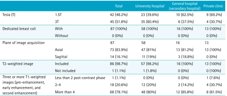
All hospitals used MRI of 1.5T or more (100%); 3T MRI and 1.5T MRI were used in 45 cases (51.8%) and 42 cases (48.2%), respectively (Table 4). Most cases (35 of 45 cases) of 3T MRI were taken at tertiary hospitals with a statistically significant difference (P = 0.04). Breast MRI was taken in the prone position using dedicated breast coil in all 87 cases (100%). The plane of image acquisition based on the T1WI with contrast enhancement was the axial plane in 73 cases (83.9%) and sagittal plane in 14 cases (16.1%). Checking the positions, breast MRI images taken in 2012-2013 (55 of 62, 88%) had a higher proportion of axial planes than those in 2010-2011 (18 of 25, 72%) had, but there was no statistically significant difference (P = 0.10). T2WI sequences were included in 86 cases out of 87 (98.7%). Three or more T1-weighted pre- and post-contrast images were included in all but one case (98.7%). Most cases in the primary, secondary, and tertiary hospitals were up to 98.7% in compliance with the essential elements.
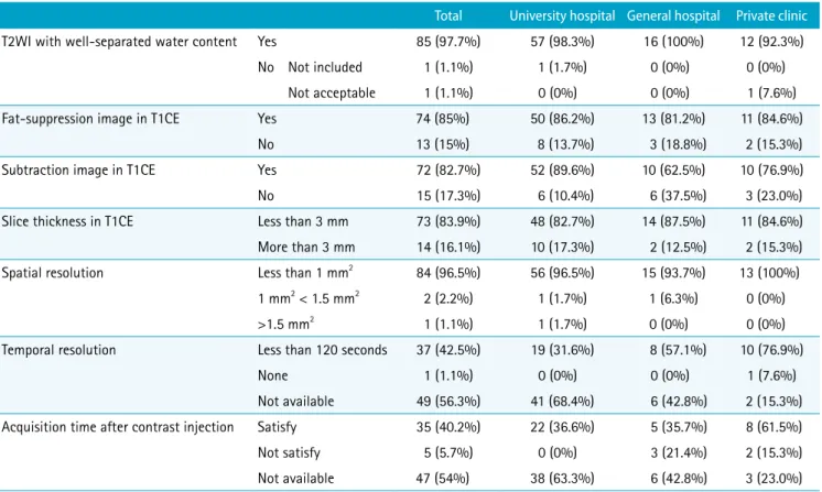
2. Elements to Consider When Evaluating Imaging Quality
There were 85 out of 87 cases (97.7%) in which the water Table 3. Number of Hospitals Included and Number of MRI Cases
Number of hospitals included Number of MRI cases Tertiary hospitals 28 (66.6%) 58 (66.6%) Secondary hospital 10 (23.8%) 16 (18.3%) Primary hospital 4 (9.5%) 13 (14.9%)
content was well separated in the T2WI; only 2 out of 87 cases (2.2%) did not satisfy the elements. Of the 87 cases, all had either T1-weighted fat-suppression images (n = 74, 85%), T1-weighted subtraction images (n = 72, 82%), or both (n = 59, 67.8%) in dynamic contrast-enhancement series (Table 5). There were 73 of the 87 cases (83.9%) that satisfied the recommendation for slice thickness, which should be ≤ 3.0 mm; 14 cases (16.1%) had over 3 mm of slice thickness. Spatial resolution was less than 1 mm2 in
84 of 87 cases (96.5%); 2 of 87 cases (2.2%) had 1 mm2
or more or less than 1.5 mm2. Only 1 of 87 cases (1.1%)
had a spatial resolution greater than 1.5 mm2. The degree
of compliance with imaging quality factors in all three different groups of hospitals was similar. The category of 'elements to consider when evaluating imaging quality' also satisfied over 96% of the criteria except for the slice thickness (83.9%).
3. Optional Elements for Breast MRI Protocol
Breast MRIs were taken in all of the 87 cases (100%) following the first recommendations shown in Table 2 (both breasts and chest walls are examined with a breast-specific bilateral breast coil (Table 6). In the prone position, the raised breast was positioned well in the center of the breast coil, and both arms were raised sufficiently to incorporate the axilla into the breast coil as much as possible (11). Of the 87 cases, 58 (66.6%) included sufficient axilla sequences (sufficient FOV included neck ~ nipple); 57 of the 87 cases (65.5%) included the DWI sequences. Of the
57 cases that included DWI sequences, 50 cases (57.4%) included 750-1000 high b-value images. The remaining 7 cases showed less than 750 as high b values: 700 in 3 cases, 600 in 2 cases, and 500 in 2 cases. The breast MRIs taken at the tertiary hospitals (74.1%) rather than at the primary or secondary hospitals (48.3%) contained more DWI sequences, and there were statistically significant differences (P = 0.03) between them. There were 15 of 87 cases (17.2%) that included the MRP in early contrast enhanced T1 weighted image (T1CE) images; 67 of the 87 cases (77.0%) included the MIPs in early T1CE images. Only 14 of the 87 cases (16.0%) contained both images, and 6 cases (6.8%) included both sagittal MRP images in early T1CEs and MIPs in early T1CE images. The patients’ menstrual cycles was not included in the analysis, because of absence of the data.
DISCUSSION
These days, breast MRI is an essential complementary modality with mammography and US, because breast MRI is more sensitive than is mammography for cancer detection (> 90%). Its main indications are staging of known cancer, screening for breast cancer in women at high risk, and evaluation of response to neo-adjuvant chemotherapy (7, 12). But most MRI protocols were multiparametric (13, 14). A breast MRI certification program was administered by the American College of Radiology (ACR) in the United States.
Table 4. Essential Elements for Breast MRI Protocol
Total University hospital (secondary hospital)General hospital Private clinic
Tesla (T) 1.5T 42 (48.2%) 23 (39.6%) 10 (62.5%) 9 (69.2%)
3T 45 (51.8%) 35 (60.4%) 6 (37.5%) 4 (30.7%)
Dedicated breast coli With 87 (100%) 58 (100%) 16 (100%) 13 (100%)
Without 0 (0%) 0 (0%) 0 (0%) 0 (0%)
Plane of image acquisition 87 58 16 13
Axial 73 (83.9%) 47 (81%) 13 (81.2%) 13 (100%)
Sagittal 14 (16.1%) 11 (19%) 3 (18.8%) 0 (0%)
T2-weighted image Included 86 (98.7%) 57 (98.2%) 16 (100%) 13 (100%)
Not included 1 (1.1%) 1 (1.8%) 0 (0%) 0 (100%)
Three or more T1-weighted images (pre-enhancement, early enhancement, and second enhancement)
Less than 2 post-contrast phase 1 (1.1%) 0 (0%) 0 (0%) 1 (7.6%)
2-4 18 (20.6%) 12 (20%) 2 (14.2%) 4 (30.7%)
Recommendations were also published in 2010, including the breast MRI protocols and indications by the European Society of Breast Cancer Specialists (EUSOMA) (15). The use of breast MRI has been rapidly growing in Korea (10), so a group of Korean domestic breast-imaging experts was recruited to establish standard protocols for breast MRI examinations suitable for a domestic medical environment based on foreign recommendations in 2018.
In this study, we evaluated the adequacy and performance of breast MRI in terms of the 2018 recommendation. We found that over 98% of the criteria for 'essential elements for breast MRI protocol' were satisfied when compared with the 2018 Recommendation. For the category of 'elements to consider when evaluating imaging quality', over 96% of the criteria were satisfied except for the slice thickness (83.9%). Most of the examinations were done in tertiary or secondary hospitals (85.0%) rather than in primary hospitals (15%) in our results. When these 87 cases were being conducted, MRI was considered to be a special medical device, but there were no specific recommendations or guidelines for breast MRI in Korea. According to the 2018 recommendation suggestions, it is best to use a field strength of at least 1.5T
to acquire images at a sufficiently high spatial resolution (16). Also, Utilizing a dedicated breast coil is mandatory in order to obtain images of diagnostic quality (17). Women lie in the prone position with the breasts hanging free in the recesses of the coil (18). In all cases (100%), MRI was done to satisfy a 1.5T or higher device that acquired images in the prone position using a breast-dedicated coil: 3T scanners in 45 cases (51.8%) and 1.5T scanners in 42 cases (48.2%). More MR exams were taken at the tertiary hospitals using 3T MRI with statistical significance than at other groups. Images were more frequently acquired in the axial plane (83.9%) than sagittal plane (16.1%). Axial scans not only have faster acquisition times of scan, but also provide a better overview of both breasts to distinguish asymmetry from breast parenchymal enhancement. Most breast cancers show peak contrast-enhancement at 60-90 seconds, so it is essential to obtain an image approximately 60-90 seconds after contrast-material administration (16). A persistent increase of the contrast-enhancement is mostly seen in benign lesions, whereas a decrease of the contrast-enhancement in the late phase is common in malignant lesions (19). A dynamic sequence
Table 5. Elements to Consider When Evaluating Imaging Quality
Total University hospital General hospital Private clinic
T2WI with well-separated water content Yes 85 (97.7%) 57 (98.3%) 16 (100%) 12 (92.3%)
No Not included 1 (1.1%) 1 (1.7%) 0 (0%) 0 (0%)
Not acceptable 1 (1.1%) 0 (0%) 0 (0%) 1 (7.6%)
Fat-suppression image in T1CE Yes 74 (85%) 50 (86.2%) 13 (81.2%) 11 (84.6%)
No 13 (15%) 8 (13.7%) 3 (18.8%) 2 (15.3%)
Subtraction image in T1CE Yes 72 (82.7%) 52 (89.6%) 10 (62.5%) 10 (76.9%)
No 15 (17.3%) 6 (10.4%) 6 (37.5%) 3 (23.0%)
Slice thickness in T1CE Less than 3 mm 73 (83.9%) 48 (82.7%) 14 (87.5%) 11 (84.6%)
More than 3 mm 14 (16.1%) 10 (17.3%) 2 (12.5%) 2 (15.3%)
Spatial resolution Less than 1 mm2 84 (96.5%) 56 (96.5%) 15 (93.7%) 13 (100%)
1 mm2 < 1.5 mm2 2 (2.2%) 1 (1.7%) 1 (6.3%) 0 (0%)
>1.5 mm2 1 (1.1%) 1 (1.7%) 0 (0%) 0 (0%)
Temporal resolution Less than 120 seconds 37 (42.5%) 19 (31.6%) 8 (57.1%) 10 (76.9%)
None 1 (1.1%) 0 (0%) 0 (0%) 1 (7.6%)
Not available 49 (56.3%) 41 (68.4%) 6 (42.8%) 2 (15.3%)
Acquisition time after contrast injection Satisfy 35 (40.2%) 22 (36.6%) 5 (35.7%) 8 (61.5%)
Not satisfy 5 (5.7%) 0 (0%) 3 (21.4%) 2 (15.3%)
Not available 47 (54%) 38 (63.3%) 6 (42.8%) 3 (23.0%)
demands at least three time points to be measured, that is, one before the administration of contrast medium, one approximately 2 min later to capture the peak, and one in the late phase to evaluate whether a lesion continues to contrast-enhancement, shows a plateau, or shows early wash-out of the contrast agent (16). Accordingly, the guideline recommends including three or more T1WI (pre-enhancement and post-contrast images). Also, contrast-enhanced T1WI should include images taken between 60 and 120 seconds after the administration of contrast medium, and images taken after 4 minutes. In this study, dynamic images were included in almost hospitals (98.7%), but in this survey, while analyzing the dynamic images, we found many cases where only post-enhancement images that lacked time information or temporal resolution were obtained. Therefore, it was difficult to accurately evaluate whether the dynamic images were taken at the time intervals suggested by the recommendation; 86 cases out of 87 had one pre-enhancement image and more than two post-enhancement images. We considered these images to be pre-enhancement, early enhancement, and second enhancement images.
The guideline also recommends including T2WI to increase the specificity for differentiation of benign and malignant lesions (15). Most of the cases (85 of 87 cases, 97.7%), except for two cases, had good separation of water content on T2WI, satisfying the recommendation for imaging quality. Subtraction images with fat suppression are often helpful for differentiating truly contrast-enhancing structures
from natively high signal intensity lesions at T1 (20). Hence contrast-enhanced T1-weighted fat-saturation images or subtraction images are included as essential elements (1). In our study, all cases (100%) had contrast-enhanced T1-weighted fat-saturation images or subtraction images. Subtraction does not require extra acquisition time and is not influenced by magnetic field inhomogeneity, and thus is the preferred type of 'fat suppression' for dynamic bilateral imaging (21). Our data also showed that the tertiary hospitals usually conducted breast MRIs with subtraction images (90%), whereas primary and secondary hospitals had only 63-77% of subtraction images in the data, which is a comparably low statistical significance (P = 0.032). The tertiary hospitals tend to have radiologists specialized in breast images, and breast imaging is the specialized area that uses subtraction images, because it is difficult to recognize the contrast-enhancing breast lesion in the background of abundant fat tissue in a breast in a short time. By convention, breast MRI should depict all contrast-enhancing cancers that are 5 mm or larger. The ACR Accreditation Breast MRI and Breast Imaging Study Group of the Korean Society of Magnetic Resonance recommends that the acquired slice thickness must be ≤ 3.0 mm (10, 12). Of the 87 cases, 73 (83.9%) satisfied the recommendation of slice thickness, which should be ≤ 3.0 mm. Another 14 cases were greater than 3 mm of slice thickness, which led to a partial volume averaging, a structure with a partial portion of imaging section, pixel, or voxel. Consequently, the signals of the structure and the adjacent or surrounding Table 6. Optional Elements for Breast MRI Protocol
Total University hospital General hospital Private clinic
Center Yes 87 (100%) 58 (100%) 16 (100%) 13(100%)
No 0 (0%) 0 (0%) 0 (0%) 0 (0%)
Inclusion of axilla (neck - nipple) Yes 58 (66.6%) 36 (62%) 14 (87.5%) 8 (61.5%)
No 29 (33.4%) 22 (38%) 2 (12.5%) 5 (38.4%)
DWI high b value Yes 750-1000 50 (57.4%) 39 (67.2%) 5 (31.2%) 6 (46.1%)
Less than 750 7 (8%) 4 (6.8%) 2 (12.5%) 1 (7.6%)
No 30 (34.4%) 15 (25.8%) 9 (56.2%) 6 (46.1%)
MRP in early T1CE Yes Axial 8 (9.1%) 8 (13.7%) 0 (0%) 0 (0%)
Sagittal 7 (8.0%) 5 (8.6%) 2 (12.5%) 0 (0%)
No 72 (82.7%) 45 (77.5%) 14 (87.5%) 13 (100%)
MIP in early T1CE Yes 67 (77.0%) 41 (70.6%) 15 (93.7%) 11 (84.6%)
No 20 (23.0%) 17 (29.3%) 1 (6.2%) 2 (13.4%)
structures present in the section, pixel, or voxel are averaged, with a possibility of erroneous pixel or voxel signals, which could lead to diagnostic misinterpretations. Because morphologic evaluation requires much finer details, the plane pixel size should be 1 × 1 mm or smaller (15, 16). So the guideline recommends that spatial resolution should be less than 1.0 mm2 or should be less than 1.5 mm2 (10).
Most cases satisfied the recommendations except for one case with more than 1.5 mm2 of spatial resolution. There
was information on temporal resolution in 38 cases (43.7%). For the optional elements, all hospitals satisfied the appropriate position by using breast coil, and 58 of the 87 cases (66.6%) included sufficient axilla sequences. Breast cancer has significantly lower ADCs than do benign breast lesions or normal tissue. DWI used with DCE-MRI increases the specificity for cancer detection, but if used alone still may be useful for widespread cancer screening (22). Of the 87 cases, 57 (65.5%) included the DWI. In particular, tertiary hospitals conducted significantly more DWIs than did the primary or secondary hospitals. Previously, DWI was often used for research purposes rather than for actual readings. Recently, there has been rapidly growing evidence of the potential value of DWI for improving breast cancer detection and characterization. Also, with technique standardization and clear interpretation guidelines, the role of DWI for clinical breast imaging is expected to grow in the future (23). Most hospitals preferred MIP images (67 of the 87 cases [77.0%]) over MRP images (15 of 87 cases [17.2%]), because MIP images provide a better glimpse of breast lesions and the structure of entire breasts and their surroundings (24), not only helping surgeons and patients to understand well but also helping surgeons to plan surgery preoperatively. It is recommended that premenopausal women should ideally be scanned during the second week of the menstrual cycle, possibly avoiding the fourth week, which is a luteal phase (21). Benign hormonal effects on breasts during the luteal phase of the menstrual cycle can render images somewhat difficult to interpret, particularly in high-risk women with dense, fibrocystic breasts (25). In this study, patients’ menstrual cycles were not analyzed, so breast MRI examinations on premenopausal women done during the fourth week of the menstrual cycle were difficult to diagnose with high accuracy.
This study has several limitations. First, there is a possibility of selection bias. Only MRI information on patients referred to our institution was collected. Almost breast MRIs were taken in secondary or tertiary hospitals, which may not have reflected the situation in primary
hospitals, because the absolute number of MRIs in primary hospitals is smaller than that in secondary or tertiary hospitals. Second, this study excluded MRI scans taken after surgery and screening and included only MRI scans for preoperative staging of breast cancer. In the future, we expect that further research can be conducted. Third, we analyzed retrospectively the copy of external breast MRIs, so there may have been restrictions that reflected the actual situations. Also, there were many cases without outside MRI readings, which limited adequate evaluation of outside MRI readings. How to use each sequence is also unknown in reading breast MRIs.
Despite these limitations, this study could be an important stepping stone to look at trends of changes of breast MRI protocols before and after the recommendation was published.
In conclusion, we described how breast MRI protocols were applied before the breast MRI recommendation was published in South Korea in 2018. In our study, most of the essential elements of the recommendation were satisfied. Exceptionally, the most inadequate element of all was the slice thickness. Optional elements for Breast MRI protocols were satisfied at various percentages. In addition, there were no differences between the groups of tertiary, secondary, and primary hospitals in most elements. However, the 3T of MRI, subtraction images and DWI were used more in the tertiary hospitals than in the other groups. In the future, we expect that the results of this study could be a stepping stone to looking at trends of changes made in recommendations provided by the Breast Imaging Study Group of the Korean Society of Magnetic Resonance.
REFERENCES
1. Lehtimaki T, Lundin M, Linder N, et al. Long-term prognosis of breast cancer detected by mammography screening or other methods. Breast Cancer Res 2011;13:R134
2. Kolb TM, Lichy J, Newhouse JH. Comparison of the performance of screening mammography, physical examination, and breast US and evaluation of factors that influence them: an analysis of 27,825 patient evaluations. Radiology 2002;225:165-175
3. Oh CM, Won YJ, Jung KW, et al. Cancer statistics in Korea: incidence, mortality, survival, and prevalence in 2013. Cancer Res Treat 2016;48:436-450
4. Hwang JY, Han BK, Ko EY, Shin JH, Hahn SY, Nam MY. Screening ultrasound in women with negative
mammography: outcome analysis. Yonsei Med J 2015;56:1352-1358
5. Youn I, Choi S, Kook SH, Choi YJ. Mammographic breast density evaluation in Korean women using fully automated volumetric assessment. J Korean Med Sci 2016;31:457-462 6. Kim S, Kang BJ, Kim SH, Lee J, Park GE. Computer-aided
detection with automated breast ultrasonography for suspicious lesions detected on breast MRI. Investig Magn Reson Imaging 2019;23:46-54
7. Plana MN, Carreira C, Muriel A, et al. Magnetic resonance imaging in the preoperative assessment of patients with primary breast cancer: systematic review of diagnostic accuracy and meta-analysis. Eur Radiol 2012;22:26-38 8. Hylton NM, Gatsonis CA, Rosen MA, et al. Neoadjuvant
chemotherapy for breast cancer: functional tumor volume by MR imaging predicts recurrence-free survival-results from the ACRIN 6657/CALGB 150007 I-SPY 1 TRIAL. Radiology 2016;279:44-55
9. Healthcare Bigdata Hub. Number of breast MRI examinations 2021. Available at. http://opendata.hira.or.kr/ op/opc/olapDiagBhvInfo.do. Accessed January 13, 2021. 10. Choi SH, Kang BJ, Jung SE. Breast magnetic resonance
image (MRI) guideline: breast imaging study group of Korean Society of Magnetic Resonance in Medicine recommendations. Investig Magn Reson Imaging 2018;22:205-208
11. Jeong EH, Choi EJ, Choi H, Park EH, Song JS. Prediction of axillary lymph node metastasis in early breast cancer using dynamic contrast-enhanced magnetic resonance imaging and diffusion-weighted imaging. Investig Magn Reson Imaging 2019;23:125-135
12. Cho N, Han W, Han BK, et al. Breast cancer screening with mammography plus ultrasonography or magnetic resonance imaging in women 50 years or younger at diagnosis and treated with breast conservation therapy. JAMA Oncol 2017;3:1495-1502
13. Marino MA, Helbich T, Baltzer P, Pinker-Domenig K. Multiparametric MRI of the breast: a review. J Magn Reson Imaging 2018;47:301-315
14. Rahbar H, Partridge SC. Multiparametric MR imaging of breast cancer. Magn Reson Imaging Clin N Am 2016;24:223-238
15. Sardanelli F, Boetes C, Borisch B, et al. Magnetic resonance imaging of the breast: recommendations from the EUSOMA working group. Eur J Cancer 2010;46:1296-1316
16. Mann RM, Kuhl CK, Kinkel K, Boetes C. Breast MRI: guidelines from the European Society of Breast Imaging. Eur Radiol 2008;18:1307-1318
17. Konyer NB, Ramsay EA, Bronskill MJ, Plewes DB. Comparison of MR imaging breast coils. Radiology 2002;222:830-834
18. Yeh ED, Georgian-Smith D, Raza S, Bussolari L, Pawlisz-Hoff J, Birdwell RL. Positioning in breast MR imaging to optimize image quality. Radiographics 2014;34:E1-17 19. Westra C, Dialani V, Mehta TS, Eisenberg RL. Using
T2-weighted sequences to more accurately characterize breast masses seen on MRI. AJR Am J Roentgenol 2014;202:W183-190
20. Cheung HS, Tse GM, Lai SY, Yeung DK. Relationship between lesion size and signal enhancement on subtraction fat-suppressed MR imaging of the breast. Magn Reson Imaging 2004;22:1259-1264
21. Kuhl C. The current status of breast MR imaging. Part I. Choice of technique, image interpretation, diagnostic accuracy, and transfer to clinical practice. Radiology 2007;244:356-378
22. Partridge SC, McDonald ES. Diffusion weighted magnetic resonance imaging of the breast: protocol optimization, interpretation, and clinical applications. Magn Reson Imaging Clin N Am 2013;21:601-624
23. Partridge SC, Nissan N, Rahbar H, Kitsch AE, Sigmund EE. Diffusion-weighted breast MRI: clinical applications and emerging techniques. J Magn Reson Imaging 2017;45:337-355
24. Kim HS, Kang BJ, Kim SH, Choi JJ, Lee JH. Usefulness of three-dimensional maximal intensity projection (MIP) reconstruction image in breast MRI. Investig Magn Reson Imaging 2009;13:183-189
25. Dontchos BN, Rahbar H, Partridge SC, Lehman CD, DeMartini WB. Influence of menstrual cycle timing on screening breast MRI background parenchymal enhancement and diagnostic performance in premenopausal women. J Breast Imaging 2019;1:205-211