Development of neural stem cell line
using hypoxia-inducible gene
expression system
Sung Sam Jung
Department of Medicine
Development of neural stem cell line
using hypoxia-inducible gene
expression system
Directed by Professor Keung Nyun Kim
Doctoral Dissertation
submitted to the Department of Medicine,
the Graduate School of Yonsei University
in partial fulfillment of the requirements for the degree
of Doctor of Philosophy
Sung Sam Jung
This certifies that the
Doctoral Dissertation of
Sung Sam Jung is approved.
---
Thesis Supervisor : Keung Nyun Kim
---
Kook In Park: Thesis Committee Member#1
---
Jong Eun Lee: Thesis Committee Member#2
---
Yoon Ha: Thesis Committee Member#3
---
Ho Gyun Ha: Thesis Committee Member#4
The Graduate School
Yonsei University
ACKNOWLEDGEMENTS
It would not have been possible to write this doctoral thesis without the
help and support of the kind people around me, to only some of whom it
is possible to give particular mention here. Above all, this thesis would
not have been possible without the help, support and patience of my
principal supervisor, Prof. Keung Nyun Kim. The good advice, support
and friendship of my second supervisor, Prof. Yoon Ha, has been
invaluable on both an academic and a personal level, for which I am
extremely grateful. I also thank Prof. Kook In Park and Prof. Jong Eun
Lee for their support and assistance from the start of my work. I am most
grateful to Prof. Ho Gyun Ha for providing me with spiritual art of his
own all the time. I would also like to thank Jin Soo Oh for his kindness,
friendship and support. Especially, I would like to thank my wife Jong
Sook for her personal support and great patience at all times. My parents,
brother and sister have given me their unequivocal support throughout, as
always, for which my mere expression of thanks likewise does not
suffice.
<TABLE OF CONTENTS>
ABSTRACT ··· 1
I. INTRODUCTION ··· 3
II. MATERIALS AND METHODS ··· 5
1. Plasmids construction ··· 5
2. Establishment of stable NSC lines and in vitro neuronal differentiation ··· 6
3. Hypoxia ··· 7
4. Luciferase assay ··· 7
5. Luciferase expression imaging ··· 7
6. RT-PCR ··· 8
7. Western blot assay ··· 8
8. Rat SCI model ··· 8
9. Immunohistochemistry ··· 9
10. Statistical analysis ··· 9
III. RESULTS ··· 10
1. Construction of hypoxia-inducible gene expression double-promoter plasmids ··· 10
2. Generation of nonviral hypoxia-regulated or non-regulated NSC lines ··· 11
3. Hypoxia-responsive luciferase expression in stable NSC lines ··· 13
4. Kinetics of hypoxia-responsive luciferase expression in stable NSC lines ·· 15
5. Neural differentiation potency of stable NSC lines in vitro and in vivo ··· 18
IV. DISCUSSION ··· 22
V. CONCLUSION ··· 26
REFERENCES ··· 27
LIST OF FIGURES
Figure 1. Construction of hypoxia-responsive double-promoter
plasmids co-expressing DsRed and luciferase (Luc). ··· 11
Figure 2. Generation of the nonviral hypoxia-regulated or
non-regulated NSC lines. ··· 13
Figure 3. Transcriptional regulation of the hypoxia-regulated
luciferase expression in stable NSC line. ··· 14
Figure 4. Kinetics of the hypoxia-regulated luciferase expression
in stable NSC lines. ··· 17
Figure 5. Neural differentiation potency of stable NSC lines
in vitro. ··· 20
Figure 6. In vivo ischemic injury-specific luciferase expression
of stable NSCs transplanted in rat spinal cord injury model. ··· 22
ABSTRACT
Deveopment of neural stem cell line using
hypoxia-inducible gene expression system
Sung Sam Jung
Department of Medicine
The Graduate School, Yonsei University
(Directed by Professor Keung Nyun Kim)
Nonviral ex vivo local gene therapy systems consisting of regulated
gene expression vectors and cellular delivery platforms represent a novel
strategy for tissue repair and regeneration. We introduced a
hypoxia-regulated plasmid-based system into mouse neural stem cells
(NSCs) as an efficient gene expression and delivery platform for rapid,
robust and persistent hypoxic/ischemic-regulated gene expression in the
spinal cord. A synthetic hypoxia-responsive erythropoietin (Epo)
enhancer, the SV40 minimal promoter and the luciferase (Luc) reporter
gene were incorporated in a DsRed-expressing double-promoter plasmid
for cell lipofection and Zeocin-selection to establish a hypoxia-regulated
stable NSC line (NSC-Epo-SV-Luc). A non-hypoxia-regulated stable
NSC line (NSC-SV-Luc) was also established as a control. Under the
transcriptional regulation of the Epo enhancer, in vitro luciferase
expression in NSC-Epo-SV-Luc, but not in NSC-SV-Luc, was sensitively
augmented according to the strength and duration of the hypoxic stimulus
of the hypoxic cells. Furthermore, deoxygenation of the reoxygenated
cells clearly enhanced the luciferase activity again. After transplantation
into a rat spinal cord injury (SCI) model, only NSC-Epo-SV-Luc showed
ischemic injury-specific luciferase expression Notably, the engineered
NSC lines maintained the neural differentiation potential and retained the
hypoxia-regulated luciferase expression after differentiation. We propose
that NSCs engineered with the Epo-SV-therapeutic gene will be valuable
for developing a controllable stem cell-mediated nonviral gene therapy
for SCI or other central nervous system diseases accompanied with
chronic or episodic hypoxic/ischemic stresses.
---
Key words: Controllable gene therapy; Neural stem cell; Non-viral gene
delivery; Hypoxia; Erythropoietin enhancer; Spinal cord injury
Development of neural stem cell line using
hypoxia-inducible gene expression system
Sung Sam Jung
Department of Medicine
The Graduate School, Yonsei University
(Directed by Professor Keung Nyun Kim)
I. INTRODUCTION
Stem cells can be used to restore tissue function because they secrete paracrine trophic factors to increase cell survival, directly replace dying and dead cells, and function as vehicles that deliver therapeutic genes to injured tissue targets 1. A combination of stem cells and gene therapy is thus expected to
become a promising strategy for increasing the efficacy over stem cells or gene therapy alone 2, 3. A variety of ex vivo local gene therapy systems consisting of
cellular delivery platforms have been successfully used to increase gene delivery efficiency and tissue repair potential in animal models 4-7 and human
diseases 8-12. Until now, one of the most extensively studied candidates are
mesenchymal stem cell (MSC)-based gene and cell therapy, mainly because MSCs can be readily isolated from bone marrow or adipose tissue and are reported to possess multipotent differentiation capacity even after being genetically modified with therapeutic genes 13-15. However, the transduction of the therapeutic genes into MSCs remains difficult for sufficient and persistent expression, even when recombinant viral vectors are used as gene carriers 16,17. Meanwhile, the use of viral materials has raised safety concerns in the treatment
of human diseases 14, 18-20, and the uncontrollable expression of foreign genes
also potentially leads to serious adverse effects 18-20. Thus, the safety and
efficacy of this new therapy remain a major obstacle in its advancement to the clinical setting for tissue repair and regeneration.
The development of a controllable gene therapy system is highly desirable to increase safety and maintain efficient therapeutic gene expression specific to the defect and injured tissue 21. Using a synthetic erythropoietin (Epo) enhancer upstream of a SV promoter in a pSV plasmid, we previously established a novel hypoxia-inducible gene expression system that achieves high therapeutic gene expression specific to hypoxic conditions and avoids unwanted gene overexpression in normoxic conditions 22. In an animal spinal cord injury (SCI)
model, although the naked gene delivery efficiency was very low, but the effective regulation of gene expression by this hypoxia-inducible gene expression system for VEGF gene therapy was confirmed 23. The above results
suggest a potential application of the hypoxia-inducible gene expression system during gene therapy for regeneration of ischemic tissue. However, to improve gene delivery efficiency for this system a novel gene delivery strategy needs to be developed.
As the use of viral materials evokes many health concerns in the treatment of human diseases and since MSCs are extremely resistant to nonviral transduction
14-17, 24, 25, alternative cells must be developed as nonviral-based gene delivery
platforms to increase the efficacy of cell and gene therapy for various diseases. Neural stem cells (NSCs) possess capacity for long-term expansion in vitro and extensive functional stability and plasticity, and are amenable to genetic engineering as large-scale cell sources for neural transplantation 26, 28.
Genetically modified NSCs have been transplanted into animal models of SCI and stroke to promote functional recovery 29, 30. Compared to MSCs, NSCs can
be more easily engineered with a foreign gene and were more feasibly used as a non-viral delivery platform for a therapeutic or reporter genes. Through
lipofection with Lipofectamine 2000 and selection with Zeocin, a stable NSC line was established in our preliminary study using a double-promoter plasmid expressing a DsRed fluorescence reporter gene.
To simplify the establishment of hypoxia-regulated stable NSCs by using only a single plasmid, the Epo-SV-Luc fragment from pEpo-SV-Luc was incorporated in the DsRed-expressing double-promoter plasmid. In this construct, the hypoxia-inducible overexpression of the luciferase gene is regulated by the Epo enhancer upstream of a SV40 promoter, and the constitutive expression of the reporter DsRed, a red fluorescence protein for easy detection, is driven by an EF1α promoter as an internal expression reference. Following the engineering of stable NSCs with Epo-SV-Luc, we confirmed that this stable NSC/gene system sensitively enhanced the luciferase expression upon hypoxic stimulus, both in vitro and in vivo, and quickly down-regulated luciferase expression to a low basal level after reoxygenation of the hypoxic cells.
II. MATERIALS AND METHODS
1. Plasmids construction
The construction of pEpoSV-Luc with Epo enhancer and pSV-Luc was described previously 22. Two inverted Epo enhancer sequences were ligated at
the XbaI site and then inserted to the BglII site in pSV-Luc. To subclone pBudCE4.1-DsRed, the DsRed fragment was obtained by BamHI/HpaI digestion from a pDsRed-monomer-golgi Vector (Clontech, Palo Alto, CA, USA) and then inserted into the compatible sites (BglII/PmeI) downstream of the EF-1α promoter in pBudCE4.1 (Invitrogen, Carlsbad, CA, USA). To construct pBudSV-Luc and pBudEpoSV-Luc, the SV-Luc and EpoSV-Luc fragments (NheI/BamHI) from pSV-Luc and pEpoSV-Luc, respectively, were
inserted into pBudCE4.1-DsRed to replace the CMV promoter-containing sequence between SpeI and BamHI sites.
2. Establishment of stable NSC lines and in vitro neuronal differentiation The mouse NSC line used in this study was purchased from the American Type Culture Collection (ATCC, Catalog #CRL-2925). The cells were maintained in DMEM supplemented with 10% FBS and 1% penicillin/streptomycin in a 5% CO2 incubator. To establish stable NSC lines,
about 0.8 μg of the pBudCE4.1-DsRed, pBudSV-Luc or pBudEpoSV-Luc was lipofected into 1×10⁴ normal NSCs according to a previously described protocol 31. After 24 hours of culture in a 12-well plate and then subculturing in 100-mm
dishes, the cells were fed with culture medium containing 200 μg/ml Zeocin every two days until formation of visible colonies. Single Zeocin-resistant and DsRed-positive cell colonies were mechanically isolated and disaggregated by trypsin in a 96-well plate. Each suspended colony was then transferred into a 24-well plate and maintained in culture medium containing 50 μg/ml Zeocin. The expanded stable NSC lines with intense DsRed expression were cultured under normoxic or hypoxic conditions for screening in a luciferase activity assay.
To evaluate the differentiation potency of the established stable NSC lines, the cells were seeded into a 24-well plate (5×10³/well) or 6-well plates
(3×10⁵/well). Neuronal differentiation was induced by treatment with 1 μM
retinoic acid (RA) for 7 days. For the luciferase assay, the differentiated cells in 6-well plates were further cultured under normoxic or hypoxic conditions for 12 hours. Morphological examination of stable NSCs or the differentiated neuronal cells were examined and images were visualized using an inverted fluorescence microscope equipped with an Olympus DP71 camera and DP Controller software (Olympus, IX71, Japan).
3. Hypoxia
Hypoxic conditions were created by flushing a hypoxic gas mixture, using a Forma Series II Water Jacketed CO2 Incubator (Thermo Fisher Scientific Inc.,
USA), in which hypoxia (1% O2) in a gas phase can be achieved within 30 min after opening and closing of the door. More strict control of hypoxia, with medium pre-equilibrated under hypoxic conditions, was applied to study the kinetics of hypoxia-responsive luciferase expression. Furthermore, a comparative study of the hypoxia-responsive luciferase expression was also performed using the hypoxic-mimicking reagent, CoCl2.
4. Luciferase assay
The luciferase activities expressed by the engineered NSCs both in vitro and
in vivo rat spinal cord were measured by the Promega Luciferase Assay System
according to the manufacturer’s protocol. Briefly, the cells or tissues were lysed with 1× Reporter Lysis Buffer (Promega, Madison, WI, USA) through homogenization and one freeze-thaw cycle. Following a 15-min incubation on ice and 2-min centrifugation at maximum speed, the supernatant lysates were transferred to fresh tubes prechilled on ice and used immediately or stored at -80°C until use. After measuring the protein concentration with a BCA kit (Pierce, Iselin, NJ, USA), luciferase production in cell or tissue lysates was measured by luminometery. Levels are expressed as relative light units (RLU) per microgram of total protein.
5. Luciferase expression imaging
IVIS image system was used to confirm whether NSCs introduced by hypoxia inducible gene expression system show the hypoxia specific expression under hypoxia. Luciferase expression of stable NSCs was confirmed according to a previously-described method 32.
6. RT-PCR
Total RNA was extracted from normoxic or hypoxic cultures of stable NSCs using a RNA extraction kit (QIAGEN, Valencia, CA, USA). The reverse-transcribed samples were amplified by PCR using Taq polymerase with luciferase forward primer (5’-CAAATCATTCCGGATACTGCG-3’) and luciferase reverse primer (5’-GAATTACACGGCGATCTTTCC-3’).
7. Western blot assay
In addition to morphological examination to identify neural differentiation, western blot assays were conducted to detect the expression of specific marker proteins in NSC and RA-treated cultures, using mouse cortex extracts as a control. Following BCA assay, samples were electrophoresed on 12% SDS-PAGE gels and transferred onto PVDF membranes at 200 V for 1 h. Filters were blocked for 2 h at room temperature with 5% nonfat milk in PBS containing 0.05% Tween 20. The blots were probed with rabbit anti-MAP2 or β-Ⅲ-tubulin and mouse anti-nestin, SSEA-1/CD-15, GFAP, or β–actin antibodies, respectively, and bands were visualized with suitable horseradish peroxidase-conjugated reagents (Pierce, Iselin, NJ, USA) and ECL western blotting detection reagents (Amersham, GE Healthcare, UK).
8. Rat SCI model
Animal studies followed protocols approved by the Animal Care Committee of the Medical Research Institute of Yonsei University Hospital. Adult male Sprague-Dawley rats (250 g) were obtained from Orient Bio Co. (Kyungki-do, Korea). Animals were anesthetized using sodium penthobarbital (20 mg/kg). After laminectomy, SCI was performed at the T9 level by compression with a vascular clip (width, 2 mm; occlusion pressure, 50 g) for 10 minutes as previously described 22. Non-SCI rats were used as a normal control group.
the injured or non-injured epicenter of the spinal cord using an electrode microneedle. The SCI and non-SCI rats for transplantation were divided into two groups. Group 1 received NSCs-SV-Luc cells (3x10⁵ cells/5 µl DMEM)
while Group 2 received NSCs-EpoSV-Luc cells (3x10⁵ cells/5 µl DMEM). All
animals received cyclosporine (10 mg/kg, Chong Kun Dang Pharm, Seoul, Korea) every day after transplantation. Two days after transplantation, animals were anesthetized and then transcardially perfused with saline. Spinal cord tissues, including the injury epicenter and transplanted sites, were obtained and lysed as above for luciferase assay.
9. Immunohistochemistry
To test the in vivo neural differentiation of the transplanted stable NSCs, the cell transplantation was performed seven days after surgery and SCI. After transplantation for four weeks, animals were fixed by transcardial perfusion with ice-cold PBS and then 4% paraformaldehyde. Fixed spinal cord tissues, including the injury epicenter and transplanted sites, were removed and longitudinal sections of 10-µm thicknesses were mounted on glass slides. The sections were then stained overnight at 4°C with mouse anti-GFAP (1:500) and rabbit anti-MAP-2 (1:500). The secondary FITC-conjugated goat anti-mouse or rabbit IgG was used for detection. Confocal images were obtained with a Nikon Eclipse C1-plus Confocal Microscope system
10. Statistical analysis
The differences between the two groups in all experiments were analyzed using a two-tailed Student’s t-test. A P value less than 0.01 was considered statistically significant.
III. RESULTS
1. Construction of hypoxia-inducible gene expression double-promoter plasmids
The hypoxia-inducible gene expression plasmid, pEpo-SV-Luc (Fig. 1), was previously constructed using the Epo enhancer and pSV-Luc plasmid 22. This
Epo-SV-Luc reporter system could achieve efficient delivery and significant transcriptional regulation of gene expression under in vitro hypoxic conditions, but was inefficient in spinal cord in vivo. To overcome this problem, a double promoter pBudCE4.1 vector was used for ex vivo engineering of hypoxia-regulated NSCs to ensure sufficient in vivo gene delivery and expression. The plasmid, pBudCE4.1, was initially constructed to express a DsRed fluorescence gene driven by the EF1α promoter (pBudCE4.1-DsRed). Then the SV-Luc or Epo-SV-Luc fragment from pSV-Luc or pEpo-SV-Luc was further inserted to replace the CMV promoter of pBudCE4.1-DsRed, leading to the construction of pBudSV-Luc or pBudEpo-SV-Luc (Fig. 1). These constructs were confirmed by restriction analysis as well as by their expression of DsRed and/or luciferase in transfected NSCs, as described below.
Fig.1. Construction of hypoxia-responsive double-promoter plasmids co-expressing DsRed and luciferase (Luc). (A) The map of pEpoSV-Luc. Two inverted Epo enhancer sequences were ligated at the XbaI site and then inserted to the BglII site in pSV-Luc. (B) The map of pBudCE4.1-DsRed. The DeRed reporter gene expression is constitutively regulated by an EF1α promoter. (C and D) The maps of pBudSV-Luc and pBudEpoSV-Luc. The SV-Luc and EpoSV-Luc fragments generated by NheI/BamHI digestion from pSV-Luc and pEpoSV-Luc, respectively, were inserted into pBudCE4.1-DsRed at the SpeI/BamHI digestion site. Arrowheads indicate the subcloning sites.
2. Generation of nonviral hypoxia-regulated or non-regulated NSC lines To evaluate the potential use of NSCs as a non-viral gene delivery platform, the double promoter plasmids were lipofected into NSCs using Lipofectamine 2000 reagent. Following transient transfection, the NSCs grew normally, and more than 90% of the cells were red fluorescence-positive when transfected with pBudCE4.1-DsRed, pBudSV-Luc or pBudEpo-SV-Luc, as indicated by the representative image in Fig. 2A. After nonviral lipofection and a 10-day Zeocin selection, more than 1% of the lipofected NSCs formed visible colonies and
expressed the red fluorescence DsRed protein. Fig. 2B showed the formation of a typical round colony with clear boundary and intense DsRed fluorescence, which could be easily identified and isolated, and rapidly expanded into stable cell lines. Whenever the empty vector, non-hypoxia-responsive or hypoxia-responsive plasmid was transfected, there were no differences in the proliferation rate and the cell morphology of the stable NSCs (Fig. 2C and D). These results indicate that NSCs have the advantage of high gene transduction efficiency and desirable gene expression, which suggests that they can be a viable non-viral gene delivery platform for combined stem cell and gene therapy.
To test whether the luciferase expression is hypoxia-inducible in stable NSCs engineered with the plasmid constructs, a luciferase assay was performed after transient transfection and colony isolation and expansion. As shown in Fig. 2E, NSCs could achieve significant hypoxia-responsive luciferase expression after transient transfection with pBudEpoSV-Luc, but not with pBudSV-Luc, even though the basal luciferase expression of pBudEpoSV-Luc was slightly higher than that of pBudSV-Luc. Compared to transient transfection, it seemed that the levels of luciferase expression were much higher in stable NSCs. The luciferase expression levels in several representative stable NSC lines are shown in Fig. 2F. No detectable luciferase activity appeared in the stable NSC line transfected with pBudCE4.1-DsRed, an empty vector (EV) without the luciferase gene. Under hypoxic conditions, all tested stable NSC lines engineered with pBudSV-Luc showed a slight decrease in luciferase expression, but when engineered with pBudEpoSV-Luc, they showed a clear increase in luciferase expression. Noticeably, hypoxia-responsive luciferase expression could be induced up to 4-fold higher than the basal luciferase expression that remained at a relatively low level in NSC-EpoSV-Luc16#, in contrast to the NSC-SV-Luc7#, which had high normoxic expression, but low hypoxic expression of luciferase. These two stable NSC lines were therefore chosen for the comparative study in the following experiments. Taken together, these results indicate that NSCs can be efficiently engineered with a hypoxia-responsive gene expression system to achieve low basal gene expression under normoxia and high gene expression under hypoxia, which is critical for increasing both the safety and efficacy of gene therapy.
Fig.2. Generation of the nonviral hypoxia-regulated or non-regulated NSC lines. (A) Efficient transfection of the hypoxia-responsive plasmids into mouse NSC lines using Lipofectamine 2000. (B) Typical colony formation after nonviral lipofection and zeocin selection. (C and D) Characteristics of the representative stable NSC line lipofected with non-hypoxia-responsive or hypoxia-responsive plasmid. Scale bars in all images represent 50 μm. (E and F) Hypoxia-regulated luciferase expression in transiently transfected mouse NSCs or in several representative hypoxia-regulated or non-regulated stable NSC lines. Levels are expressed as relative light units (RLU) per microgram of total protein. EV, empty vector. *P<0.001, hypoxia versus normoxia. A two-tailed Student’s t-test was used (mean±s.d., n=4).
3. Hypoxia-responsive luciferase expression in stable NSC lines
Since the Epo enhancer regulates gene expression at the transcription level, RT-PCR assay was conducted to verify its transcriptional regulation of luciferase expression after engineering into stable NSCs. Following hypoxia or normoxia incubation, total mRNA was extracted from the stable NSC lines. Luciferase
mRNA levels were then analyzed and normalized to GAPDH expression levels through RT-PCR assay and then quantitative densitometric analysis. As shown in Fig. 3, the luciferase mRNA levels of NSC-EpoSV-Luc16# significantly increased in the presence of 100 μM CoCl2 or under hypoxia (1% O2), whereas
NSC-SV-Luc7# showed no clear change in luciferase mRNA levels under either condition. Therefore, hypoxia-responsive luciferase expression in stable NSCs engineered with Epo-SV system was regulated at the transcriptional level either in the presence of hypoxia-mimicking CoCl2 or under hypoxic conditions.
Expression pattern of luciferase in normoxia and hypoxia was confirmed using IVIS imaging system. NSC-EpoSV-Luc constantly showed the controlled luciferase expression compared to NSC-SV-Luc.
Fig.3. Transcriptional regulation of the hypoxia-regulated luciferase expression in stable NSC lines. (A and B) Hypoxia-regulated luciferase expression in stable NSC lines with the Epo-SV system is regulated at the transcriptional level either in the presence of hypoxia-mimicking CoCl2 (A) or under hypoxic condition (B). Luciferase mRNA levels were analyzed and normalized to GAPDH expression levels through RT-PCR assay (upper panels) and then quantitative densitometric analysis (lower panels). (C) Luciferase expression was analyzed by IVIS imaging system. *P<0.001, versus normoxia. A two-tailed Student’s
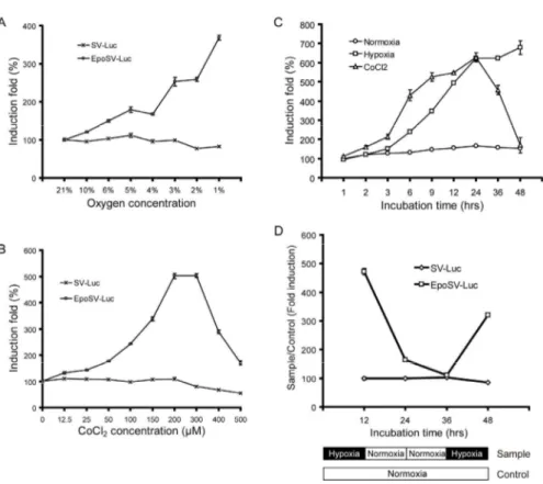
4. Kinetics of hypoxia-responsive luciferase expression in stable NSC lines To determine the sensitivity and strength of hypoxia-responsive gene expression in stable NSC-EpoSV-Luc lines, the cells were cultured in different concentrations of oxygen or CoCl2 and examined for luciferase expression by
luminometery. After reducing the oxygen concentration, luciferase expression was upregulated sensitively according to the decrease in oxygen tension. Cells exhibited less than a 2-fold induction of luciferase expression when incubated at 4-5% O2, higher than a 2-fold induction when cultured at 2-3% O2, and about a
4-fold induction at 1% O2 (Fig. 4A). Similarly, induction of luciferase
expression sensitively increased in a dose-dependent manner after the addition of 0-200 μM hypoxic-mimicking CoCl2, with a peak of induction up to 5-fold at
200-300 μM CoCl2 (Fig. 4B). Although being extensively used to mimic
hypoxia, it should be noticed that CoCl2 appeared toxic to the cells when its
concentration was higher than 300 μM. As a negative control, luciferase expression in NSC-SV-Luc7# expectedly showed a slight decrease, instead of a hypoxia-responsive increase, in all tested conditions both under hypoxia and in the presence of CoCl2.
We next examined the responsive speed and the persistence of transgene expression through analysis of the hypoxia-responsive time course in the stable NSC-EpoSV-Luc line. As shown in Fig. 4C, the engineered EpoSV-Luc in NSCs clearly responded to hypoxic stimulus after as few as 2 (1% O2) or 3 (200
μM CoCl2) hours. During the hypoxic incubation under 1% O2, the maximum
response speed appeared at about 3-6 hours and the hypoxia-inducible luciferase expression continuously increased at all tested time points during two days. When incubated in the presence of 200 μM CoCl2, the hypoxia-inducible
luciferase expression quickly increased from 3 to 24 hours, but dropped abruptly after incubation for 36 hours. This pattern implied that, in addition to high concentration of CoCl2, long incubation with CoCl2 might also potentially
the cells exhibiting hypoxia-responsive luciferase expression showed almost the same morphology and proliferative rate as normal NSCs, indicating that NSCs are potentially resistant to sublethal hypoxia and that hypoxia responsiveness of the engineered element did not affect NSC characteristics.
One advantage of the hypoxia-inducible gene expression system is that gene expression could be downregulated in normal or recovered tissue to avoid the risk of potential side effects. To confirm this effect in the stable NSC line, we further performed a reoxygenation study under mimic conditions of recovered tissue by switching NSCs from hypoxia to normoxia. Cells were first cultured under hypoxia for 12 hours, and then recovered under normoxic conditions for 12 hours plus an additional 12 hours, followed by deoxygenation under the second hypoxia for 12 hours (Fig. 4D). During the first hypoxia stage, luciferase expression in the NSC-EpoSV-Luc16# line increased dramatically, as above. In response to the increase in oxygen tension, expectedly, luciferase expression rapidly attenuated to a very low level within 12 hours, and gradually reached the basal level within an additional 12 hours. Particularly, deoxygenation of the reoxygenated cells clearly enhanced the luciferase activity. However, all these hypoxia-responsive gene expression patterns were not observed in NSC-SV-Luc7#. These results confirmed that the EPO enhancer can regulate the gene expression level in response to oxygen pressure.
Fig. 4. Kinetics of the hypoxia-regulated luciferase expression in stable NSC lines. (A and B) The sensitivity and strength of hypoxia-regulated gene expression in stable NSC lines. Hypoxia-regulated luciferase expression was demonstrated with the increase in the strength of hypoxia (A) or the concentration of hypoxia-mimicking CoCl2 (B). (C) The responsive speed and the persistence of hypoxia-regulated gene expression in stable NSC lines. Time course of the hypoxia-regulated luciferase expression was analyzed under hypoxia (1% O2) or in the presence of hypoxia-mimicking CoCl2 (200 μM). (D) Reoxygenation study after hypoxia treatment. Cells were first cultured under hypoxia for 12 hours, and then recovered under normoxic conditions for 12 hours plus an additional 12 hours, followed by deoxygenation under the second hypoxia for 12 hours. Induction folds (hypoxia/normoxia) of luciferase expression are presented in all experiments.
5. Neural differentiation potency of stable NSC lines in vitro and in vivo The therapeutic potential of NSCs is mainly attributed to their ability to differentiate into all neural cell types including neurons, astrocytes and oligodendrocytes. To confirm whether the engineered NSCs still retain their neural differentiation potency, the stable NSC lines were treated with 1 μM RA to induce differentiation. After treatment with RA for several days, neuron formation was initiated. Neurite-like processes outgrew early after RA treatment for 3 days, and elongated quickly in the following 4 days along with the formation of prominent neuronal networks (Fig. 5A). During these neuronal developmental stages, individual and grouped neurons could be identified in the RA-treated cultures. These results were similar to the neuronal differentiation patterns of the normal NSCs described in previous reports 33, 34. In addition, the
neuronal differentiation was further confirmed by immunoblot assay with neuronal markers. After RA-induction for 7 days, the cells were harvested and lysed for western blot analysis. As shown in Fig. 5B, the neural stem cell markers, nestin and SSEA-1/CD15, were expressed in the stable NSC lines with or without RA treatment, however, the neuronal markers, MAP-2 and β-Ⅲ-tubulin, were detected only in the RA-treated cultures. The astroglial marker, GFAP, could not be detected in any of the cell samples, except for the mouse cortex extracts. Together with the morphological development of neurons, these immunoblotting results strongly suggest that the engineered stable NSCs retain their neuronal differentiation potency in vitro.
Further, we performed a luciferase assay for the neuronal differentiated NSC lines after normoxic or hypoxic incubation for 12 hours. After RA induction for 7 days, the basal luciferase expression at normoxic condition decreased in NSC-SV-Luc7#, but clearly increased in NSC-EpoSV-Luc16#, which was possibly induced in response to the relatively hypoxic status in the dense and overconfluent neuronal aggregates. Nevertheless, hypoxia-inducible luciferase expression could still be detected in NSC-EpoSV-Luc16#, but not in
NSC-SV-Luc7# (Fig. 5C). Given that the basal luciferase expression in NSC-EpoSV-Luc16# increased following RA treatment, the induction fold of luciferase expression was accordingly decreased during the neuronal differentiation process of the stable NSC line, while the hypoxia-responsive characteristic of the engineered EpoSV element was maintained.
In order to examine the in vivo differentiation potential of the stable NSCs, rat spinal cord tissues transplanted with NSCs for 4 weeks were stained with neural markers including GFAP and MAP-2. As shown in Fig. 5D, the green immunofluorescence image of spinal cord tissue probed with anti-GFAP antibody clearly overlapped with some red fluorescence of the DsRed-positive NSCs, indicating that there was partial differentiation of NSCs into GFAP-positive glial cells. Colocalization with DsRed fluorescence was observed when the tissue was probed with the MAP-2 for neuronal markers. These results imply that the engineered stable NSCs retain their neural differentiation potency in vivo.
Fig.5. Neural differentiation potency of stable NSC lines in vitro. (A and B) The engineered stable NSCs retained their neuronal differentiation potency in vitro. The stable NSC lines were treated with 1 μM RA to induce differentiation. Arrows indicate individual and grouped neurons and arrowheads indicate neurites. In vitro neuronal differentiation was identified by morphological examination (A) and immunoblot assay (B) with neural markers. (C) The hypoxia-responsive characteristics of the engineered EpoSV element were maintained after neuronal differentiation. (D) The engineered stable NSCs retained their neural differentiation potency in vivo. Immunofluorescence staining with neural and glial markers was conducted to identify the in vivo neural differentiation. Scale bars in all images represent 20 μm. *P<0.001, hypoxia versus normoxia. A two-tailed Student’s t-test was used (mean±s.d., n=4). Scale bars in all images represent 20 μm.
6. In vivo ischemic injury-specific luciferase expression
To verify the in vivo hypoxia/ischemia inducible expression of luciferase, the engineered stable NSC lines, NSC-SV-Luc7# and NSC-EpoSV-Luc16#, were transplanted into injured and uninjured rat spinal cord. Two days after transplantation, spinal cord tissues were removed and lysed for luciferase assay. As shown in Fig. 6, luciferase expression could be detected in all transplanted spinal cord tissues, indicating that transplantation with both NSC-SV-Luc7# and NSC-EpoSV-Luc16# efficiently delivered the luciferase gene to the target tissues. Most importantly, transplantation of NSC-EpoSV-Luc16#, but not NSC-SV-Luc7#, showed ischemic injury-specific luciferase expression and achieved a maximal induction of approx 2.5 to 4.3-fold above the basal expression level in uninjured rat spinal cord tissues. These data were consistent with the in vitro luciferase expression patterns of the two stable NSC lines under normoxic or hypoxic conditions. Therefore, in addition to successful and efficient delivery of the gene to target tissues, the transplanted stable NSCs engineered with the EpoSV-gene fragment were capable of increasing gene expression specific to the ischemic tissues, while maintaining low gene expression levels in normal tissues.
We confirmed whether how long times continue the luciferase expression after transplantation. In this result, luciferase expression in both NSC-SV-Luc and NSC-EpoSV-Luc transplanted was maintained for 2 weeks, but expression intensity was low at 2 weeks post transplantation.
Fig.6. In vivo ischemic injury-specific luciferase expression of stable NSCs transplanted in rat spinal cord injury model. (A) Luciferase expression levels in transplanted spinal cord of rats. Injured or non-injured spinal cord tissues transplanted with stable NSC lines for two days were removed and lysed for luciferase assay. Levels are expressed as relative light units (RLU) per microgram of total protein. (B) Induction fold (SCI/normal) of luciferase expression in transplanted spinal cord of rats. (C) Luciferase expression of stable neural stem cells transplanted into the injured spinal cord. *P<0.01, SCI versus normal. A two-tailed Student’s t-test was used (mean±s.d., n=4).
IV. DISCUSSION
This study aimed to increase the safety and efficacy of gene therapy for SCI and other ischemic central nervous system (CNS) diseases through
establishment of stable therapeutic NSCs engineered with a hypoxia-inducible gene therapy system. This novel strategy focused on achieving successful and efficient gene delivery and subsequent regulatory gene expression in the target tissue. To regulate gene expression in vivo, defect or injury tissue-specific regulatory gene expression systems are highly desirable 20, 21. Because
hypoxic/ischemic stresses usually accompany SCI, various hypoxia-responsive elements, such as the Epo enhancer and RTP801 promoter have been used to develop hypoxia-inducible gene expression systems for SCI therapy 22, 23. We
have confirmed that the Epo enhancer-based system increases gene expression specifically in hypoxic cells 22, 23. Furthermore, in vivo ischemia-specific
regulated gene expression with this system has been verified in various animal models, such as ischemic rabbit myocardium 35 , 36 and injured rat spinal cord 22, 23. Moreover, only a basal level of gene expression is detected in normal tissue,
suggesting that this system may also minimize the side effects in normal or recovered tissue 22, 23. Therefore, the Epo enhancer-based system and other
hypoxia-regulated gene expression systems represent a much safer gene therapy platform with a high potential for a range of clinical applications.
Unfortunately, the in vivo gene expression exhibited by these systems is unsuitable for treatment of ischemic injury tissues due to inefficient in vivo delivery, even when very large amount of plasmid DNA are used for the injection, with or without PEI and Lipofectamine 22, 23. In order to achieve
efficient gene delivery and expression in vivo, viral-based approaches are often adopted 8, 9. However, the uncontrollable gene expression that may result from
the use of viral vectors with or even without permanent integration potentially results in serious adverse side effects 18, 19. In addition, the special properties of
viral materials have raised many other serious safety concerns 14, 18-20, such as
the immune and inflammatory responses to the viral-encoded proteins, the activation of oncogenes or inactivation of tumor-suppressor genes resulting from the integration of a foreign gene, and the potential risk of generating an
infectious, replication-competent virus. Because non-viral-based systems could circumvent these safety concerns, many efforts have focused on the development of novel and efficient nonviral ex vivo gene delivery systems.
Recently, due to significant progress in stem cell biology, varieties of stem cells including MSCs, NSCs and induced-pluripotent stem cells (iPSCs) could potentially offer valuable and feasible sources for therapeutic cells 13, 14. Patient
or disease-specific human iPSCs have become available recently and could be differentiated into transplantable NSCs, neurons or other useful therapeutic lineages for potential autologous transplant to treat patients 44. Based on the confirmed therapeutic potential of each type of stem cells 1, 13-15, 26, 27, 29, 45, a
combined stem cell and gene therapy may increase the efficacy over cell or gene therapy alone. Thus, ex vivo engineered stem cells have become a promising nonviral gene expression and delivery platform for gene therapy.
In the current study, an Epo enhancer-based gene expression system was incorporated in a DsRed-expressing double-promoter plasmid for ex vivo engineering of hypoxia-regulated stable NSCs. Because of the expression of the selectable marker Zeocin and the fluorescent DsRed gene, this plasmid allowed not only efficient ex vivo engineering of stable NSCs, but also easy detection of the engineered NSCs both in vitro and in vivo. Using this plasmid, we demonstrated that NSCs have the advantages of high gene transduction efficiency and desirable gene expression; thereby the hypoxia-regulated luciferase expression system could be efficiently lipofected into NSCs to achieve low basal gene expression under normoxia and to induce high gene expression under hypoxia. Furthermore, the engineering of hypoxia-regulated NSCs improved the kinetics of gene regulation both in vitro and in vivo. In kinetics studies, the hypoxia-regulated NSC line showed high sensitivity and rapid response to oxygen tension, and thus enabled a prominent induction of robust and persistent gene expression upon hypoxic stimulus, or a significant downregulation of gene expression to a low basal level after reoxygenation of
the hypoxic cells.
In contrast to the low delivery efficiency achieved by direct injection of the plasmid DNA 22, transplantation of the hypoxia-regulated or non-regulated
stable NSC line successfully delivered sufficient gene expression to the target spinal cord, as shown by the basal expression in normal tissue and the much higher induced expression in ischemic-injured tissue. Most importantly, only transplantation of the hypoxia-regulated NSCs achieved ischemic injury-specific luciferase expression. The hypoxia-regulated NSCs transplanted into ischemic rat spinal cord tissues led to a maximal induction of approx 2.5 to 4.3-fold above the basal expression level, consistent with in vitro data in this study and previous in vivo results from direct injection of the plasmid DNA 22, 23.
Noticeably, the engineered stable NSCs retained neural differentiation potency
in vitro, while the hypoxia-responsive characteristic of the engineered EpoSV
element was maintained after the differentiation. All of the features of hypoxia-regulated NSCs suggest that this is a highly favorable nonviral gene expression and delivery platform.
In this study, we confirmed the long time effect of luciferase expression of NSCs after transplantation. NSCs (3x10⁵ cells) were immediately transplanted into the injured site of spinal cord, and were analyzed at 1 or 2 weeks post transplantation. Luciferase expression was maintained for 2 weeks, but its intensity was decreased at 2 weeks post transplantation (data not shown). Spinal cord injury leads to the hypoxic environment, expression of HIF-1α (hypoxia inducible factor-1α) increased after spinal cord injury. Expression of HIF-1α peaked at 3 day, then slowly decreased after 1 week post injury 46. Because our
hypoxia inducible gene expression system depended on the HIF-1α expression, the expression of luciferase might be decreased by low expression of HIF-1α after 1 week post injury. This system may be useful to detect the hypoxic environment after spinal cord injury without animal sacrifice but also provide the optimal time point for stem cell transplantation.
Recently, we and others have shown that hypoxia-induction of gene expression
in vitro and in vivo could be further increased dramatically when using nonviral or
viral vectors that incorporate the Epo enhancer or hypoxia-responsive elements (HRE) concatamers together with an oxygen-dependent degradation (ODD) domain of hypoxia-inducible factor-1α 45, 47, 48. Even though the hypoxia-induction
of this ODD-fused expression system is sensitive and robust, the absolute gene expression levels appeared less sufficient for effective therapy than other mentioned systems 48. We speculate that NSCs engineered with this system could
reduce the basal expression to a minimal level, overcome the limitations of insufficient in vivo gene delivery, and implement hypoxia-inducible gene expression in ischemic tissues. In future work, we will incorporate therapeutic genes such as VEGF, GM-CSF, or other neurotrophic factors into this NSC-based hypoxia-regulated system to evaluate their therapeutic efficacy and safety for SCI and other ischemic CNS diseases.
V. CONCLUSION
We established hypoxia-regulated stable NSCs as a controlled nonviral ex
vivo stem cell-based gene therapy system. The genetically engineered
hypoxia-responsive stable NSCs can be conferred with high sensitivity and rapid response to oxygen tension, thus leading to a prominent induction of robust and persistent gene expression upon hypoxic/ischemic stimulus both in
vitro and in vivo. In addition, this system demonstrates minimal activation under
normoxia and in normal tissue, a significant down regulation of the gene expression to a low basal level once the hypoxic cells recover to normoxic conditions. Therefore, hypoxia-regulated NSCs ex vivo engineered with a therapeutic gene is good candidate for a controllable nonviral gene expression and delivery platform, and will be valuable for the treatment of SCI or other CNS diseases accompanied with chronic or episodic hypoxic/ischemic stresses.
REFERENCES
1. Daley G, Scadden D Prospects for stem cell-based therapy. Cell 2008; 132: 544-8
2. Conrad C, Gupta R, Mohan H, et al. Genetically engineered stem cells for therapeutic gene delivery. Current gene therapy 2007; 7: 249-60 3. Kumar S, Chanda D, Ponnazhagan S Therapeutic potential of
genetically modified mesenchymal stem cells. Gene Therapy 2008; 15: 711-5
4. Peng H, Wright V, Usas A, et al. Synergistic enhancement of bone formation and healing by stem cell-expressed VEGF and bone morphogenetic protein-4. Journal of Clinical Investigation 2002; 110: 751-9
5. Gysin R, Wergedal J, Sheng M, et al. Ex vivo gene therapy with stromal cells transduced with a retroviral vector containing the BMP 4 gene completely heals critical size calvarial defect in rats. Gene therapy(Basingstoke) 2002; 9: 991-9
6. Longhi L, Watson D, Saatman K, et al. Ex vivo gene therapy using targeted engraftment of NGF-expressing human NT2N neurons attenuates cognitive deficits following traumatic brain injury in mice. Journal of neurotrauma 2004; 21: 1723-36
7. Enquist I, Nilsson E, Ooka A, et al. Effective cell and gene therapy in a murine model of Gaucher disease. Proceedings of the National Academy of Sciences 2006; 103: 13819
8. Kohn D, Candotti F Gene therapy fulfilling its promise. The New England journal of medicine 2009; 360: 518
9. Parekh-Olmedo H, Ferrara L, Brachman E, et al. Gene therapy progress and prospects: targeted gene repair. Gene Therapy 2005; 12: 639-46 10. Grossmann M, Raper S, Kozarsky K, et al. Successful ex vivo gene
therapy directed to liver in a patient with familial hypercholesterolemia. Nat Genet 1994; 6: 335-41
11. Cavazzana-Calvo M, Hacein-Bey S, Basile G, et al. Gene therapy of human severe combined immunodeficiency (SCID)-X1 disease. Science 2000; 288: 669
12. KUME A Gene therapy for severe combined immunodeficiency. 13. Sasportas L, Kasmieh R, Wakimoto H, et al. Assessment of therapeutic
efficacy and fate of engineered human mesenchymal stem cells for cancer therapy. Proceedings of the National Academy of Sciences 2009;
14. Sheyn D, Pelled G, Zilberman Y, et al. Nonvirally engineered porcine adipose tissue-derived stem cells: use in posterior spinal fusion. Stem Cells 2008; 26: 1056
15. Hamada H, Kobune M, Nakamura K, et al. Mesenchymal stem cells(MSC) as therapeutic cytoreagents for gene therapy. Cancer science 2005; 96: 149-56
16. Knaan-Shanzer S, van de Watering M, van der Velde I, et al. Endowing human adenovirus serotype 5 vectors with fiber domains of species B greatly enhances gene transfer into human mesenchymal stem cells. Stem Cells 2005; 23: 1598
17. Stiehler M, Duch M, Mygind T, et al. Optimizing viral and non-viral gene transfer methods for genetic modification of porcine mesenchymal stem cells. Adv Exp Biol Med 2005
18. Schillinger K, Tsai S, Taffet G, et al. Regulatable atrial natriuretic peptide gene therapy for hypertension. Proceedings of the National Academy of Sciences of the United States of America 2005; 102: 13789 19. Gafni Y, Pelled G, Zilberman Y, et al. Gene therapy platform for bone
regeneration using an exogenously regulated, AAV-2-based gene expression system. Molecular Therapy 2004; 9: 587-95
20. Guo Z, Li Q, Bartlett D, et al. Gene transfer: the challenge of regulated gene expression. Trends in molecular medicine 2008; 14: 410-8
21. Kim H, Mahato R, Lee M Hypoxia-specific gene expression for ischemic disease gene therapy. Advanced Drug Delivery Reviews 2009; 61: 614-22 22. Lee M, Lee E, Kim Y, et al. Ischemic injury-specific gene expression in
the rat spinal cord injury model using hypoxia-inducible system. Spine 2005; 30: 2729
23. Choi B, Ha Y, Huang X, et al. Hypoxia-inducible expression of vascular endothelial growth factor for the treatment of spinal cord injury in a rat model. Journal of Neurosurgery: Spine 2007; 7: 54-60
24. Manilla P, Rebello T, Afable C, et al. Regulatory considerations for novel gene therapy products: a review of the process leading to the first clinical lentiviral vector. Human gene therapy 2005; 16: 17-25
25. Dave U, Jenkins N, Copeland N Gene therapy insertional mutagenesis insights. Science 2004; 303: 333
26. Daniela F, Vescovi A, Bottai D The stem cells as a potential treatment for neurodegeneration. METHODS IN MOLECULAR BIOLOGY-CLIFTON THEN TOTOWA- 2007; 399: 199
27. Gage F Mammalian neural stem cells. Science 2000; 287: 1433
28. Gottlieb D L ARGE-S CALE S OURCES OF N EURAL S TEM C ELLS. Annual review of neuroscience 2002; 25: 381-407
29. Kim H, Hwang D, Lee J, et al. Ex Vivo VEGF Delivery by Neural Stem Cells Enhances Proliferation of Glial Progenitors, Angiogenesis, and Tissue Sparing after Spinal Cord Injury. PLoS ONE 2009; 4
30. Lee H, Kim K, Park I, et al. Human neural stem cells over-expressing VEGF provide neuroprotection, angiogenesis and functional recovery in mouse stroke model. PLoS ONE 2007; 2
31. Liu M, Liu M, Shen Y, et al. Omi is a mammalian heat-shock protein that selectively binds and detoxifies oligomeric amyloid-{beta}. Journal of Cell Science 2009; 122: 1917
32. Okada S, Ishii K, Yamane J, et al. In vivo imaging of engrafted neural stem cells: its application in evaluating the optimal timing of transplantation for spinal cord injury. The FASEB journal 2005: 05 33. Schlett K, Herberth B, Madarasz E In vitro pattern formation during
neurogenesis in neuroectodermal progenitor cells immortalized by p53-deficiency. International Journal of Developmental Neuroscience 1997; 15: 795-804
34. Schlett K, Madarasz E Retinoic acid induced neural differentiation in a neuroectodermal cell line immortalized by p53 deficiency. Journal of neuroscience research 1997; 47: 405-15
35. Lee M, Rentz J, Bikram M, et al. Hypoxia-inducible VEGF gene delivery to ischemic myocardium using water-soluble lipopolymer. Gene Therapy 2003; 10: 1535-42
36. Su H, Arakawa-Hoyt J, Kan Y Adeno-associated viral vector-mediated hypoxia response element-regulated gene expression in mouse ischemic heart model. Proceedings of the National Academy of Sciences of the United States of America 2002; 99: 9480
37. Takahashi K, Yamanaka S Induction of pluripotent stem cells from mouse embryonic and adult fibroblast cultures by defined factors. Cell 2006; 126: 663-76
38. Takahashi K, Tanabe K, Ohnuki M, et al. Induction of pluripotent stem cells from adult human fibroblasts by defined factors. Cell 2007
39. Yamashima T, Tonchev A, Yukie M Adult hippocampal neurogenesis in rodents and primates: endogenous, enhanced, and engrafted. Reviews in the neurosciences 2007; 18: 67
40. Zhao C, Deng W, Gage F Mechanisms and functional implications of adult neurogenesis. Cell 2008; 132: 645-60
41. Ormerod B, Palmer T, Caldwell M Neurodegeneration and cell replacement. Philosophical Transactions B 2008; 363: 153
43. Trujillo C, Schwindt T, Martins A, et al. Novel perspectives of neural stem cell differentiation: From neurotransmitters to therapeutics. System 1927; 2008
44. Yamanaka S A fresh look at iPS cells. Cell 2009; 137: 13-7
45. Kim H, Kim K, Kim S, et al. Transcriptional and post-translational regulatory system for hypoxia specific gene expression using the erythropoietin enhancer and the oxygen-dependent degradation domain. Journal of Controlled Release 2007; 121: 218-24
46. Xiaowei H, Ninghui Z, Wei X, et al. The experimental study of hypoxia-inducible factor-1α and its target genes in spinal cord injury. Spinal Cord 2005; 44: 35-43
47. Fomicheva E, Turner I, Edwards T, et al. Double Oxygen-sensing Vector System for Robust Hypoxia/Ischemia-regulated Gene Induction in Cardiac Muscle In Vitro and In Vivo. Molecular therapy: the journal of the American Society of Gene Therapy 2008; 16: 1594
48. Jin H, Liu M, Kim H, et al. Role of the Oxygen-Dependent Degradation Domain in a Hypoxia-Inducible Gene Expression System in Vascular Endothelial Growth Factor Gene Therapy. Spine 2009; 34: E952
ABSTRACT (IN KOREAN)
저산소 특이적 유전자 발현 시스템을
이용한
신경줄기세포주의 개발
< 지도교수 김 긍 년 >
연세대학교 대학원 의학과
정 성 삼
유전자 발현 플라스미드와 줄기세포를 이용한 비바이러스 유전자 전달 치료 시스템은 조직 복원과 재생을 위한 새로운 전략으로서 소개된 바 있다. 본 연구에서는 손상 받은 척수 조직의 허혈성 환경에서 효율적인 유전자 발현 및 전달을 위해 저산소 특이적으로 반응하는 erythropoietin enhancer와 SV40 프로모터 그리고 luciferse 유전자를 신경줄기세포에 도입하여 저산소 특이적으로 luciferase 유전자를 발현하는 신경 줄기 세포주를 제작하였다. 시험관 조건에서, 허혈 특이적 유전자 발현 신경줄기세포주는 저산소 조건에서만 특이적으로 luciferase 유전자를 발현하였고, 정상산소 조건으로 회복되었을 때는 해당 유전자의 발현이 감소하였다. 손상 받은 척수로 허혈 특이적 유전자 발현 줄기세포주를 이식 후 luciferase 유전자의 발현을 확인하였을 때 허혈 특이적 유전자 발현 줄기세포주는 저산소 특이적 유전자 발현 양상을 보였다. 뿐만 아니라 허혈 특이적 유전자 발현 시스템이도입된 신경줄기세포주는 신경세포로의 분화능을 여전히 유지하고 있었으며 분화 후에도 저산소 특이적으로 해당 유전자의 발현을 조절 하였다. 본 연구에서 허혈 특이적 유전자 발현 시스템이 도입된 신경줄기세포주가 줄기세포를 기반으로 한 비바이러스성 유전자 전달 치료에 이용될 수 있는 가능성을 보였으며 척수 손상 이외에 다른 허혈성 중추신경 질환 치료에서도 본 시스템이 적용 될 수 있을 것으로 기대한다.