Research Article
Detection of Fusion Genes Using a Targeted RNA Sequencing
Panel in Gastrointestinal and Rare Cancers
Su Jin Lee,
1,2Jung Yong Hong,
1Kyung Kim,
1Kyoung-Mee Kim,
3So Young Kang,
3Taeyang Lee,
1Seung Tae Kim ,
1Se Hoon Park,
1Young Suk Park,
1Ho Yeong Lim,
1Won Ki Kang,
1Jeeyun Lee ,
1and Joon Oh Park
11Division of Hematology-Oncology, Department of Medicine, Sungkyunkwan University School of Medicine, Seoul,
Republic of Korea
2Division of Hematology-Oncology, Department of Internal Medicine, Ewha Womans University College of Medicine, Seoul,
Republic of Korea
3Department of Pathology, Samsung Medical Center, Sungkyunkwan University School of Medicine, Seoul, Republic of Korea Correspondence should be addressed to Jeeyun Lee; jyunlee@skku.edu and Joon Oh Park; joonoh.park@samsung.com Received 9 August 2019; Revised 9 December 2019; Accepted 19 December 2019; Published 22 January 2020
Academic Editor: Thomas E. Adrian
Copyright © 2020 Su Jin Lee et al. This is an open access article distributed under the Creative Commons Attribution License, which permits unrestricted use, distribution, and reproduction in any medium, provided the original work is properly cited. Successful identification and targeting of oncogenic gene fusion is a major breakthrough in cancer treatment. Here, we investigate the therapeutic implications and feasibility of using a targeted RNA sequencing panel to identify fusion genes in gastrointestinal and rare cancers. From February through December 2017, patients with gastrointestinal, hepatobiliary, gynecologic, sarcoma, or rare cancers were recruited for a clinical sequencing project at Samsung Medical Center (NCT #02593578). The median age of the patients was 58 years (range, 31–81 years), and the male-to-female ratio was 1.3 : 1. A total of 118 patients passed the quality control process for a next-generation sequencing- (NGS-) based targeted sequencing assay. The NGS-based targeted sequencing assay was performed to detect gene fusions in 36–53 cancer-implicated genes. The following cancer types were included in this study: 28 colorectal cancers, 27 biliary tract cancers, 25 gastric cancers, 18 soft tissue sarcomas, 9 pancreatic cancers, 6 ovarian cancers, and 9 other rare cancers. Strong fusion was detected in 25 samples (21.2%). We found that 5.9% (7/118) of patients had known targetable fusion genes involving NTRK1 (n � 3), FGFR (n � 3), and RET (n � 1), and 10.2% (12/118) of patients had potentially targetable fusion genes involving RAF1 (n � 4), BRAF (n � 2), ALK (n � 2), ROS1 (n � 1), EGFR (n � 1), and CLDN18 (n � 2). Thus, we successfully identified a substantial proportion of patients harboring fusion genes by RNA panel sequencing of gastrointestinal/ rare cancers. Targetable and potentially targetable involved fusion genes were NTRK1, RET, FGFR3, FGFR2, BRAF, RAF1, ALK,
ROS1, and CLDN18. Detection of fusion genes by RNA panel sequencing may be beneficial in refractory patients with
gas-trointestinal/rare cancers.
1. Introduction
Successful identification and targeting of oncogenic gene fusion has been one of the major breakthroughs in cancer treatment in recent decades [1–3]. Generally, the prevalence of gene fusion is lower than that of oncogenic somatic mutations in solid cancers. However, techniques for fusion detection revealed that up to 17% of solid cancers harbored at least one gene fusion [3]. Oncogenic gene fusions fre-quently involve tyrosine kinases and can cause constitutive
activation of tyrosine kinases, augmentation of downstream survival signal, and progression of cancer. Remarkable success has been achieved by targeting oncogenic gene fu-sions including diverse tyrosine kinase inhibitors against fusions involving ALK, ROS1, RET, FGFR1/2/3, and NTRK1/ 2/3 in non-small-cell lung cancer and across a wide spectrum of cancer types [4–7].
Gene fusions can be formed by various types of chro-mosomal breakage and rejoining events, including trans-locations, inversions, deletions, and duplications [1–3].
Volume 2020, Article ID 4659062, 8 pages https://doi.org/10.1155/2020/4659062
Common methods for detecting fusions in the clinic include break-apart fluorescence in situ hybridization (FISH), re-verse transcription polymerase chain reaction (RT-PCR), and next-generation sequencing (NGS) [1–3]. The first two methods show high sensitivity for fusion detection but typically test for a single fusion gene and cannot detect novel fusion partners or complex structural rearrangements; they are also less sensitive for detecting intrachromosomal fusion genes. Whole genome sequencing (WGS) and whole tran-scriptome sequencing (RNA sequencing) are two major NGS technologies used for fusion gene detection [3]. WGS provides the most comprehensive characterization of ge-nomic alterations in cancer genomes. However, WGS re-quires greater sequencing effort and intensive analysis. Additionally, the significance of fusion genes discovered by WGS must be re-evaluated to determine whether fusion RNA transcripts are produced. RNA sequencing only se-quences regions of the genome that are transcribed and spliced into mature mRNA. Thus, RNA sequencing is less costly and time-consuming and can detect multiple alter-native fusion variants. Most recent studies that discovered novel gene fusions have used RNA sequencing platforms. Here, we investigated the therapeutic implications and feasibility of using a targeted RNA sequencing panel to identify fusion genes in gastrointestinal and rare cancers.
2. Materials and Methods
2.1. Patients. From February through December 2017, 122 patients with gastrointestinal, hepatobiliary, gynecologic, sarcoma, or other rare cancers participated in the clinical sequencing project for evaluation with the NGS-based
tar-geted sequencing assay (Archer
®
FusionPlex, ArcherDx,Boulder, CO, USA) at Samsung Medical Center (NCT #02593578). In brief, patients with metastatic solid cancers in whom standard chemotherapy had failed or rare cancers who were not treated by standard chemotherapy were enrolled in the study. All patients signed informed consent forms to participate in the study, and the study protocol was approved by the institutional review board of Samsung Medical Center.
2.2. Targeted RNA Panel Sequencing. We used the NGS-based targeted sequencing assay to detect gene fusion in
36–53 cancer-implicated genes (Archer
®
FusionPlex).An-chored multiplex PCR was performed for targeted RNA
sequencing using the ArcherDx fusion assay (Archer
®
FusionPlex Comprehensive Thyroid & Lung (CTL) kit or Solid Tumor kit). Thirty-six genes in the CTL kit and 53 genes in the solid tumor kit are listed in Supplementary Tables 1 and 2. Formalin-fixed, paraffin-embedded tumor samples were microdissected to enrich the sample to ≥20% tumor nuclei, and total nucleic acid was extracted from the FFPE patient sample using AllPrep DNA/RNA FFPE kit according to the manufacturer’s recommended protocol (Qiagen, Valencia, CA). First- and second-strand comple-mentary DNA (cDNA) synthesis was performed. Unidi-rectional gene-specific primers were used to enrich target regions, followed by NGS with the Illumina MiSeq platform
(San Diego, CA, USA). The produced libraries were analyzed for the presence of relevant fusions. Reads matching a da-tabase of known fusions and other oncogenic isoforms (Quiver database, ArcherDx) as well as novel isoforms or fusions with high reads (>10% of total reads) and high confidence after bioinformatic filtering were analyzed. Samples with <4,000 unique RNA reads were reported as indeterminate and excluded from analysis. All analyzed fusions were in-frame and predicted to have preserved ki-nase domains. Fusions among the >11,000 fusions known to be present in normal tissues were excluded [8]. The clinical literature was reviewed to determine the therapeutic im-plications of the identified fusions.
2.3. Fish. To validate the NTRK1 gene rearrangements by FISH, we used the ZytoLight SPEC NTRK1 Dual Color
Table 1: Patient characteristics.
Variables Total N �122 No % Sex Male 68 55.7 Female 54 44.3 Age, years
Median (range), years 58 (31–81)
Primary cancer site and histology
Colorectal cancer (ADC) 28 23.0
Biliary tract cancer (ADC) 27 22.1
Gastric cancer (ADC) 25 20.5
Soft tissue sarcoma 18 14.8
Pancreatic cancer 9 7.4
GY cancer (ADC) 9 7.4
Ovarian cancer 6 4.9
Uterine cervical cancer 1 0.8
Fallopian tube cancer 1 0.8
Skin cancer 3 2.5
Melanoma 1 0.8
Skin squamous cell carcinoma 1 0.8
Trichilemmal carcinoma 1 0.8
Adenoid cystic carcinoma 1 0.8
Hepatocellular carcinoma 1 0.8 Pseudomyxoma peritonei 1 0.8 Urachal cancer 1 0.8 Initial stage Locoregional disease 67 54.9 Metastatic 55 45.1
Number of prior systemic treatment regimens
1 32 26.2
2 31 25.4
3 24 19.7
≥4 26 21.3
Site of distant metastasis
Liver 38 31.4 Peritoneal seeding 33 27.3 Lung 27 22.3 Lymph node 27 22.3 Bone 13 10.7 Ovary 9 7.4 Pleura 7 5.8
Break Apart Probe (ZytoVision, Bremerhaven, Germany) according to the operating instructions [9]. Using appro-priate filter sets, the interphases of normal cells or cells without a translocation involving the 1q23.1 band show two green/orange fusion signals. A 1q23.1 locus affected by a translocation is indicated by one separate green signal and one separate orange signal. A threshold of 15% nuclei positive for break apart signals was used to establish the cutoff for positive FISH results.
3. Results
3.1. Patient Characteristics. Patient characteristics are shown in Table 1. Sixty-eight patients (55.7%) were male, and the median age of the patients was 58 years (range, 31–81 years). Patients included in this study had various types of cancer: 28 patients with colorectal cancer (CRC), 27 with biliary tract cancer (BTC), 25 with gastric cancer (GC), 18 with soft tissue sarcoma (STS), 9 with pancreatic cancer, 6 with ovarian cancer, and 9 with other rare cancers. Fifty-eight patients (45.1%) showed metastatic disease at initial pre-sentation. The most common metastatic sites were as fol-lows: liver (31.4%), peritoneal seeding (27.3%), lung (22.3%),
lymph node (22.3%), bone (10.7%), ovary (7.4%), and pleura (5.8%).
3.2. Detection of Fusion Genes. Among the 122 cases, 118 cases (96.7%) passed the quality control process for the NGS-based targeted sequencing assay. Overall, we observed 28 fusion events in 25 cases (21.2%, 25/118), and 3 cases showed 2 types of fusion transcripts. Cancer types in which a fusion was detected were CRC (n � 7), STS (n � 6), BTC (n � 5), GC (n � 5), melanoma (n � 1), and pancreatic cancer (n � 1). The detection rates of fusion genes were 25.0% in CRC (7/28), 33.3% in STS (6/18), 18.5% in BTC (5/ 27), 20.0% in GC (5/25), 100% in melanoma (1/1), and 11.1% in pancreatic cancer (1/9). No fusion genes were detected in gynecologic cancers (0/9). Patient numbers, detailed de-scriptions of the cancer types, histology, and identified fu-sion genes are shown in Table 2.
Notably, known therapeutically targetable fusions were identified in 7 cases (5.9%): two CRCs with TPM1-NTRK1 fusion, one STS with PEAR1-NTRK1 fusion, one CRC with NCOA4-RET fusion, one GC and one BTC with FGFR3-TACC3 fusion, and one BTC with FGFR2-NRAP fusion.
Table 2: Individual patients’ information and concomitant genomic alterations.
No Sex Age Cancer type Histology Strong fusion SNV/indel
1 M 48 CRC M/D ADC TPM3 ⟶ NTRK1 KRAS G12V
2 F 74 CRC M/D ADC TPM3 ⟶ NTRK1,
3 F 78 STS Angiosarcoma PEAR1 ⟶ NTRK1
4 M 68 BTC M/D ADC FGFR3 ⟶ TACC3,
5 M 58 GC M/D ADC FGFR3 ⟶ TACC3,
6 F 53 CRC M/D ADC NCOA4 ⟶ RET
7 F 32 GC P/D ADC INTERGENIC ⟶ CSMD1 ⟶ RAF1
8 F 35 Melanoma Skin melanoma MAPRE2 ⟶ RAF1
9 M 58 STS Retroperitoneal leiomyosarcoma IGH-AS ⟶ RAF1,
AXL ⟶ LOC440300
10 F 60 Pancreascancer M/D ADC IQSEC1 ⟶ RAF1 KRAS G12D
11 M 56 CRC SRCC MAP7D1 ⟶ EGFR
KRAS G13D PIK3CA H1047R
12 M 57 CRC W/D ADC LACE1 ⟶ ROS1 KRAS G12A
13 M 80 BTC P/D ADC SP6 ⟶ ALK,
THADA ⟶ VPS36
14 M 65 CRC W/D ADC AXL ⟶ ALK
15 M 66 GC P/D ADC AXL ⟶ IGH-AS
16 M 56 BTC M/D ADC FGFR2 ⟶ NRAP
17 M 56 GC SRCC CLDN18 ⟶ ARHGAP26
18 M 47 GC P/D ADC CLDN18 ⟶ ARHGAP26
19 M 67 BTC ADC BRAF ⟶ INTERGENIC ⟶ PTMA
20 M 51 STS Extraskeletal myxoid
chondrosarcoma
EWSR1 ⟶ NR4A3,
BRAF ⟶ UNALIGNED ⟶ LOC100996643
21 M 50 STS Liver leiomyosarcoma CTBP2 ⟶ NOTCH2
22 F 49 STS Uterine leiomyosarcoma ENO2 ⟶ ETV4
23 M 56 STS Retroperitoneal
Malignant SFT GNA13 ⟶ PRKCA
24 F 61 BTC P/D ADC LOC100506217 ⟶ RELA
25 M 52 CRC M/D ADC ESR1 ⟶ KIAA1731
CRC: colorectal cancer; GC: gastric cancer; BTC: biliary tract cancer; STS: soft tissue sarcoma; W/D ADC: well-differentiated adenocarcinoma; M/D ADC: moderately differentiated adenocarcinoma; P/D ADC: poorly differentiated adenocarcinoma; SRCC: signet ring cell carcinoma.
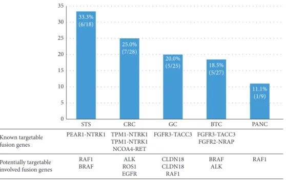
Additionally, potentially targetable fusions were found in 12 patients (10.2%). RAF1 fusion was detected in 4 cases (GC, melanoma, STS, and pancreatic cancer), BRAF fusion in 2 cases (BTC and STS), ALK fusion in 2 cases (CRC and BTC), ROS1 fusion in 1 case (CRC), and EGFR fusion in 1 case (CRC) with diverse counterparts. CLDN18-ARHGAP26 fusion was detected in two cases of GC in this study (8%, 2/ 25), which was recently reported and investigated in signet-ring GC and diffuse-type GC [10, 11]. The detection rate of fusion genes, targetable fusion genes, and potentially tar-getable-involved fusion genes according to the cancer types are illustrated in Figure 1.
3.3. STS with NTRK1 Fusion. A novel PEAR1-NTRK1 fusion was detected in a 78-year-old female patient with angiosarcoma. She initially presented with diffuse in-filtrative skin lesion in the right lower leg in December 2016 and had been administered a palliative paclitaxel, pazopanib, and ifosfamide-based combination. However, the patient showed a refractory disease course. She also underwent palliative radiotherapy to the right lower leg for wound management. Based on the PEAR1-NTRK1 fusion detection in this study, we performed immunohistochemical staining and FISH. The patient showed strong positivity for TRK immunohistochemical staining (Figure 2(a)), and NTRK1
35 30 25 20 15 10 5 0 STS Known targetable fusion genes Potentially targetable involved fusion genes
FGFR3-TACC3 FGFR3-TACC3 FGFR2-NRAP TPM1-NTRK1
PEAR1-NTRK1
RAF1
BRAF ALK CLDN18CLDN18 BRAFALK RAF1 RAF1 ROS1 EGFR TPM1-NTRK1 NCOA4-RET CRC GC BTC PANC 33.3% (6/18) 25.0% (7/28) 20.0% (5/25) 18.5% (5/27) 11.1% (1/9)
Figure1: Detection rate of fusion genes and targetable fusion genes according to cancer types.
(a) (b) ···TGCTCCCAGCACACTAACAG··· 1 Chr1 PEAR1-NTRK1 mRNA 15 9 17 (c)
Figure2: (a) Positive immunohistochemical staining for TRK. (b) Positive FISH analysis for NTRK1 fusion. (c) PEAR-NTRK1 fusion confirmed by NGS.
fusion was confirmed by FISH analysis (Figure 2(b)). We enrolled this patient in a phase I basket trial for treatment with a TRK inhibitor. The tumor was confirmed by NGS to harbor a novel PEAR1-NTRK1 fusion with the 5′ end of NTRK1, including the kinase domain, starting at exon 9 fused to exon 15 of PEAR1 (Figure 2(c)). The primary lesion in the right lower leg responded well to the TRK inhibitor (Figure 3), but she died of sepsis due to wound infection during the second cycle of treatment.
3.4. Melanoma with MAPRE2-RAF1 Fusion. A 35-year-old female patient with melanoma showed MAPRE2-RAF1 fu-sion. She had previously been treated by surgical resection of the primary melanoma in the lower leg followed by ad-ministration of adjuvant interferon therapy. She showed lymph node, lung, liver, bone, and brain metastases and was subsequently treated as follows: pembrolizumab, ipilimu-mab, dacarbazine-based combination therapy, gamma knife surgery, craniotomy, and tumor removal from the brain. After immunotherapy and dacarbazine failed, she was en-rolled in this study, and her primary resected tissues were processed for sequencing. This study identified MAPRE2-RAF1 fusion with exon 5 of MAPRE2 fused to exon 10 of RAF1 (Figure 4), and she was administered vemurafenib for 1 month. Unfortunately, she showed progressive disease during vemurafenib administration.
4. Discussion
Our study revealed that 21.2% (25/118) of patients with gastrointestinal/rare cancers harbored at least one strong
fusion by using a targeted RNA sequencing panel. In terms of gastrointestinal cancers including only CRC, GC, BTC, and pancreatic cancer, we found that 20.2% (18/89) of pa-tients harbored at least one fusion gene. Notably, we identified 5.9% (7/118) patients with known targetable fu-sion genes involving NTRK1 (n 3), FGFR (n 3), and RET (n 1) and 10.2% (12/118) of patients with potentially targetable fusion genes involving RAF1 (n 4), BRAF (n 2), ALK (n 2), ROS1 (n 1), EGFR (n 1), and CLDN18 (n 2).
The first NTRK1-TPM3 fusion was identified in colon cancer, and NTRK fusions have been identified in approx-imately 1% of all solid cancers across diverse cancer types [12, 13]. NTRK fusions are oncogenic drivers regardless of the tissue of origin, and first-generation TRK tyrosine kinase inhibitors (larotrectinib, entrectinib, or ropotrectinib) have demonstrated very promising antitumor efficacies in both adult and pediatric patients with NTRK fusion-positive cancers [13–15]. Larotrectinib induced a 75% response rate in TRK fusion-positive cancers, regardless of the tumor type, and was recently approved by the U.S. Food and Drug Administration for solid tumors with NTRK gene fusions [6]. We successfully identified 3 NTRK fusion-positive pa-tients. Subsequently, one patient with angiosarcoma (no. 3) harboring PEAR1-NTRK1 fusion was enrolled in the clinical trial of TRK inhibitor. Recently, the TRIDENT-1 trial demonstrated 8 confirmed cases of partial remission in TKI-na¨ıve or TKI-pretreated ROS1 + /NTRK + patients at various dose levels [16].
FGFR fusions with multiple partners have been de-scribed in numerous cancer types. After the first report of FGFR3-TACC3 fusion in glioblastoma, these fusions were
(a) (b) (c)
Figure3: (a) Right lower leg lesion before treatment of TRK inhibitor. (b and c) Right lower leg lesion after 1 cycle of TRK inhibitor.
···AGAAGAACACAGGCCTCGTG···
1
Chr 18 Chr 3
MAPRE2-RAF1 mRNA 5 10 17
reported in numerous solid cancers, including urothelial carcinoma, non-small-cell lung cancer, thyroid cancer, and uterine cervical carcinoma. Notably, FGFR2 fusions are also present in 13–17% of intrahepatic cholangiocarcinomas [17–19]. A phase I trial of erdafitinib, an oral pan-fibroblast growth factor receptor (FGFR) inhibitor, demonstrated that urothelial carcinoma and cholangiocellular carcinoma were most responsive to erdafitinib showing objective response rates of 46.2% (12/26) and 27.3% (3/11), respectively, in patients with FGFR mutations and fusions [20]. Other FGFR inhibitors, such as BGJ398, showed an objective response rate of 14.8% (18.8% FGFR2 fusions only) and disease control rate of 75.4% (83.3% FGFR2 fusions only) in patients with FGFR-altered advanced cholangiocarcinoma [21]. This study successfully identified 3 patients with FGFR fusions (2 patients with FGFR3-TACC3 fusion (nos. 4 and 5) and 1 patient with FGFR2-NRAP fusion (no. 16)); importantly, 2 of 3 patients harboring FGFR fusion had BTC, which may be responsive to erdafitinib or BGJ398 according to recent reports [20, 21].
RET fusions have been described in up to one-third of papillary thyroid cancers and in 2% of lung adenocarcinoma cases; CCDC6-RET and NCOA4-RET are the most com-monly identified RET fusions [22, 23]. In CRC, Le Rolle et al. reported six RET fusion kinases among 3,117 advanced cases (0.2%) through comprehensive genomic profiling and identified NCOA4-RET fusion, which was consistent with the result for the patient with CRC in this study (no. 6) [24]. A recent study comparing RET fusion-positive and RET fusion-negative CRCs revealed that right-sided and MSI-high tumors are more likely to have RET fusion, and RET fusion is an independent poor prognostic factor in overall survival [25]. Confirmed responses to multikinase inhibitors with activity against RET, such as cabozantinib and van-detanib, can be achieved in some patients with lung cancer harboring RET-rearrangement or RET-mutation [26, 27]. Studies of RET-specific inhibitors such as BLU-667 have begun to show promising responses in early-phase clinical trials [6, 28].
RAF kinase fusions, such as of BRAF or RAF1 (also known as CRAF), have been reported in various tumor types, including prostate cancer, GC, melanoma, and papillary thyroid cancer [1, 29]. RAF fusions activate the mitogen-activated protein kinase pathway, and a few reports have demonstrated the anticancer efficacy of MEK inhibitors in RAF fusion-positive melanoma [30, 31]. However, a recent report showed that existing RAF inhibitors cannot suppress RAF1-fusion-driven signaling pathways, and our study also showed that a melanoma patient harboring MAPRE2-RAF1 fusion did not respond to vemurafenib [32]. Novel ap-proaches to RAF1-directed targeted therapy should be explored.
Fusion between CLDN18, a tight junction gene, and ARHGAP26, a gene encoding an RHOA inhibitor, was first reported by the Cancer Genome Atlas to be enriched in the genomically stable subtype of GC [33]. Yao et al. detected CLDN18-ARHGAP26 fusion in 3% of Asian GCs, and cancer cells transfected with this fusion showed reduced cell-cell
adhesion and augmented invasiveness [11]. Recently, a Chinese group reported the largest dataset to date regarding signet ring cell carcinoma of GC; 17% of all signet ring cell carcinoma cases harbored this fusion gene and showed resistance to chemotherapy and worse survival outcomes [10]. Based on these results, CLDN18-ARHGAP26 fusion is considered as a driver that contributes to aggressive tumor behavior and is a strong candidate for targeted drugs.
Our results suggest that detection of fusion genes using a targeted RNA sequencing panel can be beneficial for various cancer subtypes, particularly CRC and BTC. In patients with CRC, gene fusion is rarely observed, but recent studies showed that a substantial proportion of patients with CRC had potentially actionable gene rearrangements involving ALK, ROS1, and NTRK [9, 34]. Interestingly, this study also showed that 6 of 28 patients with CRC (21.4%) had tar-getable or potentially tartar-getable gene rearrangements such as NTRK1 (n � 2), RET (n � 1), ALK (n � 1), ROS1 (n � 1), and EGFR (n � 1) fusions. In patients with BTC, there are several ongoing clinical trials targeting FGFR (NCT03230318, NCT02052778, NCT01948297, NCT02924376, NCT022 65341), BRAF/MET (NCT02034110), and ALK/ROS1 (NCT02374489, NCT02034981, and NCT02568267). In this study, we found that 4 of 27 patients with BTC (14.8%) had targetable or potentially targetable gene rearrangements such as FGFR3 (n � 1), FGFR2 (n � 1), BRAF (n � 1), and ALK (n � 1) fusions. Unfortunately, patients with CRC and BTC harboring targetable or potentially targetable fusion genes in this study could not be administered targeted therapy because they were ineligible or respective clinical trials in Korea were not available.
5. Conclusions
In conclusion, we successfully identified a substantial pro-portion of patients harboring targetable (5.9%) and po-tentially targetable (10.2%) fusion genes by RNA panel sequencing in gastrointestinal and rare cancers. Involved fusion genes were NTRK1, RET, FGFR3, FGFR2, BRAF, RAF1, ALK, ROS1, and CLDN18. We suggest that detection of fusion genes by RNA panel sequencing can be beneficial in refractory patients with gastrointestinal or rare cancers, particularly in those with CRC and BTC.
Data Availability
The clinicopathological data used to support the findings of this study are available from the corresponding author upon request.
Conflicts of Interest
The authors declare that they have no conflicts of interest.
Authors’ Contributions
Su Jin Lee and Jung Yong Hong contributed equally to this work.
Acknowledgments
This research was supported by Basic Science Research Program through the National Research Foundation of Korea (NRF) funded by the Ministry of Education (grant nos. 2016R1A6A3A11932444 and 2016R1D1A1A09918622), and by a grant of the Korean Health Technology R&D Project, Ministry of Health & Welfare, Republic of Korea (grant nos. HI14C2640 and HI14C3418).
Supplementary Materials
Supplementary Table 1: the list of the 36 target genes of Comprehensive Thyroid & Lung kit. Supplementary Table 2: the list of the 53 target genes of Solid Tumor kit. (Supple-mentary Materials)
References
[1] N. Stransky, E. Cerami, S. Schalm, J. L. Kim, and C. Lengauer, “The landscape of kinase fusions in cancer,” Nature
Com-munications, vol. 5, no. 1, p. 4846, 2014.
[2] F. G. Barr, “Fusion genes in solid tumors: the possibilities and the pitfalls,” Expert Review of Molecular Diagnostics, vol. 16, no. 9, pp. 921–923, 2016.
[3] A. M. Schram, M. T. Chang, P. Jonsson, and A. Drilon, “Fusions in solid tumours: diagnostic strategies, targeted therapy, and acquired resistance,” Nature Reviews Clinical
Oncology, vol. 14, no. 12, pp. 735–748, 2017.
[4] A. T. Shaw, D.-W. Kim, R. Mehra et al., “Ceritinib in ALK-rearranged non-small-cell lung cancer,” New England Journal
of Medicine, vol. 370, no. 13, pp. 1189–1197, 2014.
[5] A. T. Shaw, D.-W. Kim, K. Nakagawa et al., “Crizotinib versus chemotherapy in advanced ALK-positive lung cancer,” New
England Journal of Medicine, vol. 368, no. 25, pp. 2385–2394,
2013.
[6] V. Subbiah, V. Velcheti, B. B. Tuch et al., “Selective RET kinase inhibition for patients with RET-altered cancers,” Annals of
Oncology, vol. 29, no. 8, pp. 1869–1876, 2018.
[7] A. Drilon, Z. I. Hu, G. G. Y. Lai, and D. S. W. Tan, “Targeting RET-driven cancers: lessons from evolving preclinical and clinical landscapes,” Nature Reviews Clinical Oncology, vol. 15, no. 3, pp. 151–167, 2018.
[8] M. Babiceanu, F. Qin, Z. Xie et al., “Recurrent chimeric fusion RNAs in non-cancer tissues and cells,” Nucleic Acids Research, vol. 44, no. 6, pp. 2859–2872, 2016.
[9] S. J. Lee, G. G. Li, S. T. Kim et al., “NTRK1 rearrangement in colorectal cancer patients: evidence for actionable target using patient-derived tumor cell line,” Oncotarget, vol. 6, no. 36, pp. 39028–39035, 2015.
[10] Y. Shu, W. Zhang, Q. Hou et al., “Prognostic significance of frequent CLDN18-ARHGAP26/6 fusion in gastric signet-ring cell cancer,” Nature Communications, vol. 9, no. 1, p. 2447, 2018.
[11] F. Yao, J. P. Kausalya, Y. Y. Sia et al., “Recurrent fusion genes in gastric cancer: CLDN18-ARHGAP26 induces loss of epi-thelial integrity,” Cell Reports, vol. 12, no. 2, pp. 272–285, 2015. [12] S. Pulciani, E. Santos, A. V. Lauver, L. K. Long, S. A. Aaronson, and M. Barbacid, “Oncogenes in solid human tumours,” Nature, vol. 300, no. 5892, pp. 539–542, 1982. [13] E. Cocco, M. Scaltriti, and A. Drilon, “NTRK fusion-positive
cancers and TRK inhibitor therapy,” Nature Reviews Clinical
Oncology, vol. 15, no. 12, pp. 731–747, 2018.
[14] A. Drilon, S. Siena, S.-H. I. Ou et al., “Safety and antitumor activity of the multitargeted pan-TRK, ROS1, and ALK in-hibitor entrectinib: combined results from two phase I trials (ALKA-372-001 and STARTRK-1),” Cancer Discovery, vol. 7, no. 4, pp. 400–409, 2017.
[15] Entrectinib effective across NTRK fusion-positive cancers,”
Cancer Discovery, vol. 9, no. 1, p. OF4, 2019.
[16] A. E. Drilon, S.-H. I. Ou, B. C. Cho et al., “A phase 1 study of the next-generation ALK/ROS1/TRK inhibitor ropotrectinib (TPX-0005) in patients with advanced ALK/ROS1/NTRK+ cancers (TRIDENT-1),” Journal of Clinical Oncology, vol. 36, no. 15_suppl, p. 2513, 2018.
[17] D. Singh, J. M. Chan, P. Zoppoli et al., “Transforming fusions of FGFR and TACC genes in human glioblastoma,” Science, vol. 337, no. 6099, pp. 1231–1235, 2012.
[18] R. Costa, B. A. Carneiro, T. Taxter et al., “FGFR3-TACC3 fusion in solid tumors: mini review,” Oncotarget, vol. 7, no. 34, pp. 55924–55938, 2016.
[19] S. V. Williams, C. D. Hurst, and M. A. Knowles, “Oncogenic FGFR3 gene fusions in bladder cancer,” Human Molecular
Genetics, vol. 22, no. 4, pp. 795–803, 2013.
[20] R. Bahleda, A. Italiano, C. Hierro et al., “Multicenter phase I study of erdafitinib (JNJ-42756493), oral pan-fibroblast growth factor receptor inhibitor, in patients with advanced or refractory solid tumors,” Clinical Cancer Research, vol. 25, no. 16, pp. 4888–4897, 2019.
[21] M. Javle, M. Lowery, R. T. Shroff et al., “Phase II study of BGJ398 in patients with FGFR-altered advanced chol-angiocarcinoma,” Journal of Clinical Oncology, vol. 36, no. 3, pp. 276–282, 2018.
[22] Y. S. Ju, W.-C. Lee, J.-Y. Shin et al., “A transforming KIF5B and RET gene fusion in lung adenocarcinoma revealed from whole-genome and transcriptome sequencing,” Genome
Re-search, vol. 22, no. 3, pp. 436–445, 2012.
[23] K. Takeuchi, M. Soda, Y. Togashi et al., “RET, ROS1 and ALK fusions in lung cancer,” Nature Medicine, vol. 18, no. 3, pp. 378–381, 2012.
[24] A. F. Le Rolle, S. J. Klempner, C. R. Garrett et al., “Identi-fication and characterization of RET fusions in advanced colorectal cancer,” Oncotarget, vol. 6, no. 30, pp. 28929– 28937, 2015.
[25] F. Pietrantonio, F. Di Nicolantonio, A. B. Schrock et al., “RET fusions in a small subset of advanced colorectal cancers at risk of being neglected,” Annals of Oncology, vol. 29, no. 6, pp. 1394–1401, 2018.
[26] A. Drilon, N. Rekhtman, M. Arcila et al., “Cabozantinib in patients with advanced RET -rearranged non-small-cell lung cancer: an open-label, single-centre, phase 2, single-arm trial,”
The Lancet Oncology, vol. 17, no. 12, pp. 1653–1660, 2016.
[27] K. Yoh, T. Seto, M. Satouchi et al., “Vandetanib in patients with previously treated RET-rearranged advanced non-small-cell lung cancer (LURET): an open-label, multicentre phase 2 trial,” The Lancet Respiratory Medicine, vol. 5, no. 1, pp. 42–50, 2017.
[28] V. Subbiah, J. F. Gainor, R. Rahal et al., “Precision targeted therapy with BLU-667 for RET-driven cancers,” Cancer
Discovery, vol. 8, no. 7, pp. 836–849, 2018.
[29] N. Palanisamy, B. Ateeq, S. Kalyana-Sundaram et al., “Rearrangements of the RAF kinase pathway in prostate cancer, gastric cancer and melanoma,” Nature Medicine, vol. 16, no. 7, pp. 793–798, 2010.
[30] A. M. Menzies, I. Yeh, T. Botton, B. C. Bastian, R. A. Scolyer, and G. V. Long, “Clinical activity of the MEK inhibitor trametinib in metastatic melanoma containing BRAF kinase
fusion,” Pigment Cell & Melanoma Research, vol. 28, no. 5, pp. 607–610, 2015.
[31] K. B. Kim, T. Semrad, A. B. Schrock et al., “Significant clinical response to a MEK inhibitor therapy in a patient with met-astatic melanoma harboring an RAF1 fusion,” JCO Precision
Oncology, no. 2, pp. 1–6, 2018.
[32] P. Jain, T. M. Fierst, H. J. Han et al., “CRAF gene fusions in pediatric low-grade gliomas define a distinct drug response based on dimerization profiles,” Oncogene, vol. 36, no. 45, pp. 6348–6358, 2017.
[33] The Cancer Genome Atlas Research Network, “Compre-hensive molecular characterization of gastric adenocarci-noma,” Nature, vol. 513, no. 7517, pp. 202–209, 2014. [34] F. Pietrantonio, F. Di Nicolantonio, A. B. Schrock et al., “ALK,
ROS1, and NTRK rearrangements in metastatic colorectal cancer,” JNCI: Journal of the National Cancer Institute, vol. 109, no. 12, 2017.
Stem Cells
International
Hindawi www.hindawi.com Volume 2018 Hindawi www.hindawi.com Volume 2018 INFLAMMATIONEndocrinology
International Journal ofHindawi www.hindawi.com Volume 2018 Hindawi www.hindawi.com Volume 2018
Disease Markers
Hindawi www.hindawi.com Volume 2018 BioMed Research InternationalOncology
Journal of Hindawi www.hindawi.com Volume 2013 Hindawi www.hindawi.com Volume 2018Oxidative Medicine and Cellular Longevity
Hindawi
www.hindawi.com Volume 2018
PPAR Research
Hindawi Publishing Corporation
http://www.hindawi.com Volume 2013 Hindawi www.hindawi.com
The Scientific
World Journal
Volume 2018 Immunology Research Hindawi www.hindawi.com Volume 2018 Journal ofObesity
Journal of Hindawi www.hindawi.com Volume 2018 Hindawi www.hindawi.com Volume 2018 Computational and Mathematical Methods in Medicine Hindawi www.hindawi.com Volume 2018Behavioural
Neurology
Ophthalmology
Journal of Hindawi www.hindawi.com Volume 2018Diabetes Research
Journal ofHindawi
www.hindawi.com Volume 2018
Hindawi
www.hindawi.com Volume 2018 Research and Treatment
AIDS
Hindawi
www.hindawi.com Volume 2018 Gastroenterology Research and Practice
Hindawi www.hindawi.com Volume 2018