원위부 근이양증
증례보고
-연세대학교 의과대학 재활의학교실 및 근육병재활연구소, -연세대학교 의과대학 병리학교실1류중선ㆍ김윤진ㆍ이윤정ㆍ박사윤ㆍ박윤길ㆍ문재호∙노태웅
1 – Abstract –Distal Myopathy
– A case report –
Joong Sun Ryu, M.D., Yoon Jin Kim, M.D., Yoon Jeong Lee, M.D., Sa Yoon Park, M.D.,
Yoon Ghil Park, M.D., and Jae Ho Moon, M.D., Tae Woong Rho, M.D.
1Department of Rehabilitation Medicine and Research Institute of Rehabilitation, Yonsei University College of Medicine
Department of Pathology, Yonsei University College of Medicine1
Although muscle disease classically presents with proximal extremity weakness, some myopathic disor-ders, including several types of muscular dystrophy, result in predominantly, or exclusively, distal muscle involvement. Accurate diagnosis of these relatively uncommon conditions can be challenging for the clini-cian, because of unusual phenotype and significant overlap in the clinical features of theses entities. The clinical characteristics of this patient were slowly progressive symmetrical muscle weakness and wasting of all 4 extremities in distal part. Serum muscle enzymes were slightly increased. The prominent EMG findings were myopathic changes, but reduced recruitment was occasionally found in some distal muscles. The muscle biopsy finding of right gastrocnemius showed the non-specific finding of myopathy. Among various perviously described distal myopathies, several diseases have been established as clinically and genetically distinct entities. The most representative diseases are dominantly inherited Welander distal myopathy and tibial muscular dystrophy, and the recessively inherited distal myopathy with rimmed vac-uoles (Nonaka myopathy) and distal muscular dystrophy (Miyoshi myopathy). In this case clinical charac-teristic was compatible with Welander distal myopathy, but genetically not consistent with autosomal dom-inant inherited disease. Because this case showed overlap in the clinical feature of these disease entity we concluded this case was nontypical early adult onset distal myopathy.
Key Words: Distal muscular dystrophy, Distal myopathy
Address reprint requests to Yoon Ghil Park, M.D.
Department of Rehabilitation Medicine & Rehabilitation Institute of Muscular Disease, Yonsei University College of Medicine #146-92 Dogok-dong, Gangnam-gu, Seoul 135-270, Korea
TEL : 82-2-3497-3493, FAX : 82-2-3463-7585, E-mail : [email protected]
서
론
일반적으로 근이양증은 대부분 근위부 근육을 먼저 침
범하지만 드물게는 원위부 근육을 먼저 침범하는 경우도 있는데 이를 원위부 근이양증(distal muscular dys-trophy)이라 한다. 이는 흔치 않은 질환으로 주로 사지 의 원위부에 근력약화를 나타내는데, 말단부위의 근위축 이라는 점에서 다발성말초신경증, 근이양증성근긴장증
(myotonic muscular dystrophy) 그리고 척수성 근 위축증(spinal muscular atrophy)의 말단형(distal type)과의 감별진단을 요한다. 이 에 본 저자 등은 원 위부 근력약화 및 근위축을 보이고 신경전도검사 및 침 근전도검사상 근육질환의 소견을 보이며 근육생검으로 원위부 근이양증을 확인할 수 있었던 젊은 남자환자 1 예를 문헌고찰과 함께 보고하는 바이다.
증
례
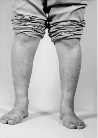
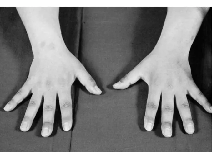
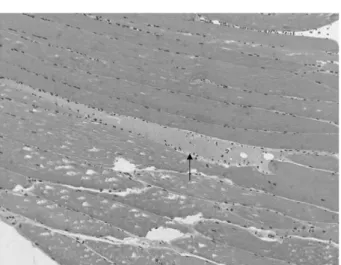
24세 남자환자로 3년 전부터 서서히 진행된 양하지의 근력약화 및 근위축을 주소로 본원 재활의학과에 내원 하였다. 환자는 1년 전부터는 계단을 오르내리기도 불 편해질 정도로 다리의 힘이 줄었다고 하였다. 가족력상 환자는 2남 1여 중 둘째 아들로 가족 모두 건강하였으 며 부모를 비롯한 다른 친척들 가운데도 유사한 증상을 가진 사람은 없었다. 신경학적 검사상 사지 모두에서 근력감퇴 및 근위축이 현저하였는데, 도수근력 검사상 양측 족저굴곡근, 배측굴곡근 및 수부내재근은 4/5이었 으며, 나머지 근육은 정상이었고 근위부보다는 원위부 에서 근위축이 현저하였다(Fig. 1~4). 속상연축(fas-ciculation)은 관찰되지 않았고 감각기능이나 뇌신경기 능도 정상이었다. 심부건반사는 상하지에서 약간 저하 되어있었다. 혈액검사 및 간기능검사에서는 혈청 CK는 678 IU/L, CK-MB 22.89 IU/L, LDH 510 IU/L로 증가되어 있었다. 갑상선기능검사를 비롯한 그 밖의 다 른 혈액 및 소변 검사 결과는 모두 정상이었다. 심전도 도 정상이었다. 방사선학적 검사상 경추부 및 요추부 자기공명영상검사상 정상이었다. 전기진단학적 검사상 양측비복신경, 정중신경 및 척골신경에서 복합근운동전 위(compound muscle action potential, CMAPs) 의 진폭이 감소되었으나 신경전도속도는 정상이었다. 침근전도검사상 양측 전경골근, 비복근 및 상완이두근 에서 안정 시 근세동전위와 양성 예각파 등의 비정상 자발전위가 관찰되었고, 자발적 수축시에 작은 진폭의 짧은 기간을 가진 다상성 운동단위 전위를 관찰할 수 있었으며, 적은 힘으로 완전 동원양식에 도달되는 소견 을 나타내었다(Fig. 5, 6). 근생검은 우측 내측 비복근 에서 시행하였는데 H & E 염색에서 비후된 섬유와 위 축된 섬유근섬유가 관찰되며, 근육세포의 크기의 변화 가 심하였고, 재생 근섬유, 증가된 내부핵, 근섬유분열Fig. 1. Photograph of the case. Distal muscle wasting in both
lower extremities: anterior compartment.
Fig. 2. Photograph of the case. Distal muscle wasting in both
(fiber splitting)등의 근이양증에 합당한 소견을 보였 다(Fig. 7, 8).
고
찰
원위부 근이양증은 매우 서서히 진행되는 원위부 근력 약화와 근위축을 임상적 특징으로 하는 질환으로서, 여 기에는 다양한 유전형태 및 임상양상과 병리소견을 보이 는 여러 질환이 포함되어있다. 이는 1)체염색체우성 Welander형 말단근병증, 2)체염색체우성 Markes-bery-Griggs형 말단근병증, 3)체염색체우성 Laing형 말단근병증 4)체염색체열성 말단근병증의 네 가지로 분 류되고, 이 네 번째 유형은 다시 Miyoshi형과 Nonaka 형으로 나누어진다(Table 1).1,2 우리 나라에서는 박영관 (1990)등이 체염색체우성 말단 근이양증 1가족 예를 보 고한 바 있으며3 체염색체열성 말단 근이양증은 일본에서 유난히 많이 보고되고 있는데 문준식(1995) 등과 전승한 (1999) 등이 각각 Nonaka형과 Miyoshi형의 말단 근이 양증을 보고한 바가 있었고 김(1993) 등이 봉입체근염과 원위부 근질환과의 감별진단이 어려웠던 1예를 보고한 바가 있었다.4-6 원위부 근질환은 1902년 Gower에 의해 처음 보고되었지만 1951년 Welander가 249예의 원위부 근질환에 대한 증례보고로 그 임상양상이 알려졌고 이 중 가장 먼저 알려진 유형인 Welander의 myopathia distalis tarda hereditaria의 특징은 상염색체우성으로 유전되고, 발병연령은 평균47세(20~77세), 남녀비가 3:2로 남자에 약간 더 많고, 사지의 원위부 근력약화 및 근육위축이 나타나는데 대부분 손에서 증상이 먼저 시작 하며 점차 전완부 및 하지의 근육도 침범하고 병은 매우 느리게 진행한다. 혈청 CK치는 정상이거나 약간 증가되 있으며 근전도 검사상 근육병성 소견을 보이며 간혹 신 경병성 소견도 보일 수 있다. 근생검상 다양한 소견을 보 인다.7,8이외에도 발병연령 및 초기 침범부위에서 차이가나는 ‘juvenile-onset hereditary distal myopathy’,
‘infantile-onset hereditary distal myopathy’ 등이 보고되었으며 유전과 관계없는 산발적인 예도 보고되고 있다.9-11Markesbery-Griggs형은 40~50대에 발병하는
Fig. 3. Photograph of the case. Distal muscle wasting in both
hands: dorsum.
Fig. 4. Photograph of the case. Distal muscle wasting in both
hands: palm.
Fig. 5. EMG findings of the right tibialis anterior muscle. Mild
to moderate spontaneous potentials are noted.
Fig. 6. EMG findings of the right tibialis anterior muscle.
Short duration, low amplitude, polyphasic potentials are noted.
체염색체우성으로 유전되는 질환으로서 발에서부터 근력 약화가 시작하여 점차 손, 어깨, 허벅지, 몸통으로 진행 하여 나중에는 걸을 수 없게 되는데, 혈청 CK치도 약간 상승하며 병리적으로 근섬유에서 현저한 공포형성을 보 인다.12 1995년 Laing 등은 전경골근과 경추부굴곡근의
원위부 근육약화와 이 후 상지의 distal finger exten-sor까지 침범하는 것을 특징으로 하는 성염색체우성 말 단근병증을 보고하였다. 발병연령은 4~25세 사이였고 혈청 CK치는 정상의 세 배까지 증가되었고 근생검상 근 육병성 소견을 보이며 공포(rimmed vacuole)는 관찰되 지 않았다.13 체염색체열성 말단근병증은 일본에서 유난 히 많이 보고되고 있는데 비교적 젊은 나이에 하지부터 증상이 시작된다는 특징을 보인다. 임상적으로는 하지에 서 현저한 근력약화 및 근위축을 보이지만 임상 및 병리 소견에 의하여 Miyoshi형과 Nonaka형으로 나누어진 다. Miyoshi형의 원위부 근이양증은 상염색체 열성으로 유전되므로 산재성으로 발생하고 15~30세 사이에 병발 한다. 대개 하지의 원위부 근육위축이 오고 전경골근 (anterior tibial muscle)보다는 비복근(gastrocne-mius muscle)을 잘 침범하며 병이 진행됨에 따라 대퇴 와 둔부 그리고 전완부 근육도 어느 정도 침범되나, 손과 발의 작은 근육은 침범하지 않는다고 하였다. 특징적으 로 혈청 CK치는 정상치보다 10에서 150배 이상으로 증 가되어있다. 진행 및 예후는 계단 오르기, 일어서기 및 걷기 등에 장애를 보이지만 거동을 못할 정도까지는 진 행하지 않는다. 근생검상 근섬유의 크기가 다양하고 괴 사와 재생소견을 관찰할 수 있으나 rimmed vacuole을 형성하지 않는다.14 Nonaka형의 근질환(distal
myopa-thy with rimmed vacuole formation)에 대한 특징 적 임상 양상을 보면 이것 역시 상염색체 열성으로 유전 되므로 산재성으로 발생하고 대개 19~27세 사이의 여자 에서 주로 병발한다. 하지의 원위부 근력약화 및 근위축 을 보이며 특히 전경골근을 잘 침범하여 계상 보행 (steppage gait) 또는 foot drop이 초기 증후로 나타날 수 있다. 병은 아급성으로 진행하며 근위부 근육까지 근 력약화가 와서 종래에는 보행이 불가능해진다. 혈청 CK 치는 정상이거나 약간 증가된 정도이고 근생검상 뚜렷한 근섬유괴사 없이 친염기성 물질로 둘러싸인 공포(ba-sophilic rimmed vacuole)를 특징적으로 보인다.15-17
본 증례는 원위부 근위축 등의 임상양상을 보여 우선 말 초신경질환 등을 의심하였으나 신경전도검사상 신경전도 속도가 정상소견을 보여 이를 감별진단 할 수 있었고, 침 근전도 검사에서 근육병증의 소견을 보여 근생검검사를 통해 원위부 근이양증임을 확인할 수 있었으나 현재 사 용되고 있는 원위부근질환의 분류에 따라 아형을 분류할 수는 없었다. 임상적 양상 및 병리소견으로는 Welander 형 말단근병증에 부합하는 소견을 보였지만 가족력상 특 이소견이 없이 산재성으로 발생하여 전형적인 Welan-der형 말단근병증과는 다른 양상을 보였다. 본 증례와 같이 가족력이 명확치 않은 경우에서 임상적 양상과 근 전도검사 및 병리소견만으로 이를 분류하기는 매우 어려 울 수밖에 없으며 이를 위해 향후 유전자분석 등의 더 상 세한 연구가 필요할 것으로 생각한다.
참고문헌
01. IIIa I: Distal myopathies. J Neurol 2000: 247(3): 169-74 02. Barohn RJ, Amato AA: Distal myopathies. Seminars in
Neurology 1999: 19(1): 25-58
03. 박영관, 선우일남, 지제근: 체염색체 우성 말단 근이양증 1
가족 예. 대한신경과학회지1990: 8(2): 349-352
04. 문준식, 선우일남, 김기환, 김태승: Rimmed Vacuole을 보이 Fig. 7. Paraffin embeded section of right gastrocnemius
mus-cle shows hypertrophic, atrophic fibers and degenerat-ing fibers (arrow)(H & E, ×100).
Fig. 8. Myopathic changes showing increased size variability,
internal nuclei and de-/regenerating fibers (arrow and inset)(H & E, ×100, inset, ×400).
는 말단근병증1가족 3례. 대한신경과학회지1995: 13(3): 665-670 05. 전승한, 장 훈, 이창훈, 김한철, 김지훈: Miyoshi 근육병증. 대한재활의학회지1999: 23(2): 425-429 06. 김성민, 이현미, 송홍기, 김상윤, 권기한, 이병철 등: 봉입체 근염? 원위부 근질환? 대한신경과학회지1993: 11(3): 427-433
07. Gower WR: A lecture on myopathy and a distal form. Br Med J 1902: 2: 89-92
08. Welander L: Myopathia distalis tarda hereditaria; 249 exa-mined cases in 72 pedigrees. Acta Med Scand 141 Suppl 1951: 265:1-124(Cited from Satoyoshi E: Distal myopa-thy. Yohoki J Exp Med 1990: 161 suppl: 1-19)
09. Miller RG, Blank NK, Layzer RB: Sporadic distal myopa-thy with early adult onset. Ann Neurol 1979: 5: 220-227 10. Mongini T, DoriguzziC, Palmucci L, Pollo B, Arnaudo E,
Bresolin N: Sporadic distal myopathy with early adult onset: Study of muscle biopsies and muscle cell cultures. Eur Neurol 1989: 29: 287-290
11. Vaccario ML, Scoppetta C, Bracaglia R, Uncini A:
Spo-radic distal myopathy. J Neurol 1981: 224: 291-295 12. Markesbery W, Griggs R: Distal myopathies. In: Engel
AG, Banker BQ des. Myology. New York, McGraw-Hill, 1994: pp 1246-1257
13. Laing NG, Laing BA, meredith C, Wilton SD, Robbins P, Honeyman K et al: Autosomal dominant distal myopathy: linkage to chromosome 14. Am J Hum Genet 1995: 56: 422-427
14. Miyoshi K, Iwasa T, Kawai H, Sasaki S, Kusaka K, Yagi-ta M et al: Autosomal recessive disYagi-tal muscular dystrophy. Nihon Rinsho 1977: 35: 3922-3928
15. Nonaka I, Sunohara N, Ishiura S, Satohoshi E: Familial distal myopathy with rimmed vacuole and lamellar(mye-loid)body formation J Neurol Sci 1981: 51: 141-55 16. Nonaka I, Sunohara N, Satoyoshi e, Terasawa K,
Yenemo-to K: AuYenemo-tosomal recessive distal muscular dystrophy: A comparative study with distal myopathy with rimmed vac-uole fromation. Ann Neurol 1985: 17: 51-9
17. Nonaka I: Distal myopathies. Current Opinion in Neurolo-gy 1999: 12: 493-9