INTRODUCTION
Hepatocellular carcinoma (HCC) is the most common
prima-ry liver malignancy and constitutes over 5% of all cancers.1,2 Its
worldwide incidence is estimated at 500000 to 1 million new cases per year.3 The development of HCC has a multi-step
car-cinogenetic process similar to other solid tumors; however, it possesses unique risk factors. Hepatitis B (HBV) or C (HCV) viral infection, heavy alcohol intake, prolonged dietary expo-sure to aflatoxin or vinyl chloride, and primary hemochroma-tosis are all associated with HCC development.4 Patients with
HCC usually have at least one of these risk factors and their prevalence varies between patients according to their geo-graphical origin.5 This HCC manifestation diversity is also
found at the genetic and epigenetic level, and a large number of these alterations have been described in the literature.6 Of
these, alterations in Wnt/β-catenin signaling have been proved to play a critical role in HCC development.7 Therefore,
comprehensive knowledge of the Wnt/β-catenin signaling
Serum Dickkopf-1 as a Biomarker
for the Diagnosis of Hepatocellular Carcinoma
Seung Up Kim
1,2,7, Jeon Han Park
3,4, Hyon-Suk Kim
5, Jae Myun Lee
3,4,8, Hyun Gyu Lee
4, Hyemi Kim
3,
Sung Hoon Choi
2,7, Shinhwa Baek
2,7, Beom Kyung Kim
1,2,7, Jun Yong Park
1,2,7, Do Young Kim
1,2,7,
Sang Hoon Ahn
1,2,7,8, Jong Doo Lee
6, and Kwang-Hyub Han
1,2,7,8Departments of 1Internal Medicine, 3Microbiology, 5Laboratory Medicine, and 6Nuclear Medicine, 2Institute of Gastroenterology,
4Institute for Immunology and Immunological Diseases, Yonsei University College of Medicine, Seoul;
7Liver Cirrhosis Clinical Research Center, Seoul;
8Brain Korea 21 PLUS Project for Medical Science, Seoul, Korea.
Purpose: Dickkopf-1 (DKK-1) is a Wnt/β-catenin signaling pathway inhibitor. We investigated whether DKK-1 is related to pro-gression in hepatocellular carcinoma (HCC) cells and HCC patients.
Materials and Methods: In vitro reverse-transcription polymerase chain reaction (RT-PCR), wound healing assays, invasion as-says, and ELISAs of patient serum samples were employed. The diagnostic accuracy of the serum DKK-1 ELISA was assessed us-ing receiver operatus-ing characteristic (ROC) curves and area under ROC (AUC) analyses.
Results: RT-PCR showed high DKK-1 expression in Hep3B and low in 293 cells. Similarly, the secreted DKK-1 concentration in the culture media was high in Hep3B and low in 293 cells. Wound healing and invasion assays using 293, Huh7, and Hep3B cells showed that DKK-1 overexpression promoted cell migration and invasion, whereas DKK-1 knock-down inhibited them. When serum DKK-1 levels were assessed in 370 participants (217 with HCC and 153 without), it was significantly higher in HCC patients than in control groups (median 1.48 ng/mL vs. 0.90 ng/mL, p<0.001). The optimum DKK-1 cutoff level was 1.01 ng/mL (AUC=0.829; sensitivity 90.7%; specificity 62.0%). Although DKK-1 had a higher AUC than alpha-fetoprotein (AFP) and des-gamma-carboxy prothrombin (DCP) (AUC=0.829 vs. 0.794 and 0.815, respectively), they were statistically similar (all p>0.05). When three bio-markers were combined (DKK-1 plus AFP plus DCP), they showed significantly higher AUC (AUC=0.952) than single marker, DKK-1 plus AFP, or DKK-1 plus DCP (all p<0.001).
Conclusion: DKK-1 might be a key regulator in HCC progression and a potential therapeutic target in HCC. Serum DKK-1 could complement the diagnostic accuracy of AFP and DCP.
Key Words: Dickkopf-1, hepatocellular carcinoma, biomarker Yonsei Med J 2015 Sep;56(5):1296-1306
http://dx.doi.org/10.3349/ymj.2015.56.5.1296 pISSN: 0513-5796 · eISSN: 1976-2437
Received: November 5, 2014 Revised: December 22, 2014 Accepted: December 23, 2014
Co-corresponding authors: Dr. Kwang-Hyub Han, Department of Internal
Medi-cine, Institute of Gastroenterology, Yonsei University College of MediMedi-cine, 50-1 Yonsei-ro, Seodaemun-gu, Seoul 120-752, Korea.
Tel: 82-2-2228-1949, Fax: 82-2-393-6884, E-mail: gihankhys@yuhs.ac and Dr. Jeon Han Park, Department of Microbiology, Institute for Immunology and Immu-nological Diseases, Yonsei University College of Medicine, 50-1 Yonsei-ro, Seodae-mun-gu, Seoul 120-752, Korea.
Tel: 82-2-2228-1815, Fax: 82-2-392-7088, E-mail: jhpark5277@yuhs.ac •The authors have no financial conflicts of interest.
© Copyright: Yonsei University College of Medicine 2015
This is an Open Access article distributed under the terms of the Creative Com-mons Attribution Non-Commercial License (http://creativecomCom-mons.org/ licenses/ by-nc/3.0) which permits unrestricted non-commercial use, distribution, and repro-duction in any medium, provided the original work is properly cited.
pathway is essential to identify potential endogenous molecu-lar targets of novel drugs.
Secreted Wnt antagonists play important roles in regulating Wnt/β-catenin signaling.8-10 Of these, dickkopf-1 (DKK-1), a
prototypical member of a secreted protein family, has been known as a potent antagonist of Wnt/β-catenin signaling.10,11
DKK-1 acts as an inhibitory ligand of the low-density lipopro-tein receptor-related prolipopro-tein 5/6 co-receptors and subsequent-ly blocks their interaction with Wnt, resulting in β-catenin deg-radation.12-14 It has been reported that DKK-1 is down-regulated
in human colon tumors, indicating that DKK-1 may act as a tumor suppressor and that high DKK-1 expression indicated favorable responses to chemotherapy in brain tumors.15,16
Conversely, DKK-1 overexpression was found in human hep-atoblastomas, Wilms’ tumors, and multiple myelomas.17,18 In
addition, Forget, et al. reported high DKK-1 expression in 21 out of 73 cases of breast cancer, particularly in hormone-resistant breast tumors, and several experimental studies have shown that DKK-1 was highly expressed in HCC.20,21 Another study
showed that DKK-1 expression was significantly higher in spec-imens with poorer pathologic grade and higher 18
F-fluorode-oxyglucose positron emission tomography uptake in HCC.22
Furthermore, a recent study reported that elevated DKK-1 ex-pression is a critical event in HCC patients, indicating poor clin-ical outcomes, and that DKK-1 is a novel prognostic predictor in these patients.23 All these studies suggest that DKK-1 is
associ-ated with tumor growth and poor prognosis in some malig-nant tumors.
The controversies regarding the molecular and clinical im-plications of DKK-1 molecules remain unresolved. In this study, therefore, we investigated the role of DKK-1 in the Wnt/ β-catenin signaling pathway using several HCC cell lines and compared the diagnostic accuracy of serum DKK-1 to the con-ventional HCC tumor markers, alpha-fetoprotein (AFP) and des-gamma-carboxy prothrombin (DCP) in human patients.
MATERIALS AND METHODS
Cell linesHep3B and 293 cells were grown in modified Eagle’s medium (MEM, GIBCO BRL, Grand Island, NY, USA). Huh7 was grown in Dulbecco’s MEM (DMEM, GIBCO BRL) supplemented with 10% fetal bovine serum (GIBCO BRL), 2 mM glutamine, 100 U/ mL penicillin and 100 μg/mL streptomycin. All cells were cul-tured at 37°C in a 5% CO2 atmosphere.
Patients and specimens
All serum samples were collected from Yonsei Liver Blood Bank (YLBB) system (approval number, 4-2009-0725), Sever-ance Hospital, Sinchon-dong, Korea. This study was approved by the independent Institutional Review Board (IRB) of Sever-ance Hospital and conformed to the 1975 Helsinki declaration
ethical guidelines.
Our exclusion criteria were as follows: 1) previous history of anti-HCC treatment (n=25), 2) pathological or radiological ev-idence of mixed HCC-cholangiocellular carcinoma (n=5), 3) insufficient clinical data (n=2), and 4) insufficient serum sam-ples (n=10). Serum samsam-ples from 80 HCC patients who un-derwent surgical resection, 137 HCC patients who received non-surgical anti-HCC treatments (chemotherapy, radiother-apy, ablation, and conservative care), 67 patients with liver cir-rhosis alone, 46 patients with chronic hepatitis alone, and 40 healthy subjects with no evidence of viral hepatitis [HBV sur-face antigen (HBsAg) and HCV antibody negative and normal liver biochemistry] were collected between February 2010 and April 2011.
Diagnosis and grading of HCC and liver cirrhosis HCC diagnosis was made on the following criteria: 1) patho-logical HCC diagnosis for patients who underwent surgical re-section or percutaneous biopsy or 2) clinical and radiological HCC diagnosis for patients without available HCC tissue speci-mens, based on the guidelines of the American Association for the Study of Liver Diseases.24 In the surgical resection cases,
Tumor-Node-Metastasis (TNM) staging based on the Ameri-can Joint Commission on Cancer (7th edition) was assessed postoperatively.25 Otherwise, HCC stage was clinically defined
according to the Barcelona Clinic Liver Cancer (BCLC) stag-ing system.26 For this study, early-stage HCC was defined as
HCCs with TNM I–II and BCLC A–B.
Liver cirrhosis was histologically or clinically diagnosed as follows: 1) platelet count <100000/μL and ultrasonographic findings suggestive of cirrhosis, including a blunted, nodular liver edge accompanied by splenomegaly (>12 cm), 2) esoph-ageal or gastric varices, or 3) overt liver cirrhosis complications, including ascites, variceal bleeding, and hepatic encephalop-athy.27,28 Liver function was graded according to the
Child-Pugh classification.
Microscopic assessment of HCC and background liver histology
During histological examinations, tumor differentiation was classified according to the Edmondson grading system. Mi-crovascular invasion was defined as a tumor within the vascu-lar space of a capilvascu-lary in the tumor capsule or the sinusoidal space of a non-tumoral liver that was visible only upon micros-copy. Portal vein invasion was defined as a tumor within the vascular space of a portal vein in the portal tract of a non-tu-moral liver that was visible only upon microscopy.29 Satellite
nodules were defined as HCC nodules that existed in the same segment of the main tumor, but remained separated by an in-terval of non-tumoral liver parenchyma.30 Background liver
histology was evaluated semiquantitatively according to the Batts and Ludwig31 scoring system. Fibrosis was staged on a 0
portal fibrosis with a few septa; F3, numerous septa without cirrhosis; and F4, cirrhosis. Activity was graded as A0, none; A1, minimal; A2, mild; A3, moderate; and A4, severe activity. Reverse transcription-polymerase chain reaction (RT-PCR)
Total RNA was isolated using an RNeasy kit (Qiagen, Santa Clarita, CA, USA). cDNA was synthesized by reverse transcrip-tion with 1μg of total RNA, 0.5 μg of random hexamers (Pro-mega, Madison, WI, USA), 1.25 mM dNTP (Boehringer-Man-nheim, Man(Boehringer-Man-nheim, Germany), and 200 U Moloney murine le-ukemia virus reverse transcriptase (MMLV-RT, Invitrogen, Carl-sbad, CA, USA) in a 20 μL reaction volume. Polymerase chain reaction (PCR) was performed with 3 μL of cDNA, 10 pmol of the primer sets, 0.25 mM dNTP, and 2 U of Taq polymerase (Perkin-Elmer, Norwalk, CT, USA). PCR cycling conditions were as follows: 23–35 cycles of denaturation at 94°C for 30 s, annealing at 56–60°C for 30 s, and extension at 72°C for 30 s. The primer sequences used were as follows: DKK-1-forward, 5’-TCACACCAAAGGACAAGAAG-3’; DKK-1-reverse, 5’-ATCT TGGACCAGAAGTGTCTAGC-3’; β-catenin-forward, 5’-ACAA ACTGTTTTGAAAATCCA-3’; β-catenin-reverse, 5’-CGAGT CATTGCATACTGTCC-3’; GAPDH-forward, 5’-GCACTCAG GCTGAGAAC-3’; and GAPDH-reverse, 5’-GGTGAAGACGC CAGTGGA-3’. PCR products were analyzed by agarose gel electrophoresis.
Luciferase assay
To evaluate TCF-4/β-catenin transcriptional activity, the lucif-erase reporter assay was performed using the luciflucif-erase re-porter constructs, TOPflash and FOPflash. TOPflash contains three copies of the TCF-4 binding site and FOPflash contains mutated TCF-4 binding sites. Hep3B and 293 cells were cul-tured to 70% confluence in 24-well plates. Then, TOPflash and FOPflash plasmids were transfected using Fugene HD Re-agent (Promega) according to the manufacturer’s instructions. Following transfection, the luciferase activity was determined using the dual-luciferase reporter assay system (Promega). All luciferase activities were normalized to renilla luciferase activ-ity (internal control) using a luminometer (Centro XS3 LB960, Berthold.Tec, Bad Wildbad, Germany). Luciferase assay data from three independent experiments were analyzed.
DKK-1 over-expression and knock-down
For DKK-1 overexpression, 293 and Huh7 cells were transfect-ed with either pcDNA3.1-Luciferase (control) or the same vector containing the DKK-1 coding sequence. Briefly, cells were seeded into plates and grown for 24 h until they reached 50–60% confluence, followed by transfection with the DKK-1 or control vector using the Fugene HD transfection reagent. DKK-1 overexpression was confirmed using ELISA (R&D Sys-tems, Minneapolis, MN, USA). Small interfering RNA (siRNA) were transfected when the cells reached 70% confluence. The
DKK-1 siRNAs (siDKK-1, NM012242) and GFP siRNA (siGFP, SP-2011) were purchased from Bioneer (Daejeon, Korea). Ex-periments were conducted using Fugene HD transfection re-agent and 100 nM siRNA. The DKK-1 knockdown was con-firmed using ELISA and measured at 450 nm using a spectrophotometric microplate reader (Molecular Devices, Toronto, Canada).
Wound healing and invasion assays
For the wound healing assay, Huh7 or 293 cells were seeded into 35 mm dishes (Corning, NY, USA). Once the cells reached 100% confluence, wound healing assays were performed by scratching a sterile pipette tip through the confluent mono-layer. The cells were then washed, and cultured for another 24 h before microscopic observation (Olympus America, Mel-ville, NY, USA) at 40× magnification. The wound healing assay was performed in triplicate.
For the trans-well invasion assay, 1×10⁵ Huh7 cells in
se-rum-free medium were placed into the upper chamber of the trans-well insert (Corning Costar, Cambridge, MA, USA) with Matrigel (BD Biosciences, San Jose, MA, USA). After 24 h of incubation at 37°C, the cells in the upper chamber and mem-brane were removed, the remaining cells stained with hema-toxylin (Dako, Carpinteria, CA, USA) and eosin (Sigma, ST. Louis, MO, USA), and the number of cells adhering to the lower membrane of the inserts was counted. For each experi-mental group, the invasion assay was performed in triplicate. Three randomly selected fields in each replicate were chosen for cell number quantification.
Cell growth assay
Cell growth in Huh7 cells was analyzed using the 3-(4,5-di-methylthiazol-2-yl)-2,5-diphenyltetrazolium bromide (MTT) assay. Briefly, cells were seeded into a 96-well plate, left over-night to adhere, and treated with siDKK-1. Following treat-ment, 50 μL of MTT solution (2 mg/mL in PBS) was added to each well and incubated for 4 h at 37°C. The supernatant was discarded and 150 μL of dimethyl sulfoxide (Sigma) was added. The plates were agitated until the crystals dissolved. Reduced MTT was measured spectrophotometrically at 595 nm in a beam microplate reader (Molecular Devices).
Serum sample analysis
Peripheral blood samples collected into anticoagulant-free tubes were centrifuged and stored at -70°C prior to testing. An experienced researcher at Severance Hospital performed the assays for serum DKK-1. This individual had no access to pa-tients’ clinical information. ELISA (R&D Systems) was per-formed according to the manufacturer’s recommendations. Briefly, 96-well Nunc-Immunomicrotitre plates with a Maxi-Sorp surface (NalgeNunc, Penfield, NY, USA) were coated with the DKK-1 monoclonal antibody supplied with the kit and incubated overnight at 4°C. The reaction was blocked
with 1% bovine serum albumin. Sera, diluted with 10% neo-natal calf serum, were incubated for 2 h at 37°C. The biotinylat-ed goat anti-human DKK-1 detection antibody was incubatbiotinylat-ed for 2 h at 37°C, followed by streptavidin-horseradish peroxi-dase for 20 min. Colour development was initiated with 3,3, 5,5-tetramethylbenzidine and hydrogen peroxide followed by 1 M sulphuric acid to stop the reaction. The optical density was measured at 450 nm and referenced to 570 nm on a Synergy 2 multimode platereader (Biotek, Winooski, VT, USA). The DKK-1 concentrations were obtained with a four-parameter logistic curve fitted against a standard curve and multiplied by the di-lution factor. When the DKK-1 concentration was less than 0.03 ng/mL (the lower limit of the standard curve), the value was defined as zero. All measurements were performed in triplicate.
AFP concentrations were measured by a UniCel DxI 800 Ac-cessimmunoanalyzer (Beckman Coulter, Inc., Fullerton, CA, USA). Serum DCP concentrations were measured using two different assays: the commercially available EIA (Haicatch PIV-KA-II, Eisai Co., Ltd., Tokyo, Japan) and a Lumipulse G1200 au-toanalyzer (Fujirebioinc., Tokyo, Japan) according to the man-ufacturers’ instructions. All AFP and DCP measurements were performed in triplicate.
Statistical analyses
Data are expressed as mean±SD, median (range), or n (%) where appropriate. The statistical significance of differences between continuous and categorical variables was examined using the Student’s t-test or Mann-Whitney test, and the chi-squared test or Fisher’s exact test, when appropriate. Receiver operating characteristics (ROC) curves were constructed to assess sensitivity, specificity, and respective areas under the curves (AUCs) with a 95% confidence interval (CI). The statis-tical significance between AUC curves were calculated based on DeLong methods. The optimum diagnosis cutoff value was calculated to maximize the sum of sensitivity and specificity, and to minimize overall error. To determine the diagnostic
ac-curacy of DKK-1, AFP, and DCP measurements, we estimated the combined marker functions by binary logistic regression. The function values were used as one marker and subjected to ROC analysis. To assess whether the DKK-1, AFP, and DCP combination marker was better than the individual biomark-ers, we created a new variable predicted probability (p) for HCC on the basis of a binary logistic regression equation. The combination formula (DKK-1 plus AFP, DKK-1 plus DCP, and DKK-1 plus AFP plus DCP) indicated simple addition of the value of each component, not dichotomized categorization of each variable (positive vs. negative). The correlation between DKK-1 serum concentrations and clinic-pathological charac-teristics was analyzed with a Spearman’s rank correlation test. All statistical analyses were assessed using the Statistical Pack-age for Social Science (SPSS version 20.0, Armonk, NY, USA). A p-value<0.05 was considered statistically significant.
RESULTS
DKK-1 expression in Hep3B, Huh7, and 283 cells To determine the DKK-1 expression in HCC cells, reverse tran-scription-PCR (RT-PCR) and ELISA were performed in Hep3B and Huh7 cells with 293 cells as a negative control. RT-PCR analysis showed high DKK-1 mRNA expression in Hep3B cells and low in 293 cells (Fig. 1A). Consistent with these findings, the secreted DKK-1 concentration in the culture media was high in Hep3B cells, moderate in Huh7 cells, and low in 293 cells (Fig. 1B). Luciferase assays revealed that β-catenin tran-scriptional activity increased significantly in 293 cells (ap-proximately 2.5-fold) following DKK-1 overexpression. In Hep3B cells, however, the activity remained comparable be-tween mock and DKK-1 overexpression (Fig. 1C).
DKK-1 effects on wound healing and invasivity To investigate the effect of DKK-1 on cell migration and inva-Fig. 1. DKK-1 expression in Hep3B, Huh7, and 293 cells. (A) RT-PCR analysis showed high DKK-1 mRNA expression in Hep3B cells, low in 293 cells, and none in the negative control. (B) The DKK-1 concentration secreted in the culture media, determined by ELISA, was high in Hep3B cells, moder-ate in Huh7 cells, and low in 293 cells. (C) Following DKK-1 transfection, luciferase assays revealed that the β-cmoder-atenin transcriptional activity was in-duced in 293 cells. *p<0.01. DKK-1, dickkopf-1.
DKK-1 293 Hep3B (-) β-catenin GAPDH 4 3 2 1 0 Mock DKK-1 Mock DKK-1 * Hep3B 293
Relative luciferase activity
800 600 400 200 0 Huh7 293 Conc. (ng/mL ) A B Hep3B C
sion capability, DKK-1 was overexpressed or knock-downed in 293 or Huh7 cells, with low or moderate DKK-1 secretion lev-els, respectively. In this assay, Huh7 cells with DKK-1 moderate expression, instead of Hep3B with high DKK-1 secretion, were used to prevent the potential masking of knock-down influ-ence due to significantly high DKK-1 secretion in Hep3B cells.
Wound healing assays showed that DKK-1 overexpression
efficiently promoted migration of 293 cells (Fig. 2A), whereas DKK-1 knock-down significantly inhibited Huh7 cell migra-tion (Fig. 2B) compared with mock controls. Similarly, invasion assays with Huh7 cells showed that DKK-1 overexpression promoted invasion, whereas DKK-1 knock-down inhibited the invasion (Fig. 2C). MTT assays showed that the DKK-1 knock-down significantly inhibited Huh7 cell growth after 48 h, and Fig. 2. DKK-1 promotes cell migration and invasion. (A) DKK-1 overexpression promoted 293 cell migration in wound healing assays. (B) DKK-1 knock-down inhibited Huh7 cell migration in wound healing assays. (C) DKK-1 overexpression promoted Huh7 cell invasion, whereas DKK-1 knock-knock-down in-hibited it in invasion assays. *p<0.01. DKK-1, dickkopf-1.
120 100 80 60 40 20 0 Incubation time (h) 0 12 24 W ound width (%) Mock DKK-1 * 0 12 24 h DKK-1 293 Mock A 120 100 80 60 40 20 0 Incubation time (h) 0 12 24 W ound width (%) siGFP siDKK-1 * * 0 12 24 h siDKK-1 Huh7 siGFP B 250 200 150 100 50 0 250 200 150 100 50 0
Mock DKK-1 siGFP siDKK-1
Invasiveness (%) Invasiveness (%) * * Mock siGFP DKK-1 siDKK-1 Huh7 C
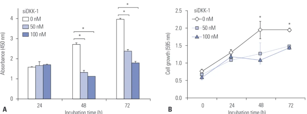
the amount of secreted DKK-1 significantly decreased (Fig. 3). Baseline study population characteristics
Baseline study population characteristics are described in Sup-plementary Table 1 (only online). The age and proportion of male participants were statistically similar between HCC pa-tients and controls (p>0.05). However, some factors were sig-nificantly higher in HCC patients when compared with con-trols, including alanine aminotransferase level (mean 54.6 IU/L vs. 32.3 IU/L), proportion of patients with liver cirrhosis (76.4% vs. 43.8%), and HBsAg positivity (85.3% vs. 59.9%) (all p<0.05). Serum DKK-1 levels in HCC patients and controls To compare serum DKK-1 levels, the DKK-1 concentration
was measured using a commercially available ELISA kit (Fig. 4). Serum DKK-1 concentrations were significantly higher in all HCC patients [patients who underwent surgical resection (n=80) and those who were treated with other modalities (n=137); total n=217; median 1.48 (range 0.03–8.88) ng/mL] than in the three control groups [healthy subjects (n=40), pa-tients with chronic hepatitis (n=46), and those with liver cir-rhosis (n=67); total n=153; median 0.85 (range 0.01–2.92); p< 0.001]. Among three control groups, DKK-1 levels were statis-tically similar [median 0.90 (range 0.01–1.91) ng/mL in healthy subjects vs. 0.72 (range 0.05–2.03) ng/mL in chronic hepatitis patients vs. 0.81 (range 0.01–2.92) ng/mL in liver cirrhosis pa-tients; all p>0.05].
We also measured serum AFP and DCP concentration. Similar to the DKK-1 results, AFP and DCP levels were signifi-cantly higher in HCC patients than in the control groups (me-dian 39.1 ng/mL vs. 3.3 ng/mL, p=0.011 and 129 mAU/mL vs. 22 mAU/mL, p=0.001 for AFP and DCP, respectively) (Table 1). Serum DKK-1 levels according to tumor stage
To investigate the correlation between serum DKK-1 concen-tration and HCC progression, patients with HCC were classified according to TNM and BCLC staging. The serum DKK-1 levels of HCC patients according to tumor stage are as shown in Table 1 and Fig. 4. The HCC patients were stratified into early- and advanced-stage HCC [TNM I–II (n=144) vs. TNM III–IV (n=73)]. DKK-1 levels in TNM I–II patients tended to be lower than TNM III–IV patients (median 1.37 ng/mL vs. 1.66 ng/mL; p= 0.093). DCP levels were significantly lower (median 61 mAU/ mL vs. 1112 mAU/mL; p=0.009), whereas AFP levels were sta-tistically similar (median 30.8 ng/mL vs. 46.4 ng/mL;
p=0.516). When the BCLC staging system was applied, DKK-1
levels were significantly lower in BCLC A–B patients (n=146) than in BCLC C–D patients (n=71) (median 1.36 ng/mL vs. 1.73 ng/mL; p=0.014). DCP level was significantly lower (me-dian 64 mAU/mL vs. 579 mAU/mL; p=0.019), whereas AFP was not statistically significant (median 46.35 ng/mL vs. 27.9 Fig. 3. Knock-down of DKK-1 inhibits cell growth. (A) DKK-1 knock-down in Huh7 cells decreased the amount of secreted DKK-1 after 48 h, deter-mined by ELISA. (B) DKK-1 knock-down inhibited Huh7 cell growth after 48 h, deterdeter-mined by MTT assays. *p<0.01. DKK-1, dickkopf-1.
4 3 2 1 0 2.5 2.0 1.5 1.0 0.5 0.0
Incubation time (h) Incubation time (h)
24 48 72 0 24 48 72
Absorbance (450 nm) Cell growth (595 nm)
0 nM siDKK-1 50 nM 100 nM ** * * * * 0 nM siDKK-1 50 nM 100 nM A B
Fig. 4. Serum DKK-1 concentration. Serum DKK-1 concentrations were significantly higher in HCC patients than those of all the control groups. DKK-1, dickkopf-1; HCC, hepatocellular carcinoma; TNM, Tumor-Node-Metastasis; BCLC, Barcelona Clinic Liver Cancer.
20 15 10 5 5 4 3 2 1 0 p<0.001 p<0.001 p=0.093 p=0.681 p=0.654 Healthy control (n=40) Chronic hepatitis (n=46) Liver cirrhosis (n=67) All HCC (n=217) TNM l–ll (n=144) TNM lll–lV (n=73) BCLC A–B (n=146) BCLC C–D (n=71) p=0.941 p=0.014 p<0.001 DKK-1 (ng/mL)
ng/mL; p=0.690).
Compared to controls (median 0.90 ng/mL), DKK-1 level in early-stage HCC patients (graded TNM I–II and BCLC A–B) were significantly higher (median 1.37 ng/mL and 1.36 ng/ mL, respectively; all p<0.001). Similarly, AFP and DCP levels were significantly different between controls and TNM I–II (p=0.038 and p<0.001) and BCLC A–B stage patients (p=0.046 and p<0.001) (Table 1).
Diagnostic accuracy of DKK-1, AFP, and DCP
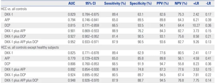
The optimum DKK-1 cutoff value was calculated as 1.01 ng/ mL (AUC=0.829; 95% CI, 0.784–0.875; sensitivity, 90.7%; speci-ficity, 62.0%), AFP as 7.5 ng/mL (AUC=0.794; 95% CI, 0.746– 0.841; sensitivity, 69.3%; specificity, 87.6%), and DCP as 40.5 mAU/mL (AUC=0.815; 95% CI, 0.771–0.859; sensitivity, 66.5%; specificity, 93.4%). The AFP and DCP cutoff values were simi-lar to our institution’s recommended clinical cutoff value (10 ng/mL and 35 mAU/mL, respectively). For ease of interpreta-tion, we adopted 1.00 ng/mL (sensitivity and specificity iden-tical to 1.01 ng/mL), 10 ng/mL (sensitivity, 65.1% and specific-ity, 89.8%), and 40 mAU/mL (sensitivity and specificity identical to 40.5 mAU/mL) as the DKK-1, AFP, and DCP cutoff values, respectively.
The diagnostic indices and corresponding ROC curves us-ing these cutoff values are shown in Table 2, Supplementary Fig. 1 (only online). In the differential diagnostic accuracy as-sessment, DKK-1 had a greater AUC for identifying HCC from the controls than AFP and DCP (AUC=0.829 vs. 0.794 and 0.815, respectively) (Table 2, Supplementary Fig. 1A, only online). However, the AUCs of three markers were statistically similar (all p>0.05). Because DKK-1, AFP, and DCP had no statistical correlations (all p>0.05 by Pearson correlation analysis), we combined these tumor markers to determine whether the di-agnostic accuracy could be enhanced. The accuracy of DKK-1 plus AFP or DKK-1 plus DCP was significantly higher than DKK-1 alone (AUC=0.901 vs. 0.829, p<0.001 and 0.927 vs. 0.829,
p<0.001, respectively). Furthermore, the accuracy of DKK-1
plus AFP plus DCP increased remarkably (AUC=0.952) which was significantly higher than any single marker and DKK- plus AFP and DKK-1 plus DCP (all p<0.001) (Table 2, Supplemen-tary Fig. 1A, only online). When healthy subjects were exclud-ed from the controls, similar findings were observexclud-ed (Table 2, Supplementary Fig. 1B, only online).
When identifying early-stage HCC patients from the con-trols, serum DKK-1 showed a higher AUC than AFP and DCP (AUC=0.818 vs. 0.772 and 0.777 for TNM I–II; AUC=0.811 vs. 0.772 and 0.783 for BCLC A–B, respectively) (Supplementary Table 2, only online, Supplementary Fig. 2A and B, only on-line). When the biomarkers were combined, the triple combi-nation (DKK-1 plus AFP plus DCP) showed the best accuracy (AUC=0.939 for TNM and 0.940 for BCLC, respectively) (Sup-plementary Table 2, only online, Sup(Sup-plementary Fig. 2A and B, only online). Following exclusion of healthy subjects from the
Table 1.
DKK-1, AFP
, and DCP Levels in the Study Population
Biomarkers Controls (n=153) Patients with HCC (n=217) p value (control vs. HCC) TNM staging BCLC staging TNM I –II (n=144) TNM III –IV (n=73) p value (control vs. TNM I –II) p value (TNM I –II vs. TNM III –IV) BCLC A –B (n=146) BCLC C –D (n=71) p value (control vs. BCLC A –B) p value (BCLC A –B vs. BCLC C –D) DKK-1 0.85 (0.01–2.92) 1.48 (0.03–8.88) <0.001 1.37 (0.03–7.53) 1.66 (0.04–8.88) <0.001 0.093 1.36 (0.03–7.53) 1.73 (0.04–0–8.88) <0.001 0.014 AFP 3.3 (0.5 –219.1) 39.1 (0.5 –765316.7) 0.011 30.8 (0.9 –765316.7) 46.4 (0.5 –217580.2) 0.038 0.516 46.35 (0.5–217580.20) 27.9 (0.9–765319.7) 0.046 0.690 DCP 22 (8–211) 129 (8–75000) 0.001 61 (8–7160) 1112 (11–75000) 0.001 0.009 64 (8–12408) 579 (11–75000) <0.001 0.019 DKK-1, dickkopf-1; AFP , alpha-fetoprotein; DCP
, des-gamma-carboxy prothrombin; HCC, hepatocellular carcinoma; TNM, T
umor
-Node-Metastasis; BCLC, Barcelona Clinic Liver Cancer
.
controls, similar findings were observed (Supplementary Ta-ble 2, only online, Supplementary Fig. 2C and D, only online). Correlation between DKK-1 and microscopic HCC findings
When correlation analysis was performed between DKK-1 and microscopic HCC characteristics using Spearman’s method, DKK-1 showed a significant correlation with necroinflamma-tory activity (coefficient 0.232, p=0.040) and a borderline statis-tical correlation to satellite nodules (coefficient 0.203, p=0.072) (Supplementary Table 3, only online). In contrast, microvessel invasion, Edmonson grade, fibrosis, tumor size, and tumor number were not significantly correlated to DKK-1 levels (all
p>0.05).
Diagnostic performance of DKK-1 in subgroups For the 173 patients with a single HCC (≤5 cm), the diagnostic performance of DKK-1 in identifying HCC from controls was better than AFP and DCP (AUC=0.781; 95% CI, 0.708–0.853 vs. AUC=0.744; 95% CI, 0.668–0.820 for AFP and AUC=0.763; 95% CI, 0.691–0.835 for DCP). In addition, the triple marker combi-nation showed the best accuracy (AUC=0.923; 95% CI, 0.886– 0.961). When patients with HBV-related chronic liver disease were selected (n=273), the diagnostic performance of DCP in identifying HCC from controls was better than DKK-1 and AFP (AUC=0.814; 95% CI, 0.764–0.864 vs. AUC=0.802; 95% CI, 0.737–0.866 for DKK-1 and AUC=0.772; 95% CI, 0.715–0.828 for AFP). Additionally, the triple marker combination showed the highest accuracy (AUC=0.942; 95% CI, 0.916–0.968).
After exclusion of 40 healthy subjects from the controls, similar findings were observed. DKK-1 was more accurate than AFP and DCP in diagnosing single HCC (≤5 cm) (AUC=0.781;
95% CI, 0.708–0.853 vs. AUC=0.744; 95% CI, 0.668–0.820 for AFP and AUC=0.763; 95% CI, 0.691–0.835 for DCP), whereas DCP was more accurate than DKK-1 and AFP in diagnosing HBV-related HCC (AUC=0.814; 95% CI, 0.764–0.864 vs. AUC= 0.802; 95% CI, 0.737–0.866 for DKK-1 and AUC=0.772; 95% CI, 0.715–0.828 for AFP). The triple marker combination showed the best accuracy in diagnosing single HCC (≤5 cm) (AUC= 0.896; 95% CI, 0.848–0.944) and HBV-related HCC (AUC= 0.942; 95% CI, 0.916–0.968).
DISCUSSION
The survival of HCC patients has improved due to tremen-dous advances of surgical techniques and perioperative man-agement; however, the mortality rate is still high. This is pri-marily due to delayed diagnosis, lack of effective therapies, and frequent recurrence and metastasis. Early HCC detection gives the opportunity to employ curative treatments such as liver transplantation, resection, or local ablative therapy, which are the best way to prolong survival.32 Although serum AFP
and ultrasound have been widely used for HCC diagnosis and surveillance, approximately 30–40% of HCC patients are se-rum AFP-negative and ultrasound detection accuracy for HCC is suboptimal.33 As a result, the widespread use of serum
AFP and ultrasound for HCC surveillance has been continu-ously questioned.
Because current diagnostic methods are insufficient, it is important to identify novel serum biomarkers with high diag-nostic accuracy to detect and screen early-stage cancers.33
DKK-1 has recently been recognized as a novel biomarker for HCC.23 Conventionally, DKK-1 is known as a secreted
glyco-Table 2. Diagnostic Accuracy of DKK-1, AFP, and DCP in Diagnosing HCC
AUC 95% CI Sensitivity (%) Specificity (%) PPV (%) NPV (%) +LR -LR HCC vs. all controls DKK-1 0.829 0.784–0.875 89.4 63.1 82.6 75.3 2.42 0.17 AFP 0.794 0.746–0.841 65.0 89.5 89.8 64.3 6.21 0.39 DCP 0.815 0.771–0.859 66.5 93.5 94.1 64.4 10.27 0.36 DKK-1 plus AFP 0.901 0.869–0.933 88.9 76.2 84.3 82.7 3.73 0.15 DKK-1 plus DCP 0.927 0.902–0.952 81.4 90.5 93.1 75.6 8.58 0.21
DKK-1 plus AFP plus DCP 0.952 0.933–0.971 87.9 90.5 93.6 82.7 9.26 0.13
HCC vs. all controls except healthy subjects
DKK-1 0.825 0.771–0.878 89.4 62.9 77.6 80.5 2.41 0.17
AFP 0.779 0.729–0.829 65.0 85.8 89.8 56.1 4.59 0.41
DCP 0.806 0.760–0.853 66.5 91.9 94.7 55.8 8.23 0.36
DKK-1 plus AFP 0.892 0.854–0.930 88.9 74.8 87.3 77.6 3.53 0.15
DKK-1 plus DCP 0.924 0.895–0.952 80.5 89.7 94.5 67.4 7.81 0.22
DKK-1 plus AFP plus DCP 0.948 0.926–0.970 87.9 88.7 94.5 76.8 7.75 0.14
DKK-1, dickkopf-1; HCC, hepatocellular carcinoma; AFP, alpha-fetoprotein; DCP, des-gamma-carboxy prothrombin; AUC, area under receiver operating character-istic curve; CI, confidence interval; PPV, positive predictive value; NPV, negative predictive value; LR, likelihood ratio.
The optimum predicted probabilities of DKK-1, AFP, DCP, and their combination were derived from their respective ROC curves by maximizing the sum of sensitiv-ity and specificsensitiv-ity and minimizing the overall error.
protein with a crucial role in limb and head development in Xenopus embryos, and as an upstream regulator of T-cell fac-tor/β-catenin that forms a negative feedback loop in the ca-nonical Wnt signaling pathway, favoring appropriate cell growth.10,34 In normal tissues DKK-1 is hardly expressed,
ex-cept in placenta and embryonic tissues.35 Paradoxically,
how-ever, DKK-1 has been shown to be secreted from some tumor into blood stream, suggesting that serum DKK-1 may be a useful biomarker for numerous cancers including HCC.36-39
Serum DKK-1 is an advantageous biomarker because it is non-invasiveness, has a simple detection protocol, and is in-expensive to quantify.40 Therefore, integration of serum
DKK-1 along with serum AFP and sonographic findings may im-prove and optimize diagnostic work-ups and surveillance strategies for HCC.39
Recently, Yu, et al.23 found that DKK-1 expression level was
associated with β-catenin staining pattern in HCC cell lines, and that DKK-1 overexpression correlated with β-catenin cy-toplasmic/nuclear accumulation in clinical HCC samples. In addition, high DKK-1 expression was an independent unfa-vorable predictor for overall and disease-free survival in HCC patients who underwent surgical resection. Our present re-sults support these observations. Hep3B cells showed high DKK-1 mRNA expression and protein secretion. Additionally, DKK-1 overexpression raised T-cell factor/β-catenin tran-scriptional activity and promoted migration of 293 cells. DKK-1 overexpression promoted invasion, whereas DKK-DKK-1 knock-down inhibited cell growth, migration, protein secretion, and invasion in Huh7 cells. These results indicate that DKK-1 ex-pression has a significant positive association with HCC pro-gression and metastatic potential. Kuang, et al.41 also observed
similar influence of DKK-1 on 293 cells. On the other hand, however, DKK-1 did not increase cellular invasiveness of SNU475 cells.42 The authors also found that DKK-1 did not
af-fect invasion-associated proteases such as matrix metallopro-tease-2 (MMP-2) that acts on collagen type IV, a major com-ponent of the Matrigel matrix and that MMP-2 levels were unaffected by DKK-1 expression in RT-PCR analysis.42
There-fore, it appears that the ability of DKK-1 to inhibit Wnt signal-ing was abrogated in HCC, most likely due to genetic altera-tions disrupting the central multi-protein complex that controls β-catenin stability. However, the exact role of DKK-1 played in migration and invasion is unclear, and the molecular mecha-nism of this controversial phenomenon needs to be further explored.
A recent large-scale multicenter study concluded that the sensitivity and specificity of serum DKK-1 were satisfactory for diagnosing HCC, including early-stage disease and in AFP-negative patients;39 DKK-1 could distinguish HCC from
non-malignant chronic hepatitis or cirrhosis, particularly in AFP-positive patients, indicating the potential of DKK-1 as a serum biomarker for HCC. The diagnostic accuracy of DKK-1, AFP, and DKK-1 plus AFP combination was similar to our findings.
In our study, the AUC of DKK-1 was higher (AUC 0.825–0.829) than AFP and DCP alone. However, when DCP, a biomarker widely used in Eastern Asia,43 was combined with DKK-1 and
AFP together, the diagnostic accuracy of HCC increased sig-nificantly with an overall AUC of approximately 0.95 and an AUC of 0.94 for early-stage HCC. However, the calculated DKK-1 cutoff value in our study was much lower (DKK-1.0DKK-1 ng/mL vs. 2.153 ng/mL).39 The exact reason for the DKK-1 cutoff value
dif-ference is unclear. It may be due to heterogeneity of carcino-genetic mechanisms because of various etiologies of chronic liver diseases of our study population. Therefore, further study is required to establish an optimal DKK-1 cutoff value.
In our study and in Shen, et al.,39 serum DKK-1 levels were
statistically similar among control groups regardless of the presence of cirrhosis. A study by Tung, et al.44 also showed
sta-tistically similar serum DKK-1 levels between controls with and without cirrhosis; however, the DKK-1 transcript level was significantly higher in patients with cirrhosis than pa-tients with chronic hepatitis. Further studies are needed to address the discrepancy in DKK-1 serum and transcript levels according to the presence of liver cirrhosis. However, it can be assumed that DKK-1 transcript level may be correlated to the degree of fibrotic burden, which is an important risk factor for HCC development. This is in contrast to serum DKK-1 levels, which are influenced by apparent HCC development rather than high fibrotic burden, an HCC risk factor. In the present study, there was, indeed, no correlation between serum DKK-1 level and fibrosis stage. This might indicate that DKK-DKK-1 se-cretion into the blood stream could be initiated upon hepato-cyte tumorigenic transformation, and that hepatohepato-cytes with external stressors favoring HCC development, such as high background fibrotic burden, still can control DKK-1 secretion despite the high DKK-1 transcript levels. As seen in our study and others, this phenomenon might enable to diagnose early-stage HCC even in the presence of confounding influences such as liver cirrhosis.39 Preferential early-stage DKK-1
overex-pression has already been reported in prostate cancer,45 which
further supports this hypothesis. However, there was a signifi-cant correlation between DKK-1 level and necroinflammatory activity in our study. The potential influence of necroinflam-matory activity should be investigated further and, if neces-sary, compensated for to enhance the accuracy of serum DKK-1 in diagnosing HCC.
Tao, et al.46 showed that elevated DKK-1 transcript levels in
human HCC tissues were significantly correlated with the presence of tumor nodules, Edmondson-Steiner grade, and the presence of vascular invasion. Furthermore, high DKK-1 transcript levels were significantly correlated with venous in-vasion.44 These data indicate the involvement of DKK-1
over-expression in HCC tissues in HCC invasion and metastasis. In our study, however, serum DKK-1 levels showed only a bor-derline statistical correlation with satellite nodules (p=0.072) and no correlation with Edmondson-Steiner grade or vascular
invasion. This may be due to the limited number of patient samples with available histological information (n=90). When we consider that no macroscopic vascular invasion was iden-tified in any of our study participants prior to surgical resec-tion, the influence of DKK-1 overexpression on vascular inva-sion might have been underestimated. However, because no significant correlation between serum DKK-1 level and ve-nous invasion was observed by Tao, et al.,46 it should be
con-firmed whether the serum DKK-1, rather than the DKK-1 transcript levels, reflect the invasive and metastatic potential of HCC. Interestingly, by Tung, et al.44 observed that tumors
with DKK-1 expression, established by injection of PLC/PRF/5 HCC cells into mice, showed an increased microvessel density score. Another study showed that DKK-1 mediated endotheli-al cell activation by suppressing the Wnt/β-catenin pathway.47
In this study, using an in vivo microvascular remodeling ani-mal model, DKK-1 significantly increased vascular density and vessel diameter in adult rats, indicating that DKK-1 may play a role in microvascular remodeling and tumor angiogen-esis activation, and possibly accounting for DKK-1-mediated cancer growth promotion in vivo. Similarly, it has been re-ported that DKK-1 promotes angiogenic and cartilaginous degradation in synovial fibroblasts, accelerating synovial an-giogenesis and cartilage destruction. These data indicate that DKK-1 could be a new therapeutic target for future cancer treatment. A molecular-targeted agent similar to sorafenib, which suppresses tumor growth and angiogenesis by inhibit-ing the Raf/MEK/ERK signalinhibit-ing pathway and receptor tyro-sine kinases,48 could potentially be developed. Based on this
concept, a very recent study demonstrated that an anti-DKK-1 monoclonal antibody suppressed cell invasion and growth in the lung cancer cell line A549.36 It is highly possible that an
anti-DKK-1 monoclonal antibody could prevent HCC metas-tasis and recurrence.
There are several issues that remain unresolved in our study. First, the HCC diagnosis obtained in our study was not completely based on histopathology. Although histopatholo-gy remains the gold standard for HCC diagnosis and evalua-tion, it is difficult to conduct in large populations. Ultrasonog-raphy and radiological examinations remain the most common approach to diagnosing HCC in clinical practices. Furthermore, an in-depth correlation analysis between DKK-1 level, HCC histological features, and surrounding normal liver parenchy-ma was not feasible. Second, our study participants were all native Korean and HBV was the most common etiology, which may limit the generalization of our findings to other ethnici-ties and chronic liver disease etiologies. Third, the cross-sec-tional nature of our study is another limitation. Although DKK-1 expression in HCC tissues has been proved to be a significant independent predictor of long-term prognosis, a prospective study investigating whether serum DKK-1 can also be used as a biomarker for surveillance and prognosis of HCC is needed.
In conclusion, our study suggests that DKK-1 may be a key
regulator in HCC progression a promising potential therapeu-tic target for HCC. We also demonstrated that serum DDK-1 could be a useful marker for diagnosing HCC, especially in combination with AFP and DCP. Further studies are needed to confirm whether surveillance program for HCC in patients with chronic liver diseases should be adjusted according to serum DKK-1 level.
ACKNOWLEDGEMENTS
This study was supported by the Liver Cirrhosis Clinical Re-search Center, in part by a grant from the Korea Healthcare technology R & D project, Ministry of Health and Welfare, Re-public of Korea (no. HI10C2020) and in part by a grant from a faculty research grant of Yonsei University College of Medicine for 2011 (6-2011-0145). The funders had no role in study design, data collection and analysis, decision to publish, or preparation of the manuscript.
REFERENCES
1. Bruix J, Sherman M, Llovet JM, Beaugrand M, Lencioni R, Bur-roughs AK, et al. Clinical management of hepatocellular carcino-ma. Conclusions of the Barcelona-2000 EASL conference. Europe-an Association for the Study of the Liver. J Hepatol 2001;35:421-30. 2. Kim SU, Kim do Y, Park JY, Ahn SH, Nah HJ, Chon CY, et al.
Hepa-tocellular carcinoma presenting with bone metastasis: clinical characteristics and prognostic factors. J Cancer Res Clin Oncol 2008;134:1377-84.
3. Wands JR, Blum HE. Primary hepatocellular carcinoma. N Engl J Med 1991;325:729-31.
4. Schütte K, Bornschein J, Malfertheiner P. Hepatocellular carcino-ma--epidemiological trends and risk factors. Dig Dis 2009;27:80-92. 5. Seeff LB, Hoofnagle JH. Epidemiology of hepatocellular carcinoma
in areas of low hepatitis B and hepatitis C endemicity. Oncogene 2006;25:3771-7.
6. Zucman-Rossi J, Laurent-Puig P. Genetic diversity of hepatocellular carcinomas and its potential impact on targeted therapies. Phar-macogenomics 2007;8:997-1003.
7. Thompson MD, Monga SP. WNT/beta-catenin signaling in liver health and disease. Hepatology 2007;45:1298-305.
8. Bouwmeester T, Kim S, Sasai Y, Lu B, De Robertis EM. Cerberus is a head-inducing secreted factor expressed in the anterior endo-derm of Spemann’s organizer. Nature 1996;382:595-601.
9. Wang S, Krinks M, Lin K, Luyten FP, Moos M Jr. Frzb, a secreted protein expressed in the Spemann organizer, binds and inhibits Wnt-8. Cell 1997;88:757-66.
10. Glinka A, Wu W, Delius H, Monaghan AP, Blumenstock C, Niehrs C. Dickkopf-1 is a member of a new family of secreted proteins and functions in head induction. Nature 1998;391:357-62. 11. Fedi P, Bafico A, Nieto Soria A, Burgess WH, Miki T, Bottaro DP, et
al. Isolation and biochemical characterization of the human Dkk-1 homologue, a novel inhibitor of mammalian Wnt signaling. J Biol Chem 1999;274:19465-72.
12. Zorn AM. Wnt signalling: antagonistic Dickkopfs. Curr Biol 2001;11:R592-5.
13. Semënov MV, Tamai K, Brott BK, Kühl M, Sokol S, He X. Head in-ducer Dickkopf-1 is a ligand for Wnt coreceptor LRP6. Curr Biol 2001;11:951-61.
14. Mao B, Wu W, Davidson G, Marhold J, Li M, Mechler BM, et al. Kremen proteins are Dickkopf receptors that regulate Wnt/beta-catenin signalling. Nature 2002;417:664-7.
15. González-Sancho JM, Aguilera O, García JM, Pendás-Franco N, Peña C, Cal S, et al. The Wnt antagonist DICKKOPF-1 gene is a downstream target of beta-catenin/TCF and is downregulated in human colon cancer. Oncogene 2005;24:1098-103.
16. Shou J, Ali-Osman F, Multani AS, Pathak S, Fedi P, Srivenugopal KS. Human Dkk-1, a gene encoding a Wnt antagonist, responds to DNA damage and its overexpression sensitizes brain tumor cells to apoptosis following alkylation damage of DNA. Oncogene 2002;21: 878-89.
17. Wirths O, Waha A, Weggen S, Schirmacher P, Kühne T, Goodyer CG, et al. Overexpression of human Dickkopf-1, an antagonist of wingless/WNT signaling, in human hepatoblastomas and Wilms’ tumors. Lab Invest 2003;83:429-34.
18. Tian E, Zhan F, Walker R, Rasmussen E, Ma Y, Barlogie B, et al. The role of the Wnt-signaling antagonist DKK1 in the develop-ment of osteolytic lesions in multiple myeloma. N Engl J Med 2003;349:2483-94.
19. Forget MA, Turcotte S, Beauseigle D, Godin-Ethier J, Pelletier S, Martin J, et al. The Wnt pathway regulator DKK1 is preferentially expressed in hormone-resistant breast tumours and in some common cancer types. Br J Cancer 2007;96:646-53.
20. Patil MA, Chua MS, Pan KH, Lin R, Lih CJ, Cheung ST, et al. An in-tegrated data analysis approach to characterize genes highly ex-pressed in hepatocellular carcinoma. Oncogene 2005;24:3737-47. 21. Yamashita T, Forgues M, Wang W, Kim JW, Ye Q, Jia H, et al. Ep-CAM and alpha-fetoprotein expression defines novel prognostic subtypes of hepatocellular carcinoma. Cancer Res 2008;68:1451-61.
22. Lee JD, Yun M, Lee JM, Choi Y, Choi YH, Kim JS, et al. Analysis of gene expression profiles of hepatocellular carcinomas with re-gard to 18F-fluorodeoxyglucose uptake pattern on positron emis-sion tomography. Eur J Nucl Med Mol Imaging 2004;31:1621-30. 23. Yu B, Yang X, Xu Y, Yao G, Shu H, Lin B, et al. Elevated expression
of DKK1 is associated with cytoplasmic/nuclear beta-catenin ac-cumulation and poor prognosis in hepatocellular carcinomas. J Hepatol 2009;50:948-57.
24. Bruix J, Sherman M; Practice Guidelines Committee, American Association for the Study of Liver Diseases. Management of hepa-tocellular carcinoma. Hepatology 2005;42:1208-36.
25. Chun YH, Kim SU, Park JY, Kim do Y, Han KH, Chon CY, et al. Prognostic value of the 7th edition of the AJCC staging system as a clinical staging system in patients with hepatocellular carcinoma. Eur J Cancer 2011;47:2568-75.
26. Forner A, Reig ME, de Lope CR, Bruix J. Current strategy for stag-ing and treatment: the BCLC update and future prospects. Semin Liver Dis 2010;30:61-74.
27. Kim do Y, Kim SU, Ahn SH, Park JY, Lee JM, Park YN, et al. Useful-ness of FibroScan for detection of early compensated liver cirrho-sis in chronic hepatitis B. Dig Dis Sci 2009;54:1758-63.
28. Kim SU, Han KH, Nam CM, Park JY, Kim do Y, Chon CY, et al. Nat-ural history of hepatitis B virus-related cirrhotic patients hospital-ized to control ascites. J Gastroenterol Hepatol 2008;23:1722-7. 29. Roayaie S, Blume IN, Thung SN, Guido M, Fiel MI, Hiotis S, et al.
A system of classifying microvascular invasion to predict outcome after resection in patients with hepatocellular carcinoma. Gastro-enterology 2009;137:850-5.
30. Fuster J, García-Valdecasas JC, Grande L, Tabet J, Bruix J, Anglada
T, et al. Hepatocellular carcinoma and cirrhosis. Results of surgi-cal treatment in a European series. Ann Surg 1996;223:297-302. 31. Batts KP, Ludwig J. Chronic hepatitis. An update on terminology
and reporting. Am J Surg Pathol 1995;19:1409-17.
32. Forner A, Llovet JM, Bruix J. Hepatocellular carcinoma. Lancet 2012;379:1245-55.
33. Wu W, Yao DF, Yuan YM, Fan JW, Lu XF, Li XH, et al. Combined serum hepatoma-specific alpha-fetoprotein and circulating al-pha-fetoprotein-mRNA in diagnosis of hepatocellular carcinoma. Hepatobiliary Pancreat Dis Int 2006;5:538-44.
34. MacDonald BT, Tamai K, He X. Wnt/beta-catenin signaling: com-ponents, mechanisms, and diseases. Dev Cell 2009;17:9-26. 35. Krupnik VE, Sharp JD, Jiang C, Robison K, Chickering TW,
Ama-ravadi L, et al. Functional and structural diversity of the human Dickkopf gene family. Gene 1999;238:301-13.
36. Sato N, Yamabuki T, Takano A, Koinuma J, Aragaki M, Masuda K, et al. Wnt inhibitor Dickkopf-1 as a target for passive cancer immu-notherapy. Cancer Res 2010;70:5326-36.
37. Shizhuo W, Tao J, Shulan Z, Bing Z. The expression and signifi-cance of Dickkopf-1 in epithelial ovarian carcinoma. Int J Biol Markers 2009;24:165-70.
38. Sheng SL, Huang G, Yu B, Qin WX. Clinical significance and prog-nostic value of serum Dickkopf-1 concentrations in patients with lung cancer. Clin Chem 2009;55:1656-64.
39. Shen Q, Fan J, Yang XR, Tan Y, Zhao W, Xu Y, et al. Serum DKK1 as a protein biomarker for the diagnosis of hepatocellular carcinoma: a large-scale, multicentre study. Lancet Oncol 2012;13:817-26. 40. Malaguarnera G, Giordano M, Paladina I, Berretta M, Cappellani
A, Malaguarnera M. Serum markers of hepatocellular carcinoma. Dig Dis Sci 2010;55:2744-55.
41. Kuang HB, Miao CL, Guo WX, Peng S, Cao YJ, Duan EK. Dick-kopf-1 enhances migration of HEK293 cell by beta-catenin/E-cad-herin degradation. Front Biosci (Landmark Ed) 2009;14:2212-20. 42. Kwack MH, Hwang SY, Jang IS, Im SU, Kim JO, Kim MK, et al.
Anal-ysis of cellular changes resulting from forced expression of Dick-kopf-1 in hepatocellular carcinoma cells. Cancer Res Treat 2007;39: 30-6.
43. Fujiyama S, Tanaka M, Maeda S, Ashihara H, Hirata R, Tomita K. Tumor markers in early diagnosis, follow-up and management of patients with hepatocellular carcinoma. Oncology 2002;62 Suppl 1:57-63.
44. Tung EK, Mak CK, Fatima S, Lo RC, Zhao H, Zhang C, et al. Clini-copathological and prognostic significance of serum and tissue Dickkopf-1 levels in human hepatocellular carcinoma. Liver Int 2011;31:1494-504.
45. Hall CL, Daignault SD, Shah RB, Pienta KJ, Keller ET. Dickkopf-1 expression increases early in prostate cancer development and decreases during progression from primary tumor to metastasis. Prostate 2008;68:1396-404.
46. Tao YM, Liu Z, Liu HL. Dickkopf-1 (DKK1) promotes invasion and metastasis of hepatocellular carcinoma. Dig Liver Dis 2013;45: 251-7.
47. Glaw JT, Skalak TC, Peirce SM. Inhibition of canonical Wnt signal-ing increases microvascular hemorrhagsignal-ing and venular remodel-ing in adult rats. Microcirculation 2010;17:348-57.
48. Wilhelm SM, Carter C, Tang L, Wilkie D, McNabola A, Rong H, et al. BAY 43-9006 exhibits broad spectrum oral antitumor activity and targets the RAF/MEK/ERK pathway and receptor tyrosine ki-nases involved in tumor progression and angiogenesis. Cancer Res 2004;64:7099-109.