Molecules and Cells
Flagellin-Stimulated Production of Interferon-β
Promotes Anti-Flagellin IgG2c and IgA Responses
Wondae Kang
1, Areum Park
1, Ji-Won Huh
1, Gihoon You
1, Da-Jung Jung
2, Manki Song
3, Heung Kyu Lee
2,
and You-Me Kim
2,*
1Division of Integrative Biosciences & Biotechnology, Pohang University of Science and Technology, Pohang 37673, Korea,
2Graduate School of Medical Science and Engineering, Korea Advanced Institute of Science and Technology, Daejeon 34141,
Korea, 3International Vaccine Institute, Seoul 08826, Korea
*Correspondence: youmekim@kaist.ac.kr https://doi.org/10.14348/molcells.2020.2300 www.molcells.org
Flagellin, a major structural protein of the flagellum found in all motile bacteria, activates the TLR5- or NLRC4 inflammasome-dependent signaling pathway to induce innate immune responses. Flagellin can also serve as a specific antigen for the adaptive immune system and stimulate anti-flagellin antibody responses. Failure to recognize commensal-derived flagellin in TLR5-deficient mice leads to the reduction in anti-flagellin IgA antibodies at steady state and causes microbial dysbiosis and mucosal barrier breach by flagellated bacteria to promote chronic intestinal inflammation. Despite the important role of anti-flagellin antibodies in maintaining the intestinal homeostasis, regulatory mechanisms underlying the flagellin-specific antibody responses are not well understood. In this study, we show that flagellin induces interferon-β (IFN-β) production and subsequently activates type I IFN receptor signaling in a TLR5- and MyD88-dependent manner in vitro and in vivo. Internalization of TLR5 from the plasma membrane to the acidic environment of endolysosomes was required for the production of IFN-β, but not for other pro-inflammatory cytokines. In addition, we found that anti-flagellin IgG2c and IgA responses were severely impaired in interferon-alpha receptor 1 (IFNAR1)-deficient mice, suggesting that IFN-β produced by the flagellin stimulation regulates anti-flagellin antibody class switching. Our findings shed a new light on the regulation of flagellin-mediated immune activation and may help find new strategies to
promote the intestinal health and develop mucosal vaccines. Keywords: anti-flagellin antibody, flagellin, IgA, interferon-β, Toll-like receptor 5
INTRODUCTION
Flagellin is a major structural protein which polymerizes to
form a flagellum of bacteria (Lowy and McDonough, 1964).
Having a highly conserved structure and being widely ex-pressed in nearly all motile bacteria, flagellin efficiently acts as a pathogen-associated molecular pattern (PAMP) to activate
innate immune responses of animals and plants (
Ciacci-Wool-wine et al., 1998; Felix et al., 1999; Gomez-Gomez and Boller, 2000). Accordingly, a large number of studies have tested flagellin for its adjuvant activity and showed that it can be utilized as a highly potent vaccine adjuvant with minimal
safety concerns (Hajam et al., 2017).
The most studied mammalian receptor for flagellin is TLR5 (Gewirtz et al., 2001; Hayashi et al., 2001). Recognition of extracellular flagellin by TLR5 on the cell surface leads to di-merization of the receptor and the subsequent recruitment
of the adaptor molecule MyD88 (Yoon et al., 2012). Immune
cells expressing TLR5 include monocytes, neutrophils, splenic
CD4+ dendritic cells (DCs), and intestinal CD103+CD11b+
lam-Received 28 November, 2019; revised 25 December, 2019; accepted 30 December, 2019; published online 4 March, 2020
eISSN: 0219-1032
©The Korean Society for Molecular and Cellular Biology. All rights reserved.
cc This is an open-access article distributed under the terms of the Creative Commons Attribution-NonCommercial-ShareAlike 3.0 Unported License. To view a copy of this license, visit http://creativecommons.org/licenses/by-nc-sa/3.0/.
Flagellin Stimulates IFN-β Production via TLR5 Wondae Kang et al.
ina propria DCs (LP-DCs) (Shibata et al., 2012). Upon
stim-ulation with flagellin, these cells secrete pro-inflammatory cytokines such as interleukin (IL)-6, IL-12, tumor necrosis
fac-tor α (TNF-α), and various chemokines via a classical
MyD88-IRAK1/4-TRAF6-IKK-NF-κB signaling pathway (Vijay-Kumar
et al., 2008). In addition, TLR5 is also highly expressed in intestinal epithelial cells and mediates secretion of
pro-in-flammatory cytokines and antimicrobial peptides (Gewirtz et
al., 2001). Expression of TLR5 in the intestinal epithelium is limited to the basolateral surface, which prevents constitutive stimulation by flagellated commensal bacteria abundant on
the apical side of the gut epithelium (Gewirtz et al., 2001).
Unlike other cell types, intestinal epithelial cells were reported to require the adaptor molecule TRIF in addition to MyD88 for TLR5-mediated activation of NF-κB, despite the in vivo
rel-evance of this finding being unclear (Choi et al., 2010).
Cer-tain bacteria such as Salmonella can deliver flagellin into the host cell cytoplasm via a type III secretion system. The intracel-lular flagellin is recognized by NLR (NOD-like receptor) family
proteins NAIP5 and NAIP6 (Kofoed and Vance, 2011; Zhao
et al., 2011). Flagellin-bound NAIP5/6 then recruits another NLR family protein NLRC4 (also called Ipaf) and induces its polymerization, resulting in the formation of NLRC4 inflam-masome, subsequent activation of caspase-1, maturation of pro-IL-1β into IL-1β, and eventual cell death via pyroptosis (Franchi et al., 2006; Halff et al., 2012; Hu et al., 2015; Miao et al., 2006; Zhang et al., 2015).
Not only can flagellin stimulate innate immune responses
via TLR5 and NLRs, it can also effectively induce specific CD4+
T cell responses and a high affinity, class-switched antibody production due to its proteinaceous nature, a characteristics which is generally absent in other bacteria-derived PAMPs
such as LPS and peptidoglycans (McSorley et al., 2002;
Sanders et al., 2006). Though flagellin-specific CD4+ T cell
activation can occur in the absence of TLR5, the endocytosis of flagellin-bound TLR5 enhances MHC class II-mediated pre-sentation of flagellin epitopes and promotes the optimal
ac-tivation of flagellin-specific CD4+ T cells (Letran et al., 2011).
Interestingly, MyD88 was not required for robust induction of
flagellin-specific CD4+ T cells. Instead, Syk activity in DCs was
essential for the optimal presentation of flagellin to CD4+
T
cells both in vitro and in vivo (Atif et al., 2015). These results
suggest the possibility that internalization of TLR5—similar to
the LPS-induced TLR4 endocytosis (Zanoni et al., 2011)—may
be more dependent on the Syk-mediated signaling pathway than the classical MyD88-mediated signaling. However, this remains to be formally tested.
In the case of flagellin-specific antibody responses, anti-flagellin IgG2c and IgA responses were TLR5- and MyD88-dependent, whereas the IgG1 isotype was induced
in the absence of TLR5 and MyD88 (Lopez-Yglesias et al.,
2014). Accordingly, deficiency of TLR5 results in the reduced
levels of anti-flagellin IgA antibodies in the gut at the steady state and causes microbial dysbiosis as well as mucosal
bar-rier breach by flagellated bacteria (Cullender et al., 2013).
Especially, the inability to control Proteobacteria was shown to promote chronic intestinal inflammation in TLR5-deficient
mice (Carvalho et al., 2012). Despite the important roles of
anti-flagellin antibodies in maintaining the intestinal
homeo-stasis, regulatory mechanisms underlying the flagellin-specific antibody responses are not well understood.
In this study, we found that the flagellin-mediated
acti-vation of TLR5 leads to interferon-β (IFN-β) production in a
MyD88-dependent manner and the subsequent type I IFN receptor signaling is necessary for anti-flagellin IgG2c and IgA responses.
MATERIALS AND METHODS
Mice
Wild-type (WT) C57BL/6 mice were purchased from Jackson Laboratory. UNC93B1 knock-out (KO) mice were obtained from the Knockout Mouse Project Repository (University of
California at Davis, USA). TLR4 KO (Hoshino et al., 1999),
TLR5 KO (Uematsu et al., 2006), MyD88 KO (Adachi et al.,
1998), TRIF KO (Yamamoto et al., 2003), IFNAR1 KO (Muller
et al., 1994), and IFN-β-YFP reporter (mob) mice (Scheu et al., 2008) were previously described. All mice were bred and housed in specific pathogen-free facilities at Pohang University of Science and Technology (POSTECH, Korea) and Korea Advanced Institute of Science and Technology (KAIST, Korea). For the flagellin immunization experiments, mice at 6 to 8 weeks of age and the littermate controls were used. All animal experiments were approved by the Institutional Ani-mal Care and Research Committees of POSTECH (POSTECH-2016-0072-R1) and KAIST (KA2018-10).
Reagents
High purity flagellin (isolated from Salmonella typhimurium strain 14028) and MALP-2 were purchased from Enzo Life Sciences (USA). For use in ELISA of anti-flagellin antibodies, flagellin was purchased from AdipoGen Life Sciences (Swit-zerland). LPS from Escherichia coli (026:B6), bafilomycin A1, depleted zymosan, and ovalbumin were purchased from Sigma-Aldrich (USA). CpG-ODN (1826) was from TIB Mol-biol (Germany). Biotinylated mouse anti-TLR5 monoclonal antibody (clone ACT5) was a gift from Kensuke Miyake (Uni-versity of Tokyo, Japan). For flow cytometry, following fluo-rophore-conjugated antibodies were used: Ly6C-PB (clone HK1.4), Ly6G-APC (clone RB6-8C5) from eBioscience (USA); CD11b-PECy7 (clone M1/70), CD11c-PE (clone HL3) from BD Biosciences (USA); B220-PB (clone RA3-6B2) from TONBO Biosciences (USA); mPDCA1-APC (clone JF05-1C2.4.1) from Miltenyi Biotec (Germany). Following antibodies were used for immunoblotting: IRF3 (clone FL-425) from Santa Cruz Bio-technology (USA); IRF7 (clone EPR4718) from Abcam (UK); phospho-IRF3 (clone 4D4G), phospho-IRF7 (polyclonal) from Cell Signaling Technology (USA). Anti-mouse CD16/32 anti-body was purchased from BioLegend (USA). Accudenz was from Accurate Chemical and Scientific Corporation (USA). Horseradish peroxidase (HRP)-conjugated goat anti-mouse Ig antibodies were purchased from Southern Biotech (USA).
Cell lines
RAW264.7 (RAW) mouse macrophage cell lines were cul-tured in DMEM media supplemented with 5% fetal bovine serum (FBS). KBM7 human chronic myeloid leukemia cell line was cultured in IMDM media supplemented with 10%
Flagellin Stimulates IFN-β Production via TLR5 Wondae Kang et al.
FBS and 55 µM 2-mercaptoethanol. Following stable cell lines expressing fluorescent protein-tagged TLR5, CD63, IFN-β reporter (IFN-β-GFP) or interferon-stimulated response element (ISRE) reporter (ISRE-GFP) were generated by trans-duction of RAW and KBM7 cells with retrovirus or lentivirus encoding the corresponding genes: RAW/TLR5-Cherry, KBM7/TLR5-Cherry, RAW/TLR5-GFP, RAW/IFN-β-GFP, RAW/
IFN-β-GFP/TLR5-Cherry, KBM7/IFN-β-GFP, KBM7/IFN-β-GFP/
TLR5-Cherry, KBM7/ISRE-GFP, KBM7/ISRE-GFP/TLR5-Cher-ry. Syk KO cells were generated by sequential transduction of RAW cells with Cas9-expressing retrovirus (pMSCV-flag-NLS-Cas9) and guide RNA-expressing lentivirus (pLX-sgRNA-mSyk). The target sequence in the exon 2 of mouse Syk is 5’-CCGGCCCCCGGGAGTACAGCCCA-3’. Single cell clones were tested for Syk expression by immunoblotting and the mutation of the targeted sequence in the selected Syk KO clone was confirmed by sequencing.
Reporter constructs
The ISRE-GFP reporter construct (pTRH1-ISRE-dscGFP) was prepared by replacing the NF-κB-binding sequence in
pTRH1-NF-κB-dscGFP with the ISRE sequence in pISRE-Luc. The
IFN-β-GFP reporter construct (pTRH1-IFN-β-dscGFP) was gen-erated by inserting the endogenous 5’-upstream sequence (–596 to –97) of human IFN-β gene in place of the PRD3 se-quence in pTRH1-PRD3-dscGFP.
Retroviral and lentiviral transduction
Preparation of retroviruses was previously described (Kim et
al, 2013). Lentiviruses were generated in HEK293T cells by co-transfection of pTRH1 or pHAGE plasmid encoding the re-porter constructs or proteins of interest along with the pack-aging plasmids for Tet, Rev, gag/pol and VSV-G. At 30 h and 50 h post-transfection, medium containing viral particles was harvested and added to RAW or KBM7 cells together with 8 µg/ml polybrene. Cells were centrifuged at 2,200 rpm for 90 min and were given fresh media on the following day.
Quantitative real-time polymerase chain reaction (PCR)
Total cellular RNA was isolated with Trizol (Qiagen, Germany) and mRNAs were reverse-transcribed into cDNAs by using a GoScript Reverse Transcription System (Promega, USA). Quantitative PCR (qPCR) analyses were performed using the SYBR Green PCR Kit (Qiagen) on Viia7 or StepOnePlus system (Applied Biosystems, USA). The results were analyzed
by the ΔΔCt method and normalized to the HPRT
expres-sion. The following primers were used: mouse HPRT_(s) 5’-CAGACTGAAGAGCTACTG-TAATGATCA-3’, mouse HPRT_ (as) 5’-TCAACAATCAAGACATTCTTTCCA-3’, mouse IFN-β_
(s) 5’-CCCTATGGAGATGACGGAGA-3’, mouse IFN-β_(as)
5’-TCCCACGTCAATCTTTCCTC-3’, mouse IL-6_(s) 5’-GAGGA-TACCACTCCCAACAGACC-3’, mouse IL-6_(as) 5’-AAGTG-CATCATCGTTGTTCATACA-3’, mouse TNF-α_(s)
5’-GCTCTGT-GAAGGGAATGGGT-3’, mouse TNF-α_(as)
5’-CAGGTCACT-GTCCCAGCATC-3’, mouse Mx1_(s) 5’-CTCAGGGTGTCGAT-GAGGTC-3’, mouse Mx1_(as) 5’-TCTGAGG-AGAGCCAGAC-GAT-3’, mouse ISG15_(s) 5’-CCCCAGCATCTTCACCTTTA-3’, mouse ISG15_(as) 5’-TGACTGTGAGAGCAAGCAGC-3’, mouse ISG56_(s) 5’-CAGAAGCACACATTGAAGAA-3’,
mouse ISG56_(as) 5’-TGTAAGTAGCCAGAGGAAGG-3’, mouse IRF7_(s) 5’-CACCCCCATCTTCGACTTCA-3’, mouse IRF7_(as) 5’-CCAAAACCCAGGTAGATGGTGTA-3’, human HPRT_(s) 5’-GCAGTATAATCC-AAAGATGGTCAA-3’, human HPRT_(as) 5’-TGGAATTTCAAATCCAACAAAGT-3’, human
IFN-β_(s) 5’-AAACTCATGAGCAGTCTGCA-3’, human IFN-β_
(as) 5’-AGGAGATCTTCAGTTTCGGAGG-3’, human Mx1_ (s) 5’-GGTGGCTGAGAACAACCTGT-3’, human Mx1_ (as) 5’-GGTCCTGCTCCACACCTAGA-3’, human ISG15_ (s) 5’-GAGAGGCAGCGAACTCATCT-3’, human ISG15_ (as) 5’-CTTCAGCTCTGACACCGACA-3’, human ISG56_ (s) 5’-AAGGCAGGCTGTCCGCTTA-3’, human ISG56_(as) 5’-TCCTGTCCTTCATCCTGAAGCT-3’, human IRF7_(s) 5’-TG-GTCCTGGTGAAGCTGGAA-3’, human IRF7_(as) 5’-GATGTC-GTCATAGAGGCTGTTGG-3’.
Ex vivo IFN-β-YFP reporter assay
Bone marrow (BM) and spleens were isolated from mob mice. Red blood cells were removed from BM cells by using ACK lysis buffer. Spleens were minced and incubated with collagenase D and DNase I at 37°C for 30 min with stirring. After stopping the enzyme reaction with EDTA, red blood cells were removed with ACK lysis buffer. Isolated BM or spleen cells were stimulated with flagellin or LPS at 37°C for indicated time periods, washed, incubated with the Fc block on ice for 15 min, and then labeled with fluorophore-conju-gated antibodies for cell surface marker staining on ice for 20 min. Following the wash with FACS buffer, cells were ana-lyzed for the YFP expression on the LSR Fortessa flow cytom-eter (BD Biosciences).
Isolation of immune cells
After the ACK lysis, CD3-negative BM cells were enriched by magnetic-activated cell sorting with CD3 MicroBeads (Miltenyi Biotec). Cells were then incubated with the Fc block on ice for 15 min and labeled with
fluorophore-conjugat-ed antibodies on ice for 15 min. Neutrophils (CD11b+
Ly6C–
Ly6G+) and monocytes (CD11b+Ly6C+Ly6G–) were sorted on
the MoFlo XDP cell sorter (Beckman Coulter, USA). LP-DCs
were isolated as previously described (Jang et al., 2006).
Analysis of in vivo flagellin-induced cytokine production
WT, TLR4 KO, TLR5 KO, MyD88 KO, TRIF KO, UNC93B1 KO,
or IFNAR1 KO mice were injected with flagellin (2 µg/mouse)
via an intraperitoneal (i.p.) or intravenous (i.v.) route. After indicated time periods, blood was collected and serum cyto-kine levels were measured with ELISA kits (IFN-β kit from PBL
Assay Science [USA], TNF-α and IL-6 kits from R&D Systems
[USA]) according to the manufacturer’s protocol.
Detection of IRF3 and IRF7 by immunoblotting
RAW/TLR5-Cherry cells were stimulated with 100 ng/ml flagellin at 37°C for 2 h or 12 h. The total cell lysates were prepared by lysing the cells in the lysis buffer (50 mM Tris, 150 mM NaCl, 1% SDS, 1% Triton X-100, 5 mM EDTA, pro-tease inhibitors and phosphatase inhibitors). For detection of phosphorylated IRF3 and IRF7 in the nuclear fraction, cells were resuspended in the ice-cold hypotonic lysis buffer (10
Flagellin Stimulates IFN-β Production via TLR5 Wondae Kang et al.
mM dithiothreitol, protease inhibitors and phosphatase inhib-itors) and incubated for 10 min on ice. The cytosolic fraction was removed by centrifugation at 1,200g for 5 min. The nu-clei in the pellet were washed with the hypotonic buffer three times, resuspended in the ice-cold extraction buffer (10 mM HEPES, 450 mM NaCl, 0.2 mM EDTA, protease inhibitors and phosphatase inhibitors), and ruptured by 5 cycles of freezing/ thawing. The nuclear extracts were obtained from the su-pernatant after centrifugation at 16,000g for 20 min. After protein quantification with the BCA protein assay kit (Pierce, USA), the nuclear extracts and the total cell lysates were sep-arated by SDS-PAGE, transferred to nitrocellulose membrane, and probed with antibodies for IRF3, IRF7, phospho-IRF3, and phospho-IRF7. Following the incubation with HRP-conju-gated anti-rabbit IgG and the subsequent incubation with an ECL solution (DoGenBio, Korea), signals were visualized on ImageQuant LAS 4000 (GE Healthcare Life Science, USA).
TLR5 internalization assay
RAW/TLR5-GFP cells were stimulated with 100 ng/ml flagel-lin at 37°C for indicated time periods. TLR5 on the cell surface was labeled with biotinylated anti-TLR5 monoclonal antibody and Alexa647-conjugated streptavidin on ice and cells were analyzed on the LSR Fortessa flow cytometer (BD Bioscienc-es). The extent of flagellin-induced TLR5 internalization was determined by comparing the cell surface TLR5 level with that of unstimulated cells.
Confocal imaging of TLR5 internalization
BM-derived dendritic cells (BM-DCs) were prepared from WT
mice as previously described (Kim et al., 2013) and retrovirally
transduced to express TLR5-GFP and CD63-Cherry. Cells were grown in 8 well chambered coverglass (Nunc, Danmark) and incubated with 100 ng/ml flagellin at 37°C. After indicated time periods, cells were imaged with a spinning-disk confocal
microscope as previously described (Kim et al., 2013).
Flagellin-specific antibody measurement
TLR5 KO, IFNAR1 KO and the littermate WT mice were i.p.
injected with flagellin (5 µg/mouse) at day 0 and 15. Serum
and feces were collected before the first immunization and at day 7, 14, and 22. The fecal extracts were prepared by suspending the fecal pellets in phosphate-buffered saline (PBS) supplemented with 0.01% sodium azide and protease inhibitors and taking the supernatant after the centrifugation at 13,000 rpm for 10 min. Flagellin-specific antibody levels in the serum and the fecal extracts were measured by ELISA. Briefly, high binding polystyrene half-area 96-well plates (CORNING) were coated with 1 µg/ml flagellin. Diluted se-rum and fecal samples were added to wells and incubated for 1 h at room temperature (RT). After wash, the plates were incubated with HRP-conjugated anti-mouse Ig antibod-ies (anti-IgM, anti-IgG, anti-IgG1, anti-IgG2c, and anti-IgA) for 1 h at RT, washed again, and developed with TMB sub-strate solution (SurModics, USA). After stopping the reaction
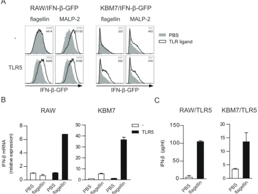
Fig. 1. Flagellin induces TLR5-dependent IFN-β production in mouse and human cell lines. (A) RAW/IFN-β-GFP and KBM7/IFN-β-GFP cells expressing or not expressing TLR5 were sti mulated with flagellin (100 ng/ml) or MALP-2 (100 ng/ml) for 24 h, and GFP expression was measured by flow cytometry. (B) RAW and KBM7 cells expressing or not expressing TLR5 were sti mulated with PBS or flagellin (100 ng/ml) for 2 h. IFN-β mRNA expression was measured by qPCR. (C) RAW and KBM7 cells expressing TLR5 were stimulated with PBS or
flagellin (100 ng/ml) for 12 h (RAW/TLR5) or 24 h (KBM7/TLR5). IFN-β in the culture supernatant was measured by ELISA.
Figure 1
IFN-β-GFP TLR5 -RAW/IFN-β-GFPA
flagellin MALP-2B
C
KBM7 RAW RAW/TLR5 KBM7/TLR5 KBM7/IFN-β-GFP flagellin MALP-2 PBS TLR ligand flagellin PBS PBSflagellin -TLR5 flagellinPBS PBSflagellin PBSflagellin PBSflagellin
0 10 20 30 40 50 0 2 4 6 8 (re la tiv e ex pr es si on ) IFN -β mRNA 0 5 10 15 20 0 50 100 150 IF N-β (p g/ ml ) IFN-β-GFP 4414 4296 8556 2956 101304296 8199 2956 320 322 550 297 493 322 476 297
Flagellin Stimulates IFN-β Production via TLR5 Wondae Kang et al.
with 0.5 M H2SO4, the absorbance was read at 450 nm with
SPECTROstar Nano (BMG Labtech, Germany).
Statistical analysis
Results are shown as the mean ± SEM. Statistical significance was evaluated with an unpaired Student’s t-test using Graph-Pad Prism 5 software (GraphGraph-Pad Software, USA). Differences
were noted as significant when P < 0.05 (*P < 0.05, **P <
0.01, ***P < 0.001).
RESULTS
Flagellin induces IFN-β production and subsequent type I
IFN receptor signaling via TLR5
Systemic immunization of flagellin induces production of
anti-flagellin IgA antibodies (Flores-Langarica et al., 2012).
Because the type I IFN signaling is implicated in the IgA class switching and several bacterial PAMPs such as LPS and MALP-2 induce IFN-β production via TLR activation, we test-ed whether flagellin can induce IFN-β production via TLR5. First, we used the mouse macrophage cell line RAW264.7 (RAW) and the human myeloid leukemia cell line KBM7, both of which express various TLRs but not TLR5. Using the IFN-β-GFP reporter assay, we found that flagellin can induce the IFN-β promoter activation in these cells only when TLR5 is
ectopically expressed (Fig. 1A). As expected, IFN-β
respons-es to MALP-2 (TLR2 ligand) were not affected by the TLR5 expression. Similarly, flagellin-stimulated induction of IFN-β
mRNA was observed in the TLR5-expressing cells (Fig. 1B).
We also detected secretion of IFN-β proteins from
TLR5-ex-pressing cells by ELISA (Fig. 1C).
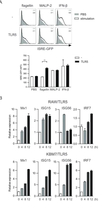
Next, we evaluated whether IFN-β, produced upon flagel-lin stimulation, can promote the type I interferon receptor signaling. Binding of IFN-α and -β to the heterodimeric re-ceptor IFNAR1/2 activates the JAK/STAT pathway and results in the subsequent induction of interferon-stimulated genes (ISGs). Many of ISGs have the interferon-stimulated response element (ISRE) in their promoter region. Thus, we first mea-sured the flagellin-stimulated ISRE activation in KBM7 cells using the ISRE-GFP reporter assay. In accordance with the
IFN-β production results shown in Fig. 1, flagellin induced
the ISRE activation only when TLR5 was expressed (Fig. 2A).
MALP-2 as well as recombinant IFN-β (used as a positive
control for the ISRE activation) stimulated the ISRE activation irrespective of the TLR5 expression. Next, we confirmed the flagellin-induced activation of the interferon receptor down-stream signaling by measuring mRNAs of several ISGs. Flagel-lin increased the expression of Mx1, ISG15, ISG56, and IRF7 mRNAs in a time-dependent manner in both RAW and KBM7
cells expressing TLR5 (Fig. 2B).
Flagellin induces IFN-β production in mouse primary cells
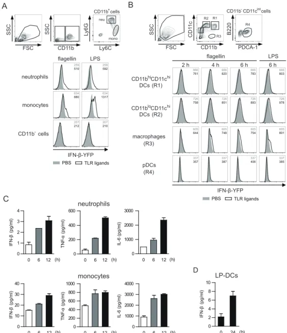
To test whether flagellin can induce IFN-β production in primary cells expressing endogenous TLR5, we used the β-YFP reporter (mob) mice in which the bi-cistronic IFN-β-IRES-YFP sequence was inserted in the endogenous IFN-β locus and therefore cells expressing IFN-β also express YFP (Scheu et al., 2008). Neutrophils and monocytes constitute the majority of TLR5-expressing cell populations in the BM
(Shibata et al., 2012). Accordingly, we found that a
signif-icant proportion of neutrophils (CD11b+
Ly6G+
) and
mono-cytes (CD11b+Ly6C+), but not other cells (CD11b–), expressed
YFP when we stimulated BM cells with flagellin ex vivo (Fig.
3A). The IFN-β promoter activation by flagellin was also
de-tected in splenic macrophages (R3) and to a lesser extent
in CD11chi DC subsets (R1 and R2), but not in plasmacytoid
Fig. 2. Flagellin promotes type I IFN receptor signaling in a TLR5-dependent manner. (A) KBM7/ISRE-GFP cells expressing or not expressing TLR5 were stimulated with flagellin (100 ng/ ml), MALP-2 (100 ng/ml), or IFN-β (20 U/ml) for 12 h and GFP expression was measured by flow cytometry. (B) RAW and KBM7 cells expressing TLR5 were stimulated with flagellin (100 ng/ml) up to 12 h. Expression of Mx1, ISG15, ISG56, and IRF7 mRNA was measured by qPCR.
Figure 2
A
TLR5
-flagellin MALP-2 IFN-β
B
KBM7/TLR5 RAW/TLR5
ISRE-GFP
Mx1 ISG15 ISG56
Mx1 ISG15 ISG56 IRF7
IRF7 PBS stimulation 0 2 4 6 8 R el at ive e xp re ss io n 0 5 10 15 0 5 10 15 0 2 4 6 8 0 2 4 6 8 10 R el at ive e xp re ss io n 0 1 2 3 0 1 2 3 0.0 0.5 1.0 1.5 2.0 0 4 812 (h) 0 4 812 0 4 812 0 4 812 0 4 812 (h) 0 4 812 0 4 812 0 4 812 226 228 411 236 371 228 367 236 566 228 463 236 -TLR5 IFN-β MALP-2 flagellin PBS 0 100 200 300 400 500 600 700 IS R E-G FP (M FI ) **
Flagellin Stimulates IFN-β Production via TLR5 Wondae Kang et al.
DCs (R4) (Fig. 3B). Furthermore, we were able to confirm the
flagellin-induced secretion of IFN-β proteins in purified BM neutrophils and monocytes as well as in small intestinal
LP-DCs (Figs. 3C and 3D).
Flagellin induces IFN-β production in a TLR5- and MyD88-
dependent manner in vivo
Next, we examined whether we could detect
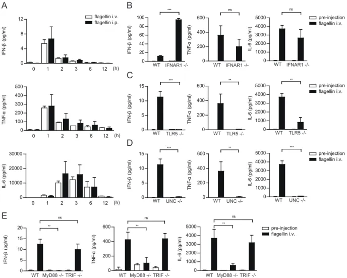
flagellin-stim-ulated IFN-β secretion in vivo. Upon i.p. or i.v. injection of
WT mice with flagellin, serum IFN-β levels peaked after an
hour and then rapidly decreased (Fig. 4A). Serum TNF-α
levels also showed a similar kinetics of induction while the peak induction of IL-6 was delayed compared to IFN-β and TNF-α (Fig. 4A). Flagellin did not increase serum IFN-α levels (Supplementary Fig. S1). Consistent with the fast increase
in the serum IFN-β, we detected YFP expression in splenic
neutrophils, monocytes, and macrophages after an hour of
flagellin injection to mob mice (Supplementary Fig. S2). Type
Fig. 3. Flagellin induces IFN-β production in mouse primary cells. (A) BM cells were isolated from the IFN-β-YFP (mob) mice and
stimulated with PBS, flagellin (100 ng/ml) or LPS (100 ng/ml) for 6 h. YFP expression was measured in CD11b+Ly6G+ neutrophils,
CD11b+
Ly6C+
monocytes, and CD11b–
cells by flow cytometry. (B) Mob mice were injected with Flt3L-expressing B16 cells to expand the DC subsets. Twenty days after injection, splenocytes were isolated and stimulated with PBS, flagellin (100 ng/ml) or LPS (100 ng/ml) for 2, 4, or 6 h. YFP expression was measured in DC subsets and macrophages by flow cytometry. (C) BM neutrophils and monocytes were
sorted by FACS and stimulated with PBS or flagellin (100 ng/ml) for 6 h and 12 h. IFN-β levels in the culture supernatant were measured
by ELISA. (D) Small intestinal LP-DCs were stimulated with PBS or flagellin (1 µg/ml) for 24 h, and IFN-β levels in the culture supernatant were measured by ELISA.
Figure 3
FSC CD11b SSC CD11c PDCA-1 B220A
IFN-β-YFP 2 h 4 h 6 h macrophages pDCsB
IFN-β-YFP flagellin LPS neutrophils monocytes FSC CD11b Ly6C SSC SSC Ly6G neu mono LPS 6 h flagellin R1 R4 R3 PBS TLR ligandsC
neutrophils monocytes 0 6 12 0 1 2 3 4 (h) 0 0 6 12 1000 2000 3000 (h) 0 6 12 0 200 400 600 (h) 0 6 12 0 10 20 30 40 (h) 0 0 6 112 1000 2000 3000 4000 (h) 0 6 12 0 200 400 600 800 1000 (h) R2D
LP-DCs PBS TLR ligands CD11bhiCD11chi DCs CD11bloCD11chi CD11b-cellsCD11b+cells CD11b-CD11cintcells
(R1) DCs (R2) (R3) (R4) TN F-α (pg/ml) IL-6 (pg/ml) IFN -β (pg/ml) TN F-α (pg/ml) IL-6 (pg/ml) IFN -β (pg/ml) IFN -β (pg/ml) 0 24 0 2 4 6 8 10 (h) 510 259 880 634 592 259 1317634 212 207 210 207 761 666 758 726 820 666 851 726 644 605 748 605 357 337 387 337 783 666 893 726 803 666 978 726 754 605 801 605 406 337 385 337
Flagellin Stimulates IFN-β Production via TLR5 Wondae Kang et al.
I IFN receptors (IFNARs) are ubiquitously expressed in various
cell populations (Langer and Pestka, 1988). We hypothesized
that the rapid decline of serum IFN-β levels might be due to the receptor-mediated consumption of IFN-β. Indeed, we found that IFNAR1 KO mice showed almost 10 times higher serum IFN-β levels compared to WT mice upon flagellin injec-tion whereas serum TNF-α and IL-6 levels were not changed
in IFNAR1 KO mice (Fig. 4B).
To test whether flagellin induces IFN-β secretion via TLR5, we compared WT and TLR5 KO mice. The flagellin-stimu-lated increases of serum IFN-β, TNF-α, and IL-6 were severely blunted in TLR5 KO mice compared to those of WT mice (Fig. 4C). In contrast, TLR4 KO mice showed no defects in the
flagellin-induced IFN-β secretion (Supplementary Fig. S3). We
previously showed that UNC93B1 is essential for the intracel-lular trafficking of TLR5 from the ER to the plasma membrane (Huh et al., 2014). In UNC93B1-deficient cells, TLR5 is re-tained in the ER and cannot transmit the flagellin-stimulated
signals. Accordingly, we found that flagellin-stimulated IFN-β secretion was defective in UNC93B1 KO mice, similar to TLR5
KO mice (Fig. 4D).
Bacteria-sensing TLRs such as TLR2 and TLR4 require both MyD88 and TRIF adaptor molecules to transmit activating
signals for the IFN-β induction (Aubry et al., 2012; Fitzgerald
et al., 2003; Kagan et al., 2008). In contrast, we found that the flagellin-stimulated induction of IFN-β, as well as TNF-α
and IL-6, required MyD88 but not TRIF (Fig. 4E). The flagellin
injection increased all three cytokines in both TRIF KO and WT mice in a similar manner, whereas almost no cytokine induc-tion was found in MyD88 KO mice. Transcripinduc-tion factors IRF3, IRF5, and IRF7 become phosphorylated by TBK1 upon TLR
activation and promote the type I IFN gene expression (Doyle
et al., 2002; Honda et al., 2004; Kawai et al., 2004; Takaoka et al., 2005). Using the TLR5-expressing RAW cells, we found that flagellin also promotes the accumulation of
phosphor-ylated IRF3 and IRF7 in the nucleus (Supplementary Fig. S4).
Fig. 4. Flagellin induces IFN-β production in a TLR5- and MyD88-dependent manner in vivo. (A) WT mice were injected i.v. or i.p. with
flagellin (2 µg/mouse). Sera were taken before and up to 12 h after injection. IFN-β, TNF-α, and IL-6 levels were measured by ELISA.
(B-E) WT, IFNAR1 KO, TLR5 KO, UNC93B1 KO, MyD88 KO, and TRIF KO mice were i.v. injected with flagellin (2 µg/mouse). Sera were taken
before and 1 h after injection. IFN-β, TNF-α, and IL-6 levels were measured by ELISA. Data are presented as mean ± SEM; **P < 0.01,
***P < 0.001. ns, statistically not significant.
Figure 4
0 1 2 3 6 12 0 100 200 300 400 500 (h) 0 1 2 3 6 12 0 10000 20000 30000 (h)A
B
pre-injection flagellin i.v.C
WT TLR5 IFNAR1 -/-WT UNC -/-WTE
pre-injection flagellin i.v. 0 20 40 60 80 100 *** 0 5 10 15 *** 0 5 10 15 *** 0 200 400 600 ** 0 200 400 600 ** 0 200 400 600 ns 0 1000 2000 3000 4000 5000 ns 0 1000 2000 3000 4000 5000 ** 0 1000 2000 3000 4000 5000 *** 0 1000 2000 3000 4000 5000 ns ** 0 200 400 600 ns ** 0 5 10 15 20 ns ** TN F-α (pg/ml) IL-6 (pg/ml) IFN -β (pg/ml) TN F-α (pg/ml) IL-6 (pg/ml) IFN -β (pg/ml) TN F-α (pg/ml) IL-6 (pg/ml) IFN -β (pg/ml) WT MyD88 TRIF -/-flagellin i.v. flagellin i.p. TN F-α (pg/ml) IL-6 (pg/ml) IFN -β (pg/ml) pre-injection flagellin i.v. IFNAR1 -/-WT IFNAR1 -/-WT TLR5 -/-WT TLR5 -/-WT UNC -/-WT UNC -/-WT TN F-α (pg/ml) IL-6 (pg/ml) IFN -β (pg/ml) WT MyD88 TRIF WT MyD88 TRIF-/-D
pre-injection flagellin i.v. 0 1 2 3 6 12 0 4 8 12 (h)Flagellin Stimulates IFN-β Production via TLR5 Wondae Kang et al.
Therefore, our data indicates that flagellin induces IFN-β pro-duction via a TLR5-MyD88-TBK1-IRF3/7 signaling pathway.
Internalization and endolysomal signaling of TLR5 is
re-quired for the flagellin-induced IFN-β production
At steady state, TLR5 is mostly localized at the plasma mem-brane where it recognizes extracellular flagellin and initiates signaling cascades leading to the production of
pro-inflam-matory cytokines (Huh et al., 2014). It was suggested that
flagellin-bound TLR5 is subsequently internalized, resulting in the MHC class II-mediated presentation of flagellin peptides
in antigen-presenting cells (Letran et al., 2011).
TLR5-depen-dent internalization of flagellin is also observed in polarized
intestinal epithelial cells (Eaves-Pyles et al., 2011). In
addi-tion, the TLR-mediated signaling for type I IFN production is believed to originate from endolysosomal compartments. For TLR3, TLR7, and TLR9, the receptors are constitutively localized in endolysosomes, and TLR2 and TLR4 enter them after the ligand-mediated endocytosis. However, the inter-nalization of TLR5 from the cell surface has not been directly demonstrated yet. Thus, we examined if TLR5 is also
internal-ized into endolysosomal compartments upon flagellin stimu-lation. First, we measured changes in the cell surface level of TLR5 in RAW cells expressing TLR5-GFP using an anti-TLR5 monoclonal antibody. The cell surface TLR5 levels gradually decreased and became ~20% of the basal level after flagellin stimulation for 12 h. In comparison, the total TLR5 levels— measured by GFP intensities—were moderately reduced by flagellin stimulation, implying that the loss of cell surfa9ce TLR5 is mainly due to receptor internalization and
sequestra-tion inside cells rather than receptor degradasequestra-tion (Fig. 5A).
Next, we visualized the subcellular localization of TLR5 in BM-DCs expressing TLR5-GFP and CD63-Cherry (as an en-dolysosomal marker) by confocal microscopy. Before flagellin stimulation, TLR5-GFP was uniformly distributed on the plas-ma membrane and was barely colocalized with CD63-Cherry (Fig. 5B). Upon stimulation with flagellin for 1 h, the decrease of TLR5-GFP signals from the plasma membrane became evident. Twelve hours after stimulation, TLR5-GFP was hardly seen on the plasma membrane and was extensively colo-calized with CD63-Cherry. These findings demonstrate that flagellin induces the internalization of TLR5 from the plasma
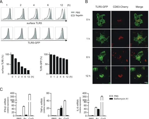
Fig. 5. Internalization and endolysosomal signaling of TLR5 is required for the flagellin-induced IFN-β induction. (A) RAW/TLR5-GFP cells were stimulated with flagellin (100 ng/ml) up to 12 h and stained with biotinylated mouse anti-TLR5 antibody and streptavidin-conjugated Alexa Fluor-647 on ice. The cell surface TLR5 (Alexa Fluor-647) and the total TLR5 (GFP) levels were analyzed by flow cytometry and compared to the ones in unstimulated cells. (B) BM-DCs expressing TLR5-GFP and CD63-Cherry were stimulated with
flagellin (100 ng/ml) for 1, 6, or 12 h. TLR5 and CD63 localization was analyzed by confocal microscopy. Scale bar = 10 µm. (C) RAW/
TLR5-Cherry cells were treated with bafilomycin A1 (1 µM) for 1 h, and then stimulated with flagellin (100 ng/ml) or CpG DNA1826 (1 µM)
for 12 h. Expression of IFN-β, TNF-α, and IL-6 mRNA was measured by qPCR.
Figure 5
A
C
1 2 4 6 12 (h)B
0 h 1 h 6 h 12 h TLR5-GFP CD63-Cherry Merge PBS flagellin surface TLR5 PBS Bafilomycin A1 0 1 2 4 6 12 0 50 100 (h) TL R5 -G FP (% ) 0 1 2 4 6 12 0 50 100 (h) su rf ac e TL R5 (% ) IFN -β mRNA TLR5-GFP PBS fla CpG 0 20 40 60 (re la tiv e ex pr es sio n) PBS fla CpG 0 20 40 60 80 300 350 400 (re la tiv e ex pr es sio n) IL-6 mRNA PBS fla CpG 0 20 40 60 80 300 350 400 (re la tiv e ex pr es sio n) TN F-α mRNAFlagellin Stimulates IFN-β Production via TLR5 Wondae Kang et al.
membrane into endolysomes.
Endolysosomal TLR signaling is dependent on the acidic environment of the organelles and can be blocked by
lyso-somotropic agents or vacuolar-type H+-ATPase inhibitors such
as bafilomycin A1. To test if TLR5 transmits IFN-β-inducing signals from the endolysomes, we pretreated the TLR5-ex-pressing RAW cells with bafilomycin A1 and then stimulated them with either flagellin or CpG DNA (a TLR9 agonist). TLR9 is constitutively localized in the endolysosomes and the TLR9-mediated production of pro-inflammatory cytokines as well as type I IFNs is sensitive to the bafilomycin A1 treatment (Hacker et al., 1998; Lund et al., 2003). As expected, we observed that the bafilomycin A1 pretreatment completely
inhibited the CpG DNA-induced production of IFN-β, TNF-α,
and IL-6 (Fig. 5C). In contrast, when the cells were stimulated
with flagellin, only IFN-β induction was inhibited by
bafilo-mycin A1. These data support the notion that TLR5 initiates
signaling for the IFN-β induction from the endolysosomes in a pH-dependent manner, whereas TLR5-mediated signaling for the pro-inflammatory cytokine production happens on the plasma membrane and is not regulated by acidity of intracel-lular organelles.
Syk is not required for the flagellin-stimulated TLR5
inter-nalization and IFN-β production
For LPS-induced endocytosis of TLR4, the signaling adaptors MyD88 and TRIF were not required, and instead the Syk
tyro-sine kinase was shown to be essential (Zanoni et al., 2011).
Therefore, we tested if Syk is also involved in the flagel-lin-stimulated TLR5 endocytosis using RAW cells where Syk was deleted by the CRISPR/Cas9 system. The extent of flagel-lin-stimulated TLR5 internalization in Syk KO cells was same
as in WT cells (Supplementary Fig. S5A). Furthermore,
flagel-lin-induced IFN-β production was also normal in the absence
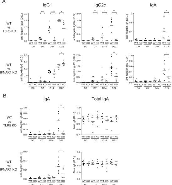
Fig. 6. Flagellin-specific IgG2c and IgA responses require the type I IFN receptor signaling. TLR5, IFNAR1 KO, and the respective
littermate control mice were i.p. immunized with flagellin (5 µg) at day 0 and 15. Sera and feces were collected at day 0, 7, 14, and 22,
and flagellin-specific antibodies in the sera (A) and fecal extracts (B) were measured by ELISA. *P < 0.05, **P < 0.01, ***P < 0.001.
Figure 6
B
IgG1 IgG2c IgA
IgA Total IgA WT vs TLR5 KO WT vs IFNAR1 KO WT vs TLR5 KO WT vs IFNAR1 KO
A
WT KO WT KO WT KO WT KO 0.0 0.5 1.0 1.5 2.0 D0 D7 D14 *** D22 * an ti-fla ge llin Ig G1 (O .D .) WT KO WT KO WT KO WT KO 0.0 0.5 1.0 1.5 2.0 D0 D7 D14 D22 ** an ti-fla ge llin Ig G2 c (O .D .) an ti-fla ge llin Ig A (O .D .) WT KO WT KO WT KO WT KO 0.0 0.5 1.0 1.5 2.0 D0 D7 D14 D22 * an ti-fla ge llin Ig G1 (O .D .) WT KO WT KO WT KO WT KO 0.0 0.5 1.0 1.5 2.0 2.5 D0 D7 D14 D22 ** an ti-fla ge llin Ig G2 c (O .D .) WT KO WT KO WT KO WT KO 0.0 0.2 0.4 0.6 0.8 1.0 D0 D7 D14 D22 ** an ti-fla ge llin Ig A (O .D .) WT KO WT KO WT KO WT KO 0.7 0.8 0.9 1.0 1.1 1.2 D0 D7 D14 D22 To ta l I gA (O .D .) WT KO WT KO WT KO WT KO 0.0 0.2 0.4 0.6 D0 D7 D14 D22 * an ti-fla ge llin Ig A (O .D .) WT KO WT KO WT KO WT KO 0.0 0.5 1.0 1.5 2.0 D0 D7 D14 D22 To ta l I gA (O .D .) WT KO WT KO WT KO WT KO 0.0 0.2 0.4 0.6 0.8 1.0 D0 D7 D14 D22 an ti-fla ge llin Ig A (O .D .) * * ** *** WT KO WT KO WT KO WT KO 0.0 0.4 0.8 1.2 D0 D7 D14 D22 *Flagellin Stimulates IFN-β Production via TLR5 Wondae Kang et al.
of Syk. As a control, we also stimulated cells with depleted zymosan, which signals via the Dectin-1/Syk pathway, and found that both IFN-β and IL-6 production were significantly blunted in Syk KO cells compared to WT cells. Therefore, we concluded that the flagellin-stimulated TLR5 endocytosis and
IFN-β production does not require Syk activation, unlike the
LPS-induced TLR4 endocytosis and IFN-β production.
Type I IFN signaling is required for flagellin-specific IgG2c and IgA antibody responses
Previous studies showed that systemic immunization with soluble flagellin induces not only IgG responses in the spleen but also IgA responses in the mesenteric lymph nodes (mLNs) (Flores-Langarica et al., 2012). Interestingly, anti-flagellin Ig-G2c and IgA responses were highly dependent on TLR5 and MyD88, whereas both TLR5 and NLRC4 inflammasome path-ways seem to activate anti-flagellin IgG1 response in a partly
redundant manner (Lopez-Yglesias et al., 2014). Because
type I IFN signaling was implicated in antigen-specific
anti-body class switching (Swanson et al., 2010; Thompson et al.,
2008) and we found that flagellin induces IFN-β production
via the TLR5/MyD88 pathway, we tested whether type I IFN signaling regulates anti-flagellin antibody responses.
WT, TLR5 KO, and IFNAR1 KO mice were immunized in-traperitoneally with flagellin, and anti-flagellin antibodies in the serum and feces were analyzed 7 and 14 days later. For detection of IgA responses, mice were boost-immunized with flagellin at day 15 and serum and fecal antibodies were measured 7 days later. Consistent with previous studies, we did not observe induction of flagellin IgG2c and IgA anti-bodies in the serum of TLR5 KO mice compared to littermate control mice. Anti-flagellin IgG1 antibodies were inhibited in the primary responses but partially recovered after the boost
immunization (Fig. 6A). In the case of IFNAR1 KO mice,
an-ti-flagellin IgG1 responses were normal and even seem to be slightly higher after the boost immunization compared to littermate control mice. In contrast, anti-flagellin IgG2c and IgA responses were significantly inhibited in IFNAR1 KO mice,
especially after the boost immunization (Fig. 6A). Similarly,
we found that fecal anti-flagellin IgA responses were severely defective in both TLR5 and IFNAR1 KO mice, whereas the total fecal IgA levels were not affected by the deficiency of
either TLR5 or IFNAR1 (Fig. 6B). In contrast to the flagellin
immunization, we found that IgG2c and IgA responses to ov-albumin immunization was not impaired in IFNAR1 KO mice (Supplementary Fig. S6). Taken together, these data suggest that IFN-β, produced upon flagellin stimulation in a TLR5-de-pendent manner, specifically promotes the flagellin-specific IgG2c and IgA class switching via type I IFN receptor signaling.
DISCUSSION
Although flagellin is known to efficiently promote induction of many pro-inflammatory cytokines, several studies reported
its inability to induce type I IFNs (Choi et al., 2010; Hemont et
al., 2013; Means et al., 2003). However, one previous study noted that flagellin exhibited an anti-osteoclastogenic effect
via IFN-β secretion during osteoclast differentiation from
BM-derived macrophages (Ha et al., 2008). Another study
showed that flagellin induces type III IFNs via TLR5 (
Oden-dall et al., 2017). In the present study, we directly demon-strate that flagellin can induce IFN-β production in various TLR5-expressing cell lines and primary cells, as well as in live mice. Notably, IFNAR1 KO mice showed a greater increase
in serum IFN-β levels upon flagellin injection compared to
WT mice, suggesting that IFN-β is rapidly bound and seques-tered by IFNAR1/2 which are ubiquitously present in many cell types. Another interpretation of the result is a possible
negative feedback regulation of IFN-β production via the
IFNAR1/2-mediated signaling. However, the TLR-mediated type I IFN production is usually positively regulated by the
IFNAR1/2-mediated signaling (Marie et al., 1998; Sato et al.,
1998). In fact, when we measured IFN-β production from
purified LP-DCs ex vivo, flagellin-stimulated IFN-β production was partially inhibited in IFNAR1 KO cells compared to WT cells (data not shown). Therefore, we believe that the higher
serum IFN-β levels found in flagellin-stimulated IFNAR1 KO
mice is likely due to the lack of IFN-β consumption by neigh-boring cells. Although we showed that flagellin injection up-regulates the IFN-β promoter activities in splenic neutrophils, monocytes, and macrophages, further studies are required to identify the major cells types that produce and consume
IFN-β in response to flagellin stimulation in vivo.
Our study also demonstrates that flagellin stimulates IFN-β production via a TLR5-MyD88-IRF3/7 signaling pathway. Sim-ilar to other bacteria-sensing TLRs such as TLR2 and TLR4, we found that flagellin-stimulated TLR5 needs to be internalized into endolysosomes to transmit signals for IFN-β induction in an acidic environment. Nonetheless, there are a few differ-ences between TLR4- and TLR5-mediated IFN-β production. In case of TLR4, the receptor endocytosis from the plasma membrane requires Syk kinase activity and the subsequent
signaling for IFN-β induction depends on the adaptor
mol-ecule TRIF (Zanoni et al., 2011). However, we found that
flagellin-induced TLR5 endocytosis and IFN-β production
occurs normally in the absence of Syk and TRIF. In this aspect, TLR5 behaves similarly to TLR7 and TLR9, inducing type I IFNs in a MyD88-, but not TRIF-, dependent manner. Of note, TLR5 also shares a similarity with TLR7 and TLR9 in that they all require UNC93B1 for the trafficking out of the ER and the proper localization inside cells.
Among various potential implications of
flagellin-stimu-lated IFN-β induction, we focused on the flagellin
anti-body production and found that anti-flagellin IgG2c and IgA —but not IgG1—responses are dependent on the type I IFN signaling. In IFNAR1 KO mice, induction of flagellin-specific IgG2c and IgA antibodies was severely impaired whereas ov-albumin-specific antibody induction was normal. Therefore, the requirement of the type I IFN signaling is specific to flagel-lin-specific antibody responses.
Upon systemic flagellin immunization, intestinal CD103+
DCs accumulate in the mLNs and mediate IgA responses (Flores-Langarica et al., 2012). Type I IFN promotes CCR7
expression in DCs for lymph node homing (Jang et al., 2006;
Parlato et al., 2001). Because we found that intestinal LP-DCs produce IFN-β upon flagellin stimulation, we hypothesized that the type I IFN signaling may regulate the accumulation
of CD103+
Flagellin Stimulates IFN-β Production via TLR5 Wondae Kang et al.
responses. However, we found that flagellin-stimulated
accu-mulation of CD103+ DCs in mLNs was normal in IFNAR1 KO
mice (data not shown). Flagellin-stimulated intestinal CD103+
DCs were also shown to promote IgA class switching by di-rectly acting on B cells in an in vitro DC-B cell co-culture assay (Uematsu et al., 2008). However, we found that the in vitro IgA induction was not defective in IFNAR1 KO B cells when
co-cultured with intestinal CD103+ DCs and flagellin equally
promoted the differentiation of IgA-secreting cells from both WT and IFNAR1 KO B cells (data not shown). Therefore, molecular mechanisms underlying the type I IFN-mediated regulation of anti-flagellin IgG2c and IgA responses are still unclear and further studies are needed. Especially, it needs
to be examined whether the IFN-β signaling modulates the
production of cytokines known to regulate the antibody class
switching, such as IL-4, IL-10, IL-21, TGF-β, APRIL, and BAFF.
Anti-flagellin antibodies, especially that of the IgA isotype, are essential for maintaining the intestinal homeostasis and keeping the epithelial barrier functions intact by limiting the
motility of flagellated bacteria in the gut (Cullender et al.,
2013). In addition, TLR5-mediated sensing of commensal
bacteria is shown to be necessary for effective antibody
re-sponses to seasonal influenza vaccination (Oh et al., 2014).
Therefore, our findings presented in this study may help de-velop strategies for promoting the intestinal health and more efficacious mucosal vaccine design.
Note: Supplementary information is available on the Mole-cules and Cells website (www.molcells.org).
Disclosure
The authors have no potential conflicts of interest to disclose.
ACKNOWLEDGMENTS
We thank Joo-Yeon Yoo for providing the IFN-β-GFP reporter plasmid and Seung Yun Chae for critical reading of the man-uscript. This work was supported by grants from the National Research Foundation of Korea (NRF-2016R1A2B3015046, NRF-2016M3A9D3918546, NRF-2017M3A9F3047085).
ORCID
Wondae Kang https://orcid.org/0000-0003-3114-5838
Areum Park https://orcid.org/0000-0002-9661-4319
Ji-Won Huh https://orcid.org/0000-0002-6169-6756
Gihoon You https://orcid.org/0000-0002-8325-8810
Da-Jung Jung https://orcid.org/0000-0003-4399-8029
Manki Song https://orcid.org/0000-0002-8279-9041
Heung Kyu Lee https://orcid.org/0000-0002-3977-1510
You-Me Kim https://orcid.org/0000-0001-8780-704X
REFERENCES
Adachi, O., Kawai, T., Takeda, K., Matsumoto, M., Tsutsui, H., Sakagami, M., Nakanishi, K., and Akira, S. (1998). Targeted disruption of the MyD88 gene results in loss of IL-1- and IL-18-mediated function. Immunity 9, 143-150. Atif, S.M., Lee, S.J., Li, L.X., Uematsu, S., Akira, S., Gorjestani, S., Lin, X., Schweighoffer, E., Tybulewicz, V.L.J., and McSorley, S.J. (2015). Rapid CD4(+) T-cell responses to bacterial flagellin require dendritic cell expression of Syk and CARD9. Eur. J. Immunol. 45, 513-524.
Aubry, C., Corr, S.C., Wienerroither, S., Goulard, C., Jones, R., Jamieson,
A.M., Decker, T., O’Neill, L.A.J., Dussurget, O., and Cossart, P. (2012). Both TLR2 and TRIF contribute to interferon-beta production during Listeria infection. PLoS One 7, e33299.
Carvalho, F.A., Koren, O., Goodrich, J.K., Johansson, M.E., Nalbantoglu, I., Aitken, J.D., Su, Y., Chassaing, B., Walters, W.A., Gonzalez, A., et al. (2012). Transient inability to manage proteobacteria promotes chronic gut inflammation in TLR5-deficient mice. Cell Host Microbe 12, 139-152. Choi, Y.J., Im, E., Chung, H.K., Pothoulakis, C., and Rhee, S.H. (2010). TRIF mediates toll-like receptor 5-induced signaling in intestinal epithelial cells. J. Biol. Chem. 285, 37570-37578.
Ciacci-Woolwine, F., Blomfield, I.C., Richardson, S.H., and Mizel, S.B. (1998). Salmonella flagellin induces tumor necrosis factor alpha in a human promonocytic cell line. Infect. Immun. 66, 1127-1134.
Cullender, T.C., Chassaing, B., Janzon, A., Kumar, K., Muller, C.E., Werner, J.J., Angenent, L.T., Bell, M.E., Hay, A.G., Peterson, D.A., et al. (2013). Innate and adaptive immunity interact to quench microbiome flagellar motility in the gut. Cell Host Microbe 14, 571-581.
Doyle, S., Vaidya, S., O’Connell, R., Dadgostar, H., Dempsey, P., Wu, T., Rao, G., Sun, R., Haberland, M., Modlin, R., et al. (2002). IRF3 mediates a TLR3/ TLR4-specific antiviral gene program. Immunity 17, 251-263.
Eaves-Pyles, T., Bu, H.F., Tan, X.D., Cong, Y.Z., Patel, J., Davey, R.A., and Strasser, J.E. (2011). Luminal-applied flagellin is internalized by polarized intestinal epithelial cells and elicits immune responses via the TLR5 dependent mechanism. PLoS One 6, e24869.
Felix, G., Duran, J.D., Volko, S., and Boller, T. (1999). Plants have a sensitive perception system for the most conserved domain of bacterial flagellin. Plant J. 18, 265-276.
Fitzgerald, K.A., Rowe, D.C., Barnes, B.J., Caffrey, D.R., Visintin, A., Latz, E., Monks, B., Pitha, P.M., and Golenbock, D.T. (2003). LPS-TLR4 signaling to IRF-3/7 and NF-kappa B involves the toll adapters TRAM and TRIF. J. Exp. Med. 198, 1043-1055.
Flores-Langarica, A., Marshall, J.L., Hitchcock, J., Cook, C., Jobanputra, J., Bobat, S., Ross, E.A., Coughlan, R.E., Henderson, I.R., Uematsu, S., et al. (2012). Systemic flagellin immunization stimulates mucosal CD103(+) dendritic cells and drives Foxp3(+) regulatory T cell and IgA responses in the mesenteric lymph node. J. Immunol. 189, 5745-5754.
Franchi, L., Amer, A., Body-Malapel, M., Kanneganti, T.D., Ozoren, N., Jagirdar, R., Inohara, N., Vandenabeele, P., Bertin, J., Coyle, A., et al. (2006). Cytosolic flagellin requires Ipaf for activation of caspase-1 and interleukin 1beta in salmonella-infected macrophages. Nat. Immunol. 7, 576-582. Gewirtz, A.T., Navas, T.A., Lyons, S., Godowski, P.J., and Madara, J.L. (2001). Cutting edge: bacterial flagellin activates basolaterally expressed TLR5 to induce epithelial proinflammatory gene expression. J. Immunol. 167, 1882-1885.
Gomez-Gomez, L. and Boller, T. (2000). FLS2: an LRR receptor-like kinase involved in the perception of the bacterial elicitor flagellin in Arabidopsis. Mol. Cell 5, 1003-1011.
Ha, H., Lee, J.H., Kim, H.N., Kwak, H.B., Kim, H.M., Lee, S.E., Rhee, J.H., Kim, H.H., and Lee, Z.H. (2008). Stimulation by TLR5 modulates osteoclast differentiation through STAT1/IFN-beta. J. Immunol. 180, 1382-1389. Hacker, H., Mischak, H., Miethke, T., Liptay, S., Schmid, R., Sparwasser, T., Heeg, K., Lipford, G.B., and Wagner, H. (1998). CpG-DNA-specific activation of antigen-presenting cells requires stress kinase activity and is preceded by non-specific endocytosis and endosomal maturation. EMBO J. 17, 6230-6240.
Hajam, I.A., Dar, P.A., Shahnawaz, I., Jaume, J.C., and Lee, J.H. (2017). Bacterial flagellin-a potent immunomodulatory agent. Exp. Mol. Med. 49, e373.
Halff, E.F., Diebolder, C.A., Versteeg, M., Schouten, A., Brondijk, T.H., and Huizinga, E.G. (2012). Formation and structure of a NAIP5-NLRC4 inflammasome induced by direct interactions with conserved N- and C-terminal regions of flagellin. J. Biol. Chem. 287, 38460-38472.
Flagellin Stimulates IFN-β Production via TLR5 Wondae Kang et al.
Hayashi, F., Smith, K.D., Ozinsky, A., Hawn, T.R., Yi, E.C., Goodlett, D.R., Eng, J.K., Akira, S., Underhill, D.M., and Aderem, A. (2001). The innate immune response to bacterial flagellin is mediated by Toll-like receptor 5. Nature 410, 1099-1103.
Hemont, C., Neel, A., Heslan, M., Braudeau, C., and Josien, R. (2013). Human blood mDC subsets exhibit distinct TLR repertoire and responsiveness. J. Leukoc. Biol. 93, 599-609.
Honda, K., Yanai, H., Mizutani, T., Negishi, H., Shimada, N., Suzuki, N., Ohba, Y., Takaoka, A., Yeh, W.C., and Taniguchi, T. (2004). Role of a transductional-transcriptional processor complex involving MyD88 and IRF-7 in Toll-like receptor signaling. Proc. Natl. Acad. Sci. U. S. A. 101, 15416-15421. Hoshino, K., Takeuchi, O., Kawai, T., Sanjo, H., Ogawa, T., Takeda, Y., Takeda, K., and Akira, S. (1999). Cutting edge: Toll-like receptor 4 (TLR4)-deficient mice are hyporesponsive to lipopolysaccharide: evidence for TLR4 as the Lps gene product. J. Immunol. 162, 3749-3752.
Hu, Z., Zhou, Q., Zhang, C., Fan, S., Cheng, W., Zhao, Y., Shao, F., Wang, H.W., Sui, S.F., and Chai, J. (2015). Structural and biochemical basis for induced self-propagation of NLRC4. Science 350, 399-404.
Huh, J.W., Shibata, T., Hwang, M., Kwon, E.H., Jang, M.S., Fukui, R., Kanno, A., Jung, D.J., Jang, M.H., Miyake, K., et al. (2014). UNC93B1 is essential for the plasma membrane localization and signaling of Toll-like receptor 5. Proc. Natl. Acad. Sci. U. S. A. 111, 7072-7077.
Jang, M.H., Sougawa, N., Tanaka, T., Hirata, T., Hiroi, T., Tohya, K., Guo, Z., Umemoto, E., Ebisuno, Y., Yang, B.G., et al. (2006). CCR7 is critically important for migration of dendritic cells in intestinal lamina propria to mesenteric lymph nodes. J. Immunol. 176, 803-810.
Kagan, J.C., Su, T., Horng, T., Chow, A., Akira, S., and Medzhitov, R. (2008). TRAM couples endocytosis of Toll-like receptor 4 to the induction of interferon-beta. Nat. Immunol. 9, 361-368.
Kawai, T., Sato, S., Ishii, K.J., Coban, C., Hemmi, H., Yamamoto, M., Terai, K., Matsuda, M., Inoue, J., Uematsu, S., et al. (2004). Interferon-alpha induction through Toll-like receptors involves a direct interaction of IRF7 with MyD88 and TRAF6. Nat. Immunol. 5, 1061-1068.
Kim, J., Huh, J., Hwang, M., Kwon, E.H., Jung, D.J., Brinkmann, M.M., Jang, M.H., Ploegh, H.L., and Kim, Y.M. (2013). Acidic amino acid residues in the juxtamembrane region of the nucleotide-sensing TLRs are important for UNC93B1 binding and signaling. J. Immunol. 190, 5287-5295.
Kofoed, E.M. and Vance, R.E. (2011). Innate immune recognition of bacterial ligands by NAIPs determines inflammasome specificity. Nature 477, 592-595.
Langer, J.A. and Pestka, S. (1988). Interferon receptors. Immunol. Today 9, 393-400.
Letran, S.E., Lee, S.J., Atif, S.M., Uematsu, S., Akira, S., and McSorley, S.J. (2011). TLR5 functions as an endocytic receptor to enhance flagellin-specific adaptive immunity. Eur. J. Immunol. 41, 29-38.
Lopez-Yglesias, A.H., Zhao, X., Quarles, E.K., Lai, M.A., VandenBos, T., Strong, R.K., and Smith, K.D. (2014). Flagellin induces antibody responses through a TLR5-and inflammasome-independent pathway. J. Immunol. 192, 1587-1596.
Lowy, J. and McDonough, M.W. (1964). Structure of filaments produced by re-aggregation of Salmonella flagellin. Nature 204, 125-127.
Lund, J., Sato, A., Akira, S., Medzhitov, R., and Iwasaki, A. (2003). Toll-like receptor 9-mediated recognition of herpes simplex virus-2 by plasmacytoid dendritic cells. J. Exp. Med. 198, 513-520.
Marie, I., Durbin, J.E., and Levy, D.E. (1998). Differential viral induction of distinct interferon-alpha genes by positive feedback through interferon regulatory factor-7. EMBO J. 17, 6660-6669.
McSorley, S.J., Ehst, B.D., Yu, Y., and Gewirtz, A.T. (2002). Bacterial flagellin is an effective adjuvant for CD4+ T cells in vivo. J. Immunol. 169, 3914-3919. Means, T.K., Hayashi, F., Smith, K.D., Aderem, A., and Luster, A.D. (2003). The toll-like receptor 5 stimulus bacterial flagellin induces maturation and
chemokine production in human dendritic cells. J. Immunol. 170, 5165-5175.
Miao, E.A., Alpuche-Aranda, C.M., Dors, M., Clark, A.E., Bader, M.W., Miller, S.I., and Aderem, A. (2006). Cytoplasmic flagellin activates caspase-1 and secretion of interleukin 1beta via Ipaf. Nat. Immunol. 7, 569-575. Muller, U., Steinhoff, U., Reis, L.F., Hemmi, S., Pavlovic, J., Zinkernagel, R.M., and Aguet, M. (1994). Functional role of type I and type II interferons in antiviral defense. Science 264, 1918-1921.
Odendall, C., Voak, A.A., and Kagan, J.C. (2017). Type III IFNs are commonly induced by bacteria-sensing TLRs and reinforce epithelial barriers during infection. J. Immunol. 199, 3270-3279.
Oh, J.Z., Ravindran, R., Chassaing, B., Carvalho, F.A., Maddur, M.S., Bower, M., Hakimpour, P., Gill, K.P., Nakaya, H.I., Yarovinsky, F., et al. (2014). TLR5-mediated sensing of gut microbiota is necessary for antibody responses to seasonal influenza vaccination. Immunity 41, 478-492.
Parlato, S., Santini, S.M., Lapenta, C., Di Pucchio, T., Logozzi, M., Spada, M., Glammarioli, A.M., Malorni, W., Fais, S., and Belardelli, F. (2001). Expression of CCR-7, MIP-3 beta, and Th-1 chemokines in type IIFN-induced monocyte-derived dendritic cells: importance for the rapid acquisition of potent migratory and functional activities. Blood 98, 3022-3029. Sanders, C.J., Yu, Y., Moore, D.A., 3rd, Williams, I.R., and Gewirtz, A.T. (2006). Humoral immune response to flagellin requires T cells and activation of innate immunity. J. Immunol. 177, 2810-2818.
Sato, M., Hata, N., Asagiri, M., Nakaya, T., Taniguchi, T., and Tanaka, N. (1998). Positive feedback regulation of type I IFN genes by the IFN-inducible transcription factor IRF-7. FEBS Lett. 441, 106-110.
Scheu, S., Dresing, P., and Locksley, R.M. (2008). Visualization of IFNbeta production by plasmacytoid versus conventional dendritic cells under specific stimulation conditions in vivo. Proc. Natl. Acad. Sci. U. S. A. 105, 20416-20421.
Shibata, T., Takemura, N., Motoi, Y., Goto, Y., Karuppuchamy, T., Izawa, K., Li, X., Akashi-Takamura, S., Tanimura, N., Kunisawa, J., et al. (2012). PRAT4A-dependent expression of cell surface TLR5 on neutrophils, classical monocytes and dendritic cells. Int. Immunol. 24, 613-623.
Swanson, C.L., Wilson, T.J., Strauch, P., Colonna, M., Pelanda, R., and Torres, R.M. (2010). Type I IFN enhances follicular B cell contribution to the T cell-independent antibody response. J. Exp. Med. 207, 1485-1500.
Takaoka, A., Yanai, H., Kondo, S., Duncan, G., Negishi, H., Mizutani, T., Kano, S., Honda, K., Ohba, Y., Mak, T.W., et al. (2005). Integral role of IRF-5 in the gene induction programme activated by Toll-like receptors. Nature 434, 243-249.
Thompson, J.M., Whitmore, A.C., Staats, H.F., and Johnston, R. (2008). The contribution of type I interferon signaling to immunity induced by alphavirus replicon vaccines. Vaccine 26, 4998-5003.
Uematsu, S., Fujimoto, K., Jang, M.H., Yang, B.G., Jung, Y.J., Nishiyama, M., Sato, S., Tsujimura, T., Yamamoto, M., Yokota, Y., et al. (2008). Regulation of humoral and cellular gut immunity by lamina propria dendritic cells expressing Toll-like receptor 5. Nat. Immunol. 9, 769-776.
Uematsu, S., Jang, M.H., Chevrier, N., Guo, Z.J., Kumagai, Y., Yamamoto, M., Kato, H., Sougawa, N., Matsui, H., Kuwata, H., et al. (2006). Detection of pathogenic intestinal bacteria by Toll-like receptor 5 on intestinal CD11c(+) lamina propria cells. Nat. Immunol. 7, 868-874.
Vijay-Kumar, M., Aitken, J.D., and Gewirtz, A.T. (2008). Toll like receptor-5: protecting the gut from enteric microbes. Semin. Immunopathol. 30, 11-21.
Yamamoto, M., Sato, S., Hemmi, H., Hoshino, K., Kaisho, T., Sanjo, H., Takeuchi, O., Sugiyama, M., Okabe, M., Takeda, K., et al. (2003). Role of adaptor TRIF in the MyD88-independent toll-like receptor signaling pathway. Science 301, 640-643.
Yoon, S.I., Kurnasov, O., Natarajan, V., Hong, M., Gudkov, A.V., Osterman, A.L., and Wilson, I.A. (2012). Structural basis of TLR5-flagellin recognition
Flagellin Stimulates IFN-β Production via TLR5 Wondae Kang et al.
and signaling. Science 335, 859-864.
Zanoni, I., Ostuni, R., Marek, L.R., Barresi, S., Barbalat, R., Barton, G.M., Granucci, F., and Kagan, J.C. (2011). CD14 controls the LPS-induced endocytosis of Toll-like receptor 4. Cell 147, 868-880.
Zhang, L., Chen, S., Ruan, J., Wu, J., Tong, A.B., Yin, Q., Li, Y., David, L., Lu, A.,
Wang, W.L., et al. (2015). Cryo-EM structure of the activated NAIP2-NLRC4 inflammasome reveals nucleated polymerization. Science 350, 404-409. Zhao, Y., Yang, J., Shi, J., Gong, Y.N., Lu, Q., Xu, H., Liu, L., and Shao, F. (2011). The NLRC4 inflammasome receptors for bacterial flagellin and type III secretion apparatus. Nature 477, 596-600.
![OPEN BID INVITATION[the Provision of Security Service]](data:image/gif;base64,R0lGODlhAQABAIAAAP///wAAACH5BAEAAAAALAAAAAABAAEAAAICRAEAOw==)