INTRODUCTION
Congenital isolated pleural effusion is a rare condition, with an incidence at about 1 in 12,000 to 1 in 15,000 pregnancies (1). The content of the effusion is usually chylous, however, a minority of cases is not chylous (2, 3). The incidence of non-chylous, isolated pleural effusion in neonate and its association of chromosome anomaly have not been exactly unknown. Only one case report has been described about congenital non-chy-lous pleural effusion with Down syndrome (4). We experienced 2 cases of non-chylous pleural effusion, one of which was asso-ciated with Down syndrome.
CASE REPORT
Case 1
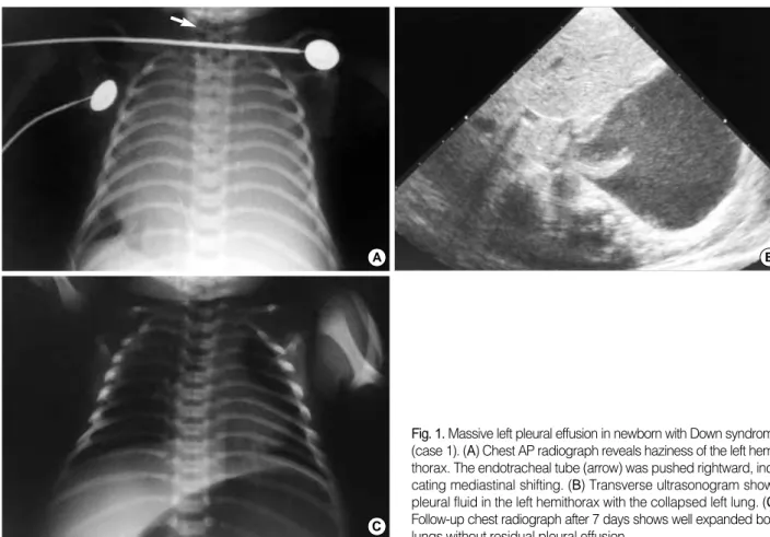
A newborn, whose mother had not received antenatal care, was transferred with respiratory distress. The initial Apgar scores were 2 at 1 min and 6 at 5 min. He had no cardiac prob-lem. He was not febrile. Chest radiograph showed haziness of the left hemithorax. The endotracheal tube was pushed right-ward, indicating mediastinal shifting (Fig. 1A). Ultrasonogram showed pleural fluid in the left hemithorax with the collapsed left lung (Fig. 1B). A chest tube was inserted for drainage of the pleural fluid which was clear. Analysis of the fluid was shown protein 34 g/L, glucose 88 mg/dL, Cl- 100 mM/L, LDH 953 IU/L, WBC 2,000/ L and RBC 1,000/ L. Bacteriologic examination demonstrated no microorganism. Subsequent analysis of the pleural fluid following total parenteral
nutri-tion revealed triglyceride (TG) 3 mg/dL, indicative of non-chy-lous pleural effusion.
His karyotype was 47,XY,+21, so he was diagnosed as Down syndrome. Follow-up chest radiograph showed well ex-panded both lungs without residual pleural effusion (Fig. 1C). Case 2
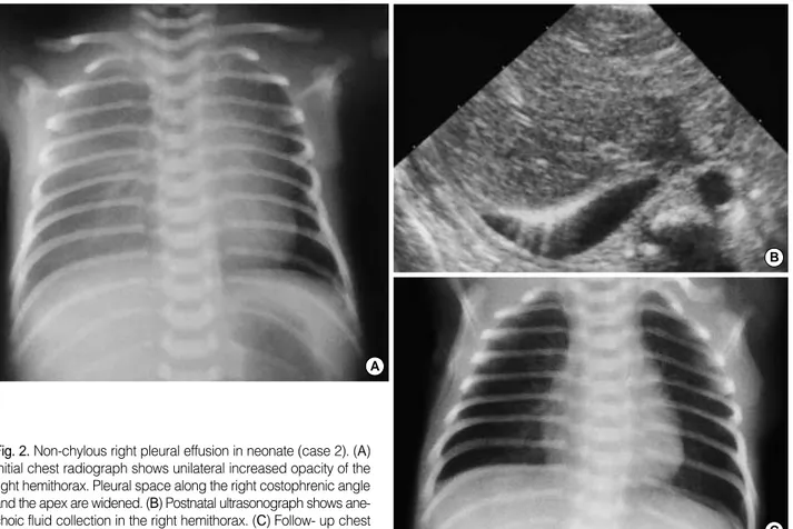
A newborn at 36 weeks of gestational age was delivered by cesarean section without respiratory distress. He had been diagnosed as fetal hydrothorax by fetal ultrasonogram at 32 weeks of gestational age (not shown). The initial chest radio-graph showed diffuse increased opacity of the right hemitho-rax with widening of pleural space (Fig. 2A). Ultrasonogram showed anechoic fluid collection in right hemithorax (Fig 2B). He underwent sono-guided thoracentesis. Analysis of pleural fluid showed protein 32 g/L, glucose 85 mg/dL, Cl- 103 mM/ L, LDH 281 IU/L, WBC 2,500/ L and RBC 220/ L. The level of TG in pleural fluid after milk feeding was 4 mg/dL. No chromosomal anomaly was detected. He had no cardiac problem. He had neither congenital mass nor infectious dis-ease. Follow-up chest radiograph showed clear resorption of pleural effusion (Fig. 2C). He did well until he was 4 yr old.
DISCUSSION
Neonatal pleural effusion may be congenital, inflammatory, iatrogenic following line placement, or secondary to congenital heart failure (5). The effusion is mostly unilateral and about 60% of cases has been found in the right hemithorax (2).
Ji Young Hwang, Jeong Hyun Yoo, Jeong Soo Suh, Chung Sik Rhee Department of Radiology, Ewha Womans University, Seoul, Korea
Received : 3 June 2002 Accepted : 1 October 2002 Address for correspondence Jeonghyun Yoo, M.D.
Department of Radiology, Ewha Womans University, Dongdaemun Hospital, 70 Chongro 6-ga, Chongro-gu, Seoul 110-783, Korea Tel : +82.2-760-5144, 5231, Fax : +82.2-760-5046 E-mail : YooLee@ewha.ac.kr
603
J Korean Med Sci 2003; 18: 603-5 ISSN 1011-8934
Copyright � The Korean Academy
of Medical Sciences
Isolated Non-chylous Pleural Effusion in Two Neonates
Isolated pleural effusion, so called primary pleural effusion denotes a pleural effusion without documented etiology such as a cardiac, inflammatory, iatrogenic problem or fetal hydrops. Chromosomal anomaly such as Down syndrome may be associ-ated with isolassoci-ated pleural effusion. The content of the isolassoci-ated pleural effusion is mostly chylous, and isolated non-chylous pleural effusion in neonate is rare. We experienced 2 cases of isolated non-chylous pleural effusion. They had neither car-diac problem nor other sign of hydrops fetalis. Imaging diagnosis was done by plain chest radiography and subsequent ultrasonogram. One of them was diagnosed to Down syndrome by karyotyping. They were fared well after diagnostic and thera-peutic thoracentesis. We describe 2 cases of non-chylous pleural effusion and re-view a few English-language case reports of this entity.
604 J.Y. Hwang, J.H. Yoo, J.S. Suh, et al.
Among the neonatal pleural effusions, isolated pleural effusion may be diagnosed when there are no other findings of hydrops fetalis nor any inflammatory, iatrogenic, and cardiac problem (5).
The content of the isolated pleural effusion is mostly chylous, resulting from a malformation or tear in the fetal thoracic duct. Chylous pleural effusion may be initially serous and turns into chylous only after milk feeding (2). Distinguishing features of chylous effusion from serous effusion are milky-white or yel-low bloody color, more than 110 mg/dL (>1.24 mMol/L) of TG level and lymphocytosis. However, lymphocytosis can be also discovered in other conditions such as tuberculosis or viral infection. Our two cases proved to non-chylous effusion after milk feeding and total parenteral nutrition containing intralipid with medium-chain TG (6).
In a minority of the cases, the content of the effusion is se-rous. Some authors reported that non-chylous or serous conge-nital pleural effusion may be associated with underlying tho-racic cause such as primary lymphangiectasia, congenital cystic adenomatoid malformation, bronchopulmonary dysplasia, diaphragmatic hernia, chest wall harmatoma, and pulmonary vein atresia (3, 7). Like our case, isolated non-chylous pleural effusion is rare. Several cases have been reported about congen-ital or fetal pleural effusion with chromosomal anomaly such as Down syndrome and Turner syndrome (8-11). Most of these
pleural effusion were chylothorax or associated hydrops fetal-is (6, 9). Hence, karyotyping fetal-is indicated in a fetus or newborn with isolated pleural effusion for the evaluation of associated chromosomal anomaly (8, 11). However, association of Down syndrome with isolated non-chylous pleural effusion has been rarely reported (4). One of our cases was diagnosed as Down syndrome.
Imaging diagnosis was done by plain chest radiograph and ultrasonogram with thoracentesis (5). Chest radiograph usually demonstrates homogeneous haziness in the involved hemitho-rax. It may be sometimes confusing. Diaphragmatic hernia filled with fluid contents, congenital chest mass, such as con-genital cystic adenomatoid malformation or pulmonary seques-tration and total atelectasis should be included in differential diagnosis. Chest ultrasonogram readily and noninvasively dis-tinguish pleural effusion from other conditions (5).
The clinical course of the isolated pleural effusion is variable. Congenital pleural effusion causing pulmonary hypoplasia or maternal polyhydroamnios by extrinsic compression of the fetal esophagus may be associated with high mortality (1). However, it is reported that some cases showed spontaneous resolution in utero or fared well with residual small amount of pleural effusion (7, 11).
Fig. 1. Massive left pleural effusion in newborn with Down syndrome (case 1). (A) Chest AP radiograph reveals haziness of the left hemi-thorax. The endotracheal tube (arrow) was pushed rightward, indi-cating mediastinal shifting. (B) Transverse ultrasonogram shows pleural fluid in the left hemithorax with the collapsed left lung. (C) Follow-up chest radiograph after 7 days shows well expanded both lungs without residual pleural effusion.
A B
Isolated Non-chylous Pleural Effusion in Two Neonates 605
REFERENCES
1. Longaker MT, Laberge JM, Dansereau J, Langer JC, Crombleholme TM, Callen PW, Golbus MS, Harrison MR. Primary fetal hydrothorax: natural history and management. J Pediatr Surg 1989; 24: 573-6. 2. Chernick V, Reed MH. Pneumothorax and chylothorax in the neonatal
period. J Pediatr 1970; 76: 624-32.
3. Laberge JM, Crombleholme TM, Longaker MT. The fetus with pleural effusions. In: Harrison MR, Golbus MS, Filly RA, editors, The unborn patient, 2nd edition, Philadelphia, WB Saunders 1990; 314-9. 4. Modi N, Cooke RW. Congenital non-chylous pleural effusion with
Down’s syndrome. J Med Genet 1987; 24: 567-8.
5. May DA, Barth RA, Yeager S, Nussbaum-Blask A, Bulas DI. Perina-tal and postnaPerina-tal chest sonography. Radiol Clin North Am 1993; 31: 499-516.
6. Yamamoto T, Koeda T, Tamura A, Sawada H, Nagata I, Nagata N, Ito T, Mio Y. Congenital chylothorax in a patient with 21 trisomy syn-drome. Acta Pediatr Jpn 1996; 38: 689-91.
7. Weber AM, Philipson EH. Fetal pleural effusion: a review and meta-analysis for prognostic indicators. Obstet Gynecol 1992; 79: 281-6. 8. Achiron R, Weissman A, Lipitz S, Mashiach S, Goldman B. Fetal
pleural effusion: the risk of fetal trisomy. Gynecol Obstet Invest 1995; 39: 153-6.
9. Ho NK, Leong NK, Lim SB. Chylothorax in Down’s syndrome asso-ciated with hydrops fetalis. J Singapore Paediatr Soc 1989; 31: 90-2. 10. Foote KD, Vickers DW. Congenital pleural effusion in Down’s
syn-drome. Br J Radiol 1986; 59: 609-10.
11. Hegay Z, Reece A, Roberts A, Hobbins JC. Isolated fetal pleural effu-sion: a prenatal management dilemma. Obstet Gynecol 1993; 81: 147-52.
C A
B
Fig. 2. Non-chylous right pleural effusion in neonate (case 2). (A) Initial chest radiograph shows unilateral increased opacity of the right hemithorax. Pleural space along the right costophrenic angle and the apex are widened. (B) Postnatal ultrasonograph shows ane-choic fluid collection in the right hemithorax. (C) Follow- up chest radiograph after 7 days shows clear resorption of pleural effusion.