Influence of Preoperative Transcatheter
Arterial Chemoembolization on Gene
Expression in the HIF-1α Pathway in Patients
with Hepatocellular Carcinoma
by
Wei Guang Xu
Major in Medicine
Department of Medical Sciences
The Graduate School, Ajou University
Influence of Preoperative Transcatheter
Arterial Chemoembolization on Gene
Expression in the HIF-1α Pathway in Patients
with Hepatocellular Carcinoma
by
Wei Guang Xu
A Dissertation Submitted to The Graduate School of
Ajou University in Partial Fulfillment of the Requirements
for the Degree of Doctor of Medicine
Supervised by
Hee-Jung Wang, M.D., Ph.D.
Major in Medicine
Department of Medical Sciences
The Graduate School, Ajou University
This certifies that the dissertation
of Wei Guang Xu is approved.
SUPERVISORY COMMITTEE
Hee-Jung Wang
Wook-Hwan Kim
Hyun-Goo Woo
Jae-Youn Cheong
Seong-Woo Hong
The Graduate School, Ajou University
November, 14th, 2014
- ABSTRACT -
Influence of Preoperative Transcatheter Arterial
Chemoembolization on Gene Expression in the HIF-1α
Pathway in patients with Hepatocellular Carcinoma
Although transcatheter arterial chemoembolization (TACE) is the most common treatment option in patients with hepatocellular carcinoma (HCC), its clinical benefits remain still controversial. Since TACE induces hypoxic necrosis in tumors, hypoxia-inducible factor 1α (HIF-1α) could critically affect biology in residual tumors after TACE treatment and subsequent prognosis. However, HIF-1α and its prognostic relevance in TACE have rarely been examined in human specimens. In the current study, we investigated the prognosis and expression of genes regulated by HIF-1α in HCC patients receiving preoperative TACE for the first time. In total, 35 patients with HCC (10 patients undergoing preoperative TACE) were retrospectively studied. The prognostic significance of TACE was analyzed using Kaplan-Meier and Cox regression models. Protein levels of HIF-1α and mRNA levels of HIF-1α –associated genes were examined using immunohistochemistry (IHC) and real-time RT-PCR, respectively. Preoperative TACE was significantly associated with increased 2-year recurrence rate (80 vs. 36%, P=0.00402) and shorter disease-free survival (DFS) time (11.9 vs. 35.7 months, p=0.0182). TACE was an independent prognostic factor for recurrence (p=0.007) and poor DFS (p=0.010) in a multivariate analysis. Immunohistochemical staining revealed in vivo activation of HIF-1α in human specimens
ii
treated with TACE. Notably, protein levels of HIF-1α were significantly increased in TACE tissues demonstrated by IHC. Transcriptional targets of HIF-1α showed mRNA expression patterns consistent with activation of HIF-1α in TACE tissues. Our findings collectively demonstrate that preoperative TACE confers poor prognosis in HCC patients through activation of HIF-1α.
Key words: Hepatocellular carcinoma. Transcatheter arterial chemoembolization. Prognosis. Hypoxia. Hypoxia-inducible factor-1α.
TABLE OF CONTENTS
ABSTRACT ··· ⅰ TABLE OF CONTENTS ··· ⅲ LIST OF FIGURES ··· ⅳ LIST OF TABLES ··· ⅴ ABBREVIATION ··· ⅵ . Ⅰ INTRODUCTION ··· 1 . Ⅱ MATERIALS AND METHODS ··· 3A. MATERIALS ··· 3
B. METHODS ··· 5
1. TRANSCATHETER ARTERIAL CHEMOEMBOLIZATION ··· 5
2. QUANTITATIVE REAL-TIME PCR ··· 6
3. IMMUNOHISTOCHEMICAL STAINING OF HIF-1α ··· 7
4. STATISTICAL ANALYSIS ··· 8 . Ⅲ RESULTS ··· 9 . Ⅳ DISCUSSION ··· 16 . Ⅴ CONCLUSION ··· 19 REFERENCES ··· 20 국문요약 ··· 26
iv
LIST OF FIGURES
Fig. 1. Kaplan-Meier curves for cumulative recurrence rate and DFS of patients. ··· 10
Fig. 2. Immunohistochemistry of HIF-1α in HCC tissues. ··· 14
LIST OF TABLES
Table 1. Clinicopathological characteristics of HCC tissues ··· 4
Table 2. Univariate Cox regression analysis for recurrence and DFS ··· 11
vi
ABBREVIATION
HCC, Hepatocellular carcinoma;
TACE, Transcatheter arterial chemoembolization; HIF-1α, Hypoxia-inducible 1α;
mRNA, Messenger ribonucleic acid; IHC, Immunohistochemistry;
Real-time RT-PCR, Real-time Reverse transcription-polymerase chain reaction; DFS, Disease-free survival;
EMT, Epithelial-mesenchymal transition; VEGF,Vascular endothelial growth factor; BCLC, Barcelona Clinic Liver Cancer; CDH1, Cadherin 1
MMP9, Matrix Metlloproteinase 9 TCE3, Transcription factor
ZEB1, Zinc-finger E-box binding homeobox 1 VHL, Von Hippel-Lindau gene
I. INTRODUCTION
Hepatocellular carcinoma (HCC) is a major malignancy of the liver with high-incidence and mortality rates worldwide (Llovet et al., 2003). Due to the complexity and heterogeneity of hepatocarcinogenesis accompanying chronic liver disease, prognosis of HCC remains poor. More than 80 % of HCC patients are diagnosed at an inoperable stage (Okuda et al., 1985), and available treatment options are limited.
Transcatheter arterial chemoembolization (TACE) is a non-curative and the most common treatment modality for HCC. TACE has been shown to improve survival and effectively suppress tumor progression (Zhang et al., 2000). In contrast, other studies have reported that TACE increases the recurrence rate and aggravates prognosis in HCC patients (Harada et al., 1996; Lee et al., 2009). The principle of TACE is to block blood vessels branching to the liver from arteries with lipiodol and/or chemo agents such as adriamycin, leading to hypoxic tumor necrosis with the aim of maximizing anti-tumor effects. Due to the limitation of TACE to stimulate angiogenesis by inducing hypoxia (Li et al., 2004; Wang et al., 2008), combining TACE with anti-angiogenic therapeutics such as sorafenib has been considered a promising strategy to improve clinical outcomes of HCC and several clinical trials including the SPACE study have been conducted (Abou-Alfa, 2011). Thus, the precise effects of TACE on tumor biology of HCC and its prognostic relevance need to be clarified.
Hypoxia is an inevitable feature of solid tumors during tumor progression. Tumor cells experience hypoxia during natural growth (Semenza, 2003), or artificial manipulation to block blood vessels, such as TACE (Bismuth et al., 1992). Although deprivation of oxygen and nutrients could kill tumor cells, the surviving tumor cells or surrounding pre-neoplastic
2
-lesions under hypoxia gain an increased capability to survive and metastasize to other organs (Maxwell et al., 1997). HIF-1α plays critical roles in cells upon oxygen deprivation. In hypoxic conditions, HIF-1α is activated to regulate the transcription of downstream effectors driving tumor angiogenesis and epithelial–mesenchymal transition (EMT) integrating cell growth, invasion, motility, and loss of cell adhesion during metastatic cancer progression (Maxwell et al., 1997; Semenza, 2012; Yang et al., 2008). As a prerequisite step for successful dissemination of tumors from primary organs and subsequent colonization in distant organs (Bastid, 2012), EMT has been closely associated with poor prognosis of HCC. HIF-1α is responsible for hypoxia-induced EMT, which contributes to poor clinical outcomes of HCC (Kim et al., 2010; Mima et al., 2013; Ogunwobi and Liu, 2012). Additionally, elevated VEGF levels concomitant with increased angiogenesis in HCC patients undergoing TACE are attributable to activation of HIF-1α signaling (Huang et al., 2005; Wang et al., 2008). However, there is a lack of studies on relationship of expression of HIF-1α and its associated EMT molecules to prognosis of HCC patients subjected to TACE treatment.
In the current study, we investigated for the first time influence of TACE on expression of HIF-1α and its target genes involved in EMT and their prognostic relevance in HCC patients. Our findings provide molecular insights that should aid in improving treatment and prognosis of HCC.
II. Materials and Methods
A. Materials
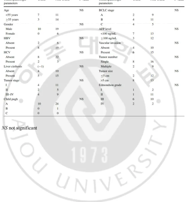
A total of 50 patients were randomized 1:1 to preoperative TACE or not before curative resection for primary HCC in Ajou Medical Centers in South Korea. Among initial 25 HCC patients undergoing preoperative TACE, we analyzed 10 patients who met inclusion criteria of both the duration from TACE to resection within 50 days and one time of TACE. The interval between TACE and surgery was an average of 26.4 ± 14.5 days ranging from 6 to 49 days. TACE tissues were taken from the viable portion of necrotic HCC tissues. All tissues were obtained with informed consent from the patients, and the study protocol was approved by the institutional review board. Table 1 summarizes the clinicopathological characteristics of the 35 HCC patients studied in the current study. We used BCLC stage and Edmondson and Steiner grade according to the traditional criteria (Patel et al., 2011; Villanueva and Llovet, 2011).
4
-Table 1. Clinicopathological characteristics of HCC tissue.
B. METHODS
1. TRANSCATHETER ARTERIAL CHEMOEMBOLIZATION
After the introduction of a selective catheter through the femoral artery using the Seldinger technique, an angiographic survey of the abdominal vessels was performed. The localization of the hepatic arteries was checked with celiac and mesenteric arteriography using selective catheterization. This was performed to define vascular anatomy. Next, indirect portography was performed to outline the portal circulation in the venous phase. A catheter was placed in the celiac trunk and advanced beyond the gastroduodenal artery. Depending on size, location, and arterial supply to the tumor, the tip of the catheter was advanced further into the segmental arteries. For superselective embolization, an infusion catheter was used. A 10 ml of iodized oil (Lipiodol Ultrafluide®, Laboratoire Andr Guerbet, Aulnay-Sous-Bois, France) and 1 mg/kg of doxorubicin hydrochloride (ADM®, Dong-A Pharm. Co. Ltd., Seoul, Korea) were mixed to be injected until stasis of the blood flow was observed. When an initial blockade of tumor feeding artery was insufficient because of the large tumor size or arterioportal shunting, an embolization was performed with gelatin sponge particles (Cutanplast®, Mascia Brunelli Spa, Viale Monza, Italy). After embolization, devascularization was confirmed with additional angiography of the hepatic artery.
6
-2. QUANTITATIVE REAL-TIME PCR
Quantitative real-time RT-PCR was carried out as previously described (Kwon et al. 2010). Total RNA was isolated from frozen tissues using an RNeasy mini kit (Qiagen, USA). The RNA integrity was evaluated by a Bioanalyzer 2100 (Agilent Technologies, USA). Reverse transcription reaction was carried out with 4 μg of total RNA and 2 μL of 10 μmol/L oligo d(T)18 primer (Genotech, Korea) at 70 °C for 7 min and then cooled on ice for 5 min. After adding the reverse transcriptase mixture to the primer-annealed total RNA, the reaction was incubated for 90 min at 42 °C. Real-time PCR (ABI PRISM 7900HT, Applied Biosystems, USA) was performed in a total volume of 10 μL (2 μL cDNA, 2 μL of 5 pmol/μ primer, 1 μL of 1 pmol/μ probe, and 5 μL Taqman master mix) according to the following 3 steps: an initial denaturation step at 95 °C for 10 min, 45 cycles of denaturation step at 95 °C for 15 s, and elongation step at 60 °C for 1 min. The primer and probe sequences were designed using Primer Express 3.0 software (Applied Biosystems, USA), and all the probes were labeled with FAM and TAMRA at the 5′ end and 3′ end, respectively. Primer and probe sequences for RT-PCR are listed in Supplementary data. The mRNA levels of genes (the threshold cycle, CT value) were measured in triplicate and then subjected to normalization with five reference genes (B2M, GAPDH, HMBS, HPRT1, and SDHA) by subtracting the average values of the mRNA levels of the reference genes as an internal control (Yang and Roberts, 2010).
3. IMMUNOHISTOCHEMICAL STAINING OF HIF-1α
Immunohistochemical staining was done on 4-μm-thick, formalin-fixed, paraffin-embedded tissue sections. Tissue sections were deparaffinized in xylene for 15 min and then rehydrated. Antigen retrieval was performed by boiling in Tris–EDTA buffer (pH 9.0) for 5 min. Slides were then incubated with anti-human HIF-1α mouse monoclonal antibody (Novus, USA) for 1 h at room temperature. The antigen–antibody reaction was detected using the DAKO REAL Detection System (LSAB+, USA) K5001 (DAKO, USA). All the immunohistochemically stained sections were evaluated in a semiquantitative fashion by two pathologists as previously described (Kwon et al., 2010). The HIF-1α expression was evaluated in 10 high-power fields (400×). Intensities were classified as 0 (negative staining), 1 (<5 % of samples stained), 2 (<25 % of samples stained), 3 (25–50 % of samples stained), and 4 (more than 50 % of samples stained).
8
-4. STATISTICAL ANALYSIS
All statistical analyses were performed with the open source statistical program R. The Cox proportional hazard regression model was used to assess prognostic significance of TACE for recurrence and disease-free survival (DFS). Kaplan–Meier survival curves were plotted using tumor recurrence (defined as the first appearance of a tumor at any site following definitive treatment) or death as the end points. The significant differences in recurrence curve or DFS curves were examined by log-rank test. 2−ΔCt values of each gene were shown in box and whisker plot, and their significant differences between TACE and non-TACE tissues were evaluated by a Student’s t test. Distribution of clinicopathologic values in TACE and non-TACE tissues was evaluated using χ2 and Fisher’s exact test. A P value <0.05 was considered statistically significant in this study.
III. RESULTS
Clinicopathological characteristics of 35 HCC patients at diagnosis were cataloged (Table 1). To exclude the effects of clinical parameters during studies on the influence of preoperative TACE on expression of genes involved in the HIF-1α pathway, chi-square and Fisher’s exact tests were performed. There were no significant differences in clinicopathological features between patients in TACE and non-TACE groups.
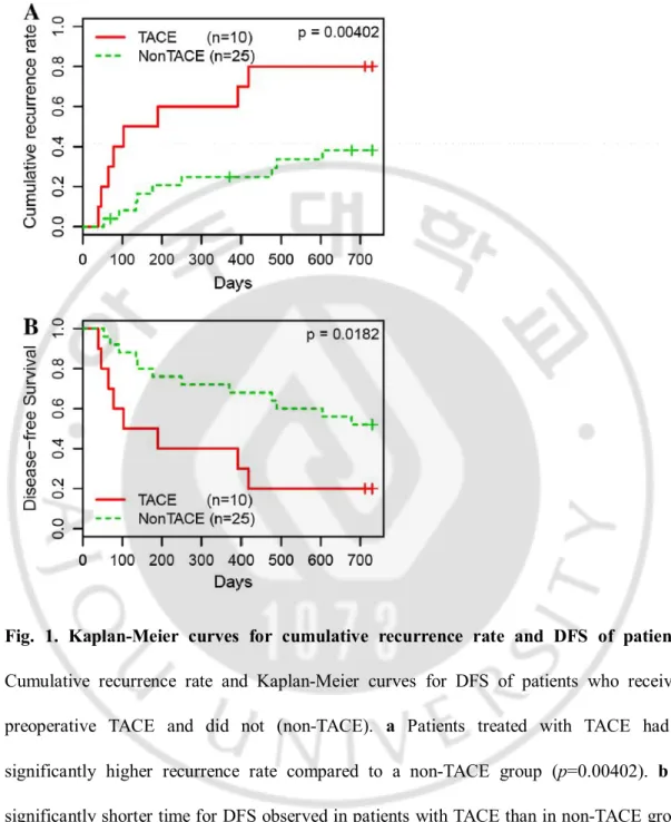
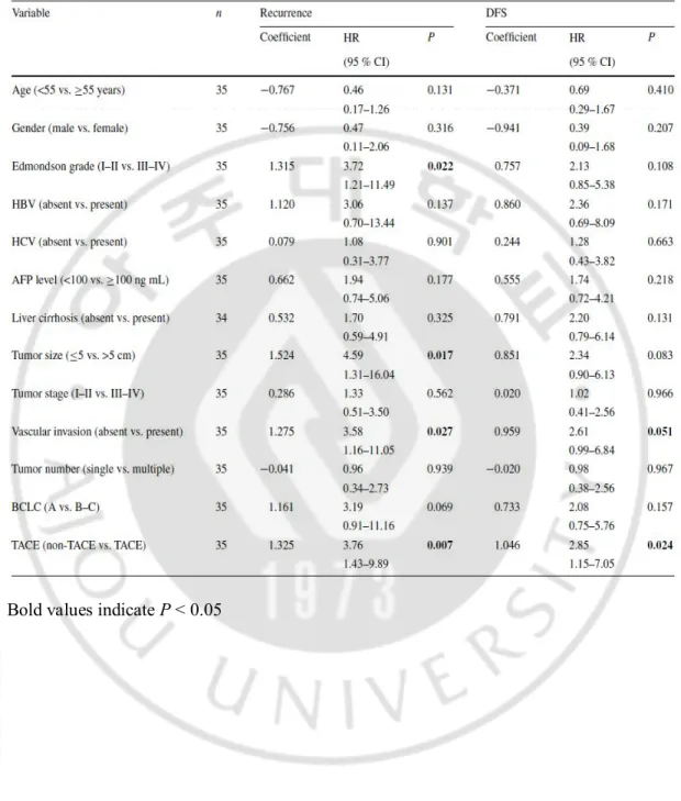
The effects of preoperative TA CE on recurrence and survival were investigated using Kaplan–Meier survival curves. At a follow-up time of 2-years, 80 % (8/10) of the HCC patients in the TA CE group displayed recurrence, whereas recurrence rate of the non-TA CE group was 36 % (9/25) (P = 0.00402; Fig. 1a). For DFS, median survival times were 11.9 months (2.1–52.2 months) and 35.7 months (1.7–136.9 months) in TACE and non-TACE groups, respectively (P = 0.0182; Fig. 1b). In contrast, the differences in overall survival time between TACE and non-TACE groups were not significant (data not shown). To confirm the prognostic significance of TACE, the Cox regression analysis was performed. In a univariate Cox regression analysis, high Edmondson grade (P = 0.022), large tumor size (P = 0.017), and vascular invasion (P = 0.027) were associated with recurrence and TACE treatment before hepatectomy was a statistically significant risk factor for earlier recurrence in HCC patients (P = 0.007). For DFS, TACE was a poor prognostic factor along with vascular invasion (vascular invasion, P = 0.051; TACE, P = 0.024) (Table 2). A multivariate Cox model demonstrated that TACE was the strongest independent poor prognostic factor for recurrence (P = 0.007), as vascular invasion was shown to have borderline significance (P = 0.054). For DFS, both TACE (P = 0.010) and vascular invasion (P = 0.041) were found to be independent poor prognostic factors (Table 3).
- 10 -
Fig. 1. Kaplan-Meier curves for cumulative recurrence rate and DFS of patients. Cumulative recurrence rate and Kaplan-Meier curves for DFS of patients who received preoperative TACE and did not (non-TACE). a Patients treated with TACE had a significantly higher recurrence rate compared to a non-TACE group (p=0.00402). b A significantly shorter time for DFS observed in patients with TACE than in non-TACE group (p=0.0182). Thin lines, patients received preoperative TACE (n=10); Broken lines, patients received only hepatectomy (n=25).
Table 2. Univariate Cox regression analysis for recurrence and DFS
- 12 -
Table 3. Multivariate Cox regression analysis for recurrence and DFS
Since HIF-1α is activated through protein stabilization via reduced proteasomal degradation under hypoxic conditions, the protein was subjected to immunohistochemcal analysis in 35 HCC tissues. Immunostaining revealed that the average intensity of nuclear and cytosolic HIF-1α was higher in TACE than non-TACE tissues and their differences were statistically significant. (1.96 vs. 2.89, P = 0.0158; Fig. 2a). Fig. 2b represents moderate and weak intensities of HIF-1α expressed in non-TACE tissues (a and b, respectively) and strong expression of HIF-1α in TA CE tissues (c).
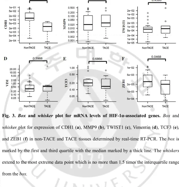
To investigate the influence of TACE on tumor biology, mRNA levels of HIF-1α target genes associated with EMT were measured using real-time RT-PCR (Fig. 3). The TACE group expressed lower levels of CDH1, a HIF-1α-regulated epithelial marker than the non-TACE group (P = 0.0003; Fig. 3d). Conversely, mRNA expression HIF-1α-regulated mesenchymal markers, such as MMP9, Twist1, and vimentin was slightly higher in TACE tissues compared to non-TACE tissues, but their differences were not statistically significant (P = 0.8140, P = 0.4586, and P = 0.3988; Fig. 3e–g). Additional mesenchymal genes TCF3 and ZEB1 were expressed at lower levels in the TACE compared to the non-TACE group (P = 0.6866 and P = 0.0468; Fig. 3h, i).
- 14 -
Fig. 2. Immunohistochemistry of 1α in HCC tissues. A Box and whisker plot for HIF-1α expression levels in HCC tissues receiving preoperative TACE or not, determined by IHC. The box is marked by the first and third quartile with the median marked by a thick line. The
whiskers extend to the most extreme data point which is no more than 1.5 times the
interquartile range from the box. Statistically significant difference in HIF-1α protein expression between TACE and non-TACE tissues was found (p=0.0158). B Representative image of HIF-1α-positive samples at ×400magnification. Moderate (a) and weak (b) staining or HIF-1α in non-TACE tissues; strong (e) staining of HIF-1α in a TACE tissues.
Fig. 3. Box and whisker plot for mRNA levels of HIF-1α-associated genes. Box and
whisker plot for expression of CDH1 (a), MMP9 (b), TWIST1 (c), Vimentin (d), TCF3 (e),
and ZEB1 (f) in non-TACE and TACE tissues determined by real-time RT-PCR. The box is marked by the first and third quartile with the median marked by a thick line. The whiskers extend to the most extreme data point which is no more than 1.5 times the interquartile range from the box.
- 16 -
Ⅳ
. DISCUSSION
TACE is the most frequently applied locoregional therapy to HCC patients in Korea. In principle, TACE accompanied by tumor ischemia plays dual roles in the treatment of HCC. Firstly, TACE induces tumor necrosis by exclusively closing off blood vessels from the hepatic artery to HCC. In this case, TACE is effective for extension of patient survival. However, TACE can promote tumor recurrence and metastasis when incomplete tumor necrosis is triggered. Poor clinical outcomes resulting from TACE have raised questions about potential tumor biology alterations triggered by TACE. Increased tumor angiogenesis and invasiveness by TACE are found to be mediated by hypoxia signaling (Gupta et al., 2006; Sergio et al., 2008), which has been effectively suppressed by anti-angiogenic therapy (Jiang et al., 2007). In this respect, many types of clinical tests on combination therapy of TACE with anti-angiogenic agents have been performed (Abou-Alfa, 2011; Lencioni, 2012). Nevertheless, recent conflicting results from two clinical trials on combination of TACE plus sorafenib (Abou-Alfa, 2011; Kudo et al., 2011; Pawlik et al., 2011) strongly imply the need to study more detailed tumor biology in human specimens treated with TACE. Accordingly, we explored the influence of preoperative TACE treatment on prognosis and status of HIF-1α and its associated genes in human HCC specimens.
In current study, HCC patients subjected to preoperative TACE exhibited significantly higher recurrence rates and shorter DFS, relative to the non-TACE group (Fig. 1). Multivariate Cox model further indicated that TACE was an independent poor prognostic factor for HCC (Table 3). These results are supported by previous reports demonstrating poor prognosis in HCC patients treated with TACE before surgery (Choi et al., 2007; Kang et al., 2010; Kishi et al., 2012; Sasaki et al., 2006; Zhou et al., 2009). Additionally, a
negative effect of TACE was reported in a mouse xenograft model of TACE (Liu et al., 2010). However, conflicting evidence has been reported with regard to the effects of preoperative TACE on HCC prognosis. For instance, Giorgio and other groups demonstrated that application of TACE before liver resection reduces HCC recurrence and improves DFS (Gerunda et al., 2000; Han et al., 1999; Majno et al., 1997; Zhang et al., 2000). The discrepancy in the prognostic effects of preoperative TACE may be attributed to differences in tumor size, liver function, borderline resectability, number of TACE treatments, or various TACE methodologies in each study. However, there is a general consensus supporting poor prognosis owing to incomplete tumor necrosis by TACE. To elucidate mechanisms underlying poor clinical outcomes after preoperative TACE in our institution, specimens used in this study were selectively derived from viable portions of HCC after hepatic resection within 50 days following only one time of TACE. Our findings confirm that imperfect TACE treatment confers dismal prognosis in HCC patients after surgery. Protein expression of HIF-1α remains low in normoxic conditions through VHL-mediated ubiquitination and subsequent proteasomal degradation. When cells are subjected to hypoxic conditions, VHL dissociates from HIF-1α leading to reduced ubiquitination and subsequent stabilization of HIF-1α with concomitant heterodimerization with HIF-1β (Semenza, 2012). Since HIF-1α is stabilized in hypoxia setting, we initially examined the effect of TACE on HIF-1α protein expression using immunohistochemical staining. As expected, HIF-1α was more strongly expressed in TACE, compared to non-TACE tissues (Fig. 2). Consistently, stabilization of HIF-1α has been previously reported in animal tissues subjected to TACE treatment (Liu et al., 2010; Rhee et al., 2007). Therefore, our results clearly demonstrated the activation of HIF-1α in tissues of HCC patients undergoing TACE. HIF-1α activates
- 18 -
transcription of genes involved in EMT. Hypoxic cells undergo EMT accompanied by loss of epithelial markers and gain of mesenchymal markers through HIF-1α activation. These gene expression changes confer increased tumor aggressiveness, invasiveness, and metastatic potentials to EMT cells (Semenza, 2012). Based on this background, clinical outcomes of TACE could be affected by expression of EMT genes concomitant with HIF-1α activation, but it has been rarely studied in patient tissues. In our results, CDH1 which is a repressive target of HIF-1α and one of epithelial markers (Krishnamachary et al., 2006) was dramatically down-regulated in tissues treated with TACE (Fig. 3d). MMP9, Twist1, and vimentin, mesenchymal markers and transcriptional target genes of HIF-1α known to increase upon hypoxia (Choi et al., 2011; Liu et al., 2010, 2012), were up-regulated in TACE tissues (Fig. 3e–g). However, mRNA expression of TCF3 and ZEB1 were down-regulated in a conflict with previous reports showing induction of TCF3 and ZEB1 by HIF-1α (Krishnamachary et al., 2006) (Fig. 3h, i). Considering that most of well-known target genes were expressed consistently with activation of HIF-1α in TACE tissues and that the tested samples were chosen with inclusion criteria within 50 days after TACE to exclude effects of HIF-1α signaling recovery from hypoxia upon TACE treatment, it may be ascribed to the small number of samples or regulation of TCF3 and ZEB1 by other unknown factors in complex physiological conditions of TACE.
.
Ⅴ
CONCLUSION
In conclusion, preoperative TACE treatment is a poor prognostic factor in HCC patients. Additionally, the biological effects of TACE are associated with HIF-1α activation and expression changes in downstream genes. Our data collectively suggest that preoperative TACE confers poor prognosis via alterations in gene expression patterns in the HIF-1α pathway. Additionally, our results confirm previous reports showing activation of hypoxia signaling upon TACE treatment and support current strategy targeting tumor biology by combining TACE and anti-angiogenic therapy for HCC treatment. The molecular evidence obtained in this study can be effectively applied to guide treatment options for HCC.
- 20 -
REFERENCES
1. Abou-Alfa GK (2011) TA CE and sorafenib: a good marriage? J Clin Oncol 29(30):3949–3952
2. Bastid J (2012) EMT in carcinoma progression and dissemination: facts, unanswered questions, and clinical considerations. Cancer Metastasis Rev 31(1–2):277–283 3. Bismuth H, Morino M, Sherlock D, Castaing D, Miglietta C, Cauquil P, Roche A
(1992) Primary treatment of hepatocellular carcinoma by arterial chemoembolization.
Am J Surg 163(4):387–394
4. Choi GH, Kim DH, Kang CM, Kim KS, Choi JS, Lee WJ, Kim BR (2007) Is preoperative transarterial chemoembolization needed for a resectable hepatocellular carcinoma? World J Surg 31(12):2370–2377
5. Choi JY, Jang YS, Min SY, Song JY (2011) Overexpression of MMP-9 and HIF-1alpha in breast cancer cells under hypoxic conditions. J Breast Cancer 14(2):88–95 6. Gerunda GE, Neri D, Merenda R, Barbazza F, Zangrandi F, Meduri F, Bisello M,
Valmasoni M, Gangemi A, Faccioli AM (2000) Role of transarterial chemoembolization before liver resection for hepatocarcinoma. Liver Transpl 6(5):619–626
7. Gupta S, Kobayashi S, Phongkitkarun S, Broemeling LD, Kan Z (2006) Effect of transcatheter hepatic arterial embolization on angiogenesis in an animal model.
Invest Radiol 41(6):516–521
8. Han YM, Park HH, Lee JM, Kim JC, Hwang PH, Lee DK, Kim CS, Choi KC (1999) Effectiveness of preoperative transarterial chemoembolization in presumed inoperable hepatoblastoma. J Vasc Interv Radiol 10(9):1275–1280
9. Harada T, Matsuo K, Inoue T, Tamesue S, Nakamura H (1996) Is preoperative hepatic arterial chemoembolization safe and effective for hepatocellular carcinoma?
Ann Surg 224(1):4–9
10. Huang GW, Yang LY, Lu WQ (2005) Expression of hypoxia-inducible factor 1alpha and vascular endothelial growth factor in hepatocellular carcinoma: impact on neovascularization and survival. World J Gastroenterol 11(11):1705–1708
11. Jiang H, Meng Q, Tan H, Pan S, Sun B, Xu R, Sun X (2007) Antiangiogenic therapy enhances the efficacy of transcatheter arterial embolization for hepatocellular carcinomas. Int J Cancer 121(2):416–424
12. Kang JY, Choi MS, Kim SJ, Kil JS, Lee JH, Koh KC, Paik SW, Yoo BC (2010) Long-term outcome of preoperative transarterial chemoembolization and hepatic resection in patients with hepatocellular carcinoma. Korean J Hepatol 16(4):383–388 13. Kim J, Hong SJ, Park JY, Park JH, Yu YS, Park SY, Lim EK, Choi KY, Lee EK, Paik SS, Lee KG, Wang HJ, Do IG, Joh JW, Kim DS (2010) Epithelial-mesenchymal transition gene signature to predict clinical outcome of hepatocellular carcinoma. Cancer Sci 101(6):1521–1528
14. Kishi Y, Saiura A, Yamamoto J, Koga R, Seki M, Morimura R, Yoshioka R, Kokudo N, Yamaguchi T (2012) Preoperative transarterial chemoembolization for hepatocellular carcinoma. Hepatogastroenterology 59(119):2295–2299
15. Krishnamachary B, Zagzag D, Nagasawa H, Rainey K, Okuyama H, Baek JH, Semenza GL (2006) Hypoxia-inducible factor- 1-dependent repression of E-cadherin in von Hippel-Lindau tumor suppressor-null renal cell carcinoma mediated by TCF3, ZFHX1A, and ZFHX1B. Cancer Res 66(5):2725–2731
- 22 -
16. Kudo M, Imanaka K, Chida N, Nakachi K, Tak WY, Takayama T, Yoon JH, Hori T, Kumada H, Hayashi N, Kaneko S, Tsubouchi H, Suh DJ, Furuse J, Okusaka T, Tanaka K, Matsui O, Wada M, Yamaguchi I, Ohya T, Meinhardt G, Okita K (2011) Phase III study of sorafenib after transarterial chemoembolisation in Japanese and Korean patients with unresectable hepatocellular carcinoma. Eur J Cancer 47(14):2117–2127
17. Kwon JH, Kim J, Park JY, Hong SM, Park CW, Hong SJ, Park SY, Choi YJ, Do IG, Joh JW, Kim DS, Choi KY (2010) Overexpression of high-mobility group box 2 is associated with tumor aggressiveness and prognosis of hepatocellular carcinoma.
Clin Cancer Res 16(22):5511–5521
18. Lee KT, Lu YW, Wang SN, Chen HY, Chuang SC, Chang WT, Shi HY, Ker CG, Chiu HC (2009) The effect of preoperative transarterial chemoembolization of resectable hepatocellular carcinoma on clinical and economic outcomes. J Surg
Oncol 99(6):343–350
19. Lencioni R (2012) Management of hepatocellular carcinoma with transarterial chemoembolization in the era of systemic targeted therapy. Crit Rev Oncol/Hematol 83(2):216–224
20. Li X, Feng GS, Zheng CS, Zhuo CK, Liu X (2004) Expression of plasma vascular endothelial growth factor in patients with hepatocellular carcinoma and effect of transcatheter arterial chemoembolization therapy on plasma vascular endothelial growth factor level. World J Gastroenterol 10(19):2878–2882
21. Liu L, Ren ZG, Shen Y, Zhu XD, Zhang W, Xiong W, Qin Y, Tang ZY (2010) Influence of hepatic artery occlusion on tumor growth and metastatic potential in a
human orthotopic hepatoma nude mouse model: relevance of epithelial-mesenchymal transition. Cancer Sci 101(1):120–128
22. Liu W, Shen SM, Zhao XY, Chen GQ (2012) Targeted genes and interacting proteins of hypoxia inducible factor-1. Int J Biochem Mol Biol 3(2):165–178
23. Llovet JM, Burroughs A, Bruix J (2003) Hepatocellular carcinoma. Lancet 362(9399):1907–1917
24. Majno PE, Adam R, Bismuth H, Castaing D, Ariche A, Krissat J, Perrin H, Azoulay D (1997) Influence of preoperative transarterial lipiodol chemoembolization on resection and transplantation for hepatocellular carcinoma in patients with cirrhosis.
Ann Surg 226(6):688–701; discussion 701–683
25. Maxwell PH, Dachs GU, Gleadle JM, Nicholls LG, Harris AL, Stratford IJ, Hankinson O, Pugh CW, Ratcliffe PJ (1997) Hypoxiainducible factor-1 modulates gene expression in solid tumors and influences both angiogenesis and tumor growth.
Proc Natl Acad Sci USA 94(15):8104–8109
26. Mima K, Hayashi H, Kuroki H, Nakagawa S, Okabe H, Chikamoto A, Watanabe M, Beppu T, Baba H (2013) Epithelial-mesenchymal transition expression profiles as a prognostic factor for disease-free survival in hepatocellular carcinoma: clinical significance of transforming growth factor-beta signaling. Oncol Lett 5(1):149–154 27. Ogunwobi OO, Liu C (2012) Therapeutic and prognostic importance of
epithelial-mesenchymal transition in liver cancers: insights from experimental models. Crit Rev
Oncol/Hematol 83(3):319–328
28. Okuda K, Ohtsuki T, Obata H, Tomimatsu M, Okazaki N, Hasegawa H, Nakajima Y, Ohnishi K (1985) Natural history of hepatocellular carcinoma and prognosis in
- 24 -
relation to treatment. Study of 850 patients. Cancer 56(4):918–928
29. Patel SH, Kneuertz PJ, Delgado M, Kooby DA, Staley CA 3rd, El-Rayes BF, Kauh JS, Sarmiento JM, Hanish S, Cohen C, Farris AB 3rd, Maithel SK (2011) Clinically relevant biomarkers to select patients for targeted inhibitor therapy after resection of hepatocellular carcinoma. Ann Surg Oncol 18(12):3384–3390
30. Pawlik TM, Reyes DK, Cosgrove D, Kamel IR, Bhagat N, Geschwind JF (2011) Phase II trial of sorafenib combined with concurrent transarterial chemoembolization with drug-eluting beads for hepatocellular carcinoma. J Clin Oncol 29(30):3960– 3967
31. Rhee TK, Young JY, Larson AC, Haines GK 3rd, Sato KT, Salem R, Mulcahy MF, Kulik LM, Paunesku T, Woloschak GE, Omary RA (2007) Effect of transcatheter arterial embolization on levels of hypoxia-inducible factor-1alpha in rabbit VX2 liver tumors. J Vasc Interv Radiol 18(5):639–645
32. Sasaki A, Iwashita Y, Shibata K, Ohta M, Kitano S, Mori M (2006) Preoperative transcatheter arterial chemoembolization reduces long-term survival rate after hepatic resection for resectable hepatocellular carcinoma. Eur J Surg Oncol 32(7):773–779
33. Semenza GL (2003) Targeting HIF-1 for cancer therapy. Nat Rev Cancer 3(10):721– 732
34. Semenza GL (2012) Hypoxia-inducible factors: mediators of cancer progression and targets for cancer therapy. Trends Pharmacol Sci 33(4):207–214
35. Sergio A, Cristofori C, Cardin R, Pivetta G, Ragazzi R, Baldan A, Girardi L, Cillo U, Burra P, Giacomin A, Farinati F (2008) Transcatheter arterial chemoembolization
(TA CE) in hepatocellular carcinoma (HCC): the role of angiogenesis and invasiveness. Am J Gastroenterol 103(4):914–921
36. Villanueva A, Llovet JM (2011) Targeted therapies for hepatocellular carcinoma.
Gastroenterology 140(5):1410–1426
37. Wang B, Xu H, Gao ZQ, Ning HF, Sun YQ, Cao GW (2008) Increased expression of vascular endothelial growth factor in hepatocellular carcinoma after transcatheter arterial chemoembolization. Acta Radiol 49(5):523–529
38. Yang JD, Roberts LR (2010) Hepatocellular carcinoma: a global view. Nat Rev
Gastroenterol Hepatol 7(8):448–458
39. Yang MH, Wu MZ, Chiou SH, Chen PM, Chang SY, Liu CJ, Teng SC, Wu KJ (2008) Direct regulation of TWIST by HIF-1alpha promotes metastasis. Nat Cell
Biol 10(3):295–305
40. Zhang Z, Liu Q, He J, Yang J, Yang G, Wu M (2000) The effect of preoperative transcatheter hepatic arterial chemoembolization on disease-free survival after hepatectomy for hepatocellular carcinoma. Cancer 89(12):2606–2612
41. Zhou WP, Lai EC, Li AJ, Fu SY, Zhou JP, Pan ZY, Lau WY, Wu MC (2009) A prospective, randomized, controlled trial of preoperative transarterial chemoembolization for resectable large hepatocellular carcinoma. Ann Surg 249(2):195–202
- 26 - - 국문요약 -
수술 전 경동맥색전술을 시행 받은 간세포암 환자들에서
HIF-1α에 관련된 유전자들의 다양한 표현경로에 대한 분석
아주대학교 대학원 의학과 허 위 광 (지도교수: 왕 희 정) 간세포암의 치료에서 경동맥색전술이 아주 흔한 치료방법이지만 그 임상적인 효과에 대해 아직도 논쟁은 많다. 경동맥색전술은 저산소로 인한 종양의 괴사를 유발하지만 hypoxia-inducible factor 1α(HIF-1α)는 남아있는 종양의 생물학적 특성에 영향을 주며 따라서 치료예후에도 영향을 주게 된다. HIF-1α와 그에 관련된 경동맥색전술의 예후의 연관성에 대한 연구는 인체조직에서 거의 진행되지 않았다. 저자들의 연구에서는, 수술 전 경동맥색전술을 시행 받은 간세포암환자들에서 HIF-1α의 유전자의 표현과 예후에 미치는 영향을 연구하였다. 전체 35 예의 간세포암환자에서(이중에 10 예의 수술 전 경동맥색전술을 시행 받은 환자를 포함) 후향적인 연구를 진행하였다. 경동맥색전술의 예후에 관하여 Kaplan-Meier and Cox regression models 을 사용하여 분석하였다. HIF-1α의 단백질 수준과 HIF-1α에 관련된 유전자의 mRNA 의 발현량은 각각 면역조직화학검사(IHC)와 Real-time RT-PCR 을 이용하여 조사하였다. 수술 전 경동맥색전술을 시행 받은 간세포암환자들에서 2 년 재발율은 유의하게 높았고(80 vs. 36%, P=0.00402), 무재발생존기간도 짧았다(11.9 vs. 35.7 months, p=0.0182). 경동맥색전술은 재발에 관련하여 독립적은 예후인자이며(p=0.007), 다변량분석에서 낮은 무재발생율을 보여줬다(p=0.010). 경동맥색전술을 시행 받은 인체조직에서 면역조직화학검사방법으로 HIF-1α에 관련하여 염색을 진행하였다. 그 결과, HIF-1α의 단백질 수준이 경동맥색전술을 시행 받은 조직에서 유의하게 증가하였다. 경동맥색전술을 시행 받은 조직에서 HIF-1α에 관련된 유전자들의 mRNA 의 발현양상이 HIF-1α의 활성과 일치하였다. 저자들의 연구결과에 따르면 수술 전 경동맥색전술을 시행 받은 간세포암 환자들은 HIF-1α의 활성으로 인하여 낮은 생존율을 보여주었다. 핵심어: 간세포암. 경동맥색전술. 예후. 저산소. Hypoxia-inducible factor-1α.