1. Introduction
Antibiotics are powerful medicines used to treat infections caused by microorganisms. However, the inappropriate use of antibiotics and their proliferation in the environment can cause toxic effects in aquatic organisms [1]. Studies have indicated that wastewater treatment facilities are one of the important point sources for anti-biotic contamination of surface waters [2-5]. The antianti-biotics in the final effluents released from these facilities are presumed to the reason for the spread of antibiotic-resistant bacteria in the environment [6-8].
Degradation of antibiotics via biological processes has been investigated to treat wastewater containing antibiotics. However, many studies have demonstrated that clinically important
anti-biotics are not completely biodegraded by conventional treatment methods, even when employing a combined anaerobic–aerobic treatment system, which has been used to treat high-strength pharmaceutical wastewater [9-11]. Advanced oxidation processes (AOPs), such as hydrogen peroxide (H2O2) or ozone (O3), catalysts (iron ions, electrodes, and metal oxides), and irradiation (UV, sunlight, ultrasound, and gamma irradiation) have shown poten-tial as alternative processes for the treatment of most industrial effluents containing toxic organic chemicals [12-19]. Ozonation can be successfully employed as a pretreatment to enhance bio-degradability of antibiotics in wastewater, although not for com-plete mineralization of the antibiotic (> 90% removal efficiencies) [13, 16, 20-22]. Removal rates have been reported to be 98% for tetracycline when used in combination with UV and TiO2
Comparison of biological and chemical assays for measuring
the concentration of residual antibiotics after treatment with
gamma irradiation
Ji-Hyun Nam
1, Ji-Hye Shin
2, Tae-Hun Kim
3, Seungho Yu
3, Dong-Hun Lee
2†1Division of Antimicrobial Resistance, Center for Infectious Diseases Research, National Institute of Health, Korea Centers for Disease Control and
Prevention, Cheongju 28160, Republic of Korea
2Department of Microbiology, Chungbuk National University, Cheongju 28644, Republic of Korea
3Research Division of Industry and Environment, Korea Atomic Energy Research Institute, Jeongeup 56212, Republic of Korea
ABSTRACT
Antibiotic pollution is one of the factors contributing to the spread of antibiotic-resistant bacteria in the environment. Advanced oxidation and irradiation processes have been introduced to eliminate antibiotics from water and wastewater. However, few studies have reported the toxic effects of residual antibiotics and their byproducts induced by a treatment system. In this study, we compared the efficacies of chemical (high-performance liquid chromatography (HPLC)) and biological (antimicrobial susceptibility test) assays for measuring the concentrations of residual antibiotics after gamma irradiation for degrading amoxicillin, cephradine, lincomycin, and tetracycline. The concentrations of residual antibiotics estimated using the two assay methods were almost identical, except cephradine. In the case of cephradine, inhibited bacterial growth was observed that was equivalent to twice the concentration measured by HPLC in the samples subjected to gamma irradiation. The observed inhibition of bacterial growth suggested the generation of potentially toxic intermediates following antibiotic degradation. These results indicate that biological and chemical assays should be used in concert for monitoring antibiotic contamination and the toxic derivatives of antibiotic degradation. The results demonstrate that these four antibiotics can be decomposed by 2.0 kGy gamma-irradiation without toxic effects of their byproducts.
Keywords: Antibiotics, Antimicrobial susceptibility test, Chemical assay, Gamma irradiation
This is an Open Access article distributed under the terms of the Creative Commons Attribution Non-Commercial License (http://creativecommons.org/licenses/by-nc/3.0/) which per-mits unrestricted non-commercial use, distribution, and reproduction in any medium, provided the original work is properly cited.
Copyright © 2020 Korean Society of Environmental Engineers
Received June 24, 2019 Accepted August 4, 2019
†Corresponding author
Email: donghun@cbnu.ac.kr
Tel: +82-43-261-3261 Fax: +82-43-264-9600 ORCID: 0000-0001-7839-3201
as a catalyst, while degradation of lincomycin was noticeably lower [22, 23]. The UV/TiO2 treatment also degrades 82% of the sulfamethoxazole [24]. The occurrence of antibiotics in the effluents of wastewater treatment facilities supports concerns regarding discharged antibiotic residues that may reside in the water supply, and thus have potentially serious environmental consequences [25]. When original medicinal modes of action disappear, degradation products should not promote formation of resistant bacterial strains [22, 26]. However, degradation com-pounds must be identified and monitored, as they may be more toxic than the parent compounds [22, 27].
Liquid chromatography coupled with mass spectrometry or tan-dem mass spectrometry has been routinely used to measure anti-biotics in wastewater, and these techniques are assumed to be sufficiently accurate and sensitive to detect these compounds [14, 28-30]. However, analytical methods require time-consuming ex-traction and concentration steps to prepare samples and are not suitable for detecting derivatives resulting from partial bio-degradation, which have the potential to induce microbial resistance to antibiotics and affect the environment [31]. Cephalosporin anti-biotics have been detected using high-performance liquid chroma-tography (HPLC), and the toxicity of the residual compound by direct and indirect photolysis has been measured using the Microtox test [32]. Similarly, Li et al. [33] reported on a toxic byproduct of oxytetracycline that was generated by ozone treatment, and which affected aquatic microbial activity as measured by a bio-luminescence assay using Vibrio fischeri.
As an alternative to chemical methods, a bioassay has been introduced to detect residual antibiotics in wastewater. Using these methods, resistance to antibiotics and antibiotic toxicity has been estimated by measuring the extent of specific gene expression [34, 35] or the inhibition of microbial activity [30, 31]. However, these methods display a relatively lower sensitivity than HPLC assays. This lower sensitivity, along with the presence of various organic compounds and their degradation intermediates in pharmaceutical wastewaters, makes such biological methods unsuitable for estimat-ing antibiotic toxicity [30].
Gamma irradiation has been shown to be effective for promoting the complete decomposition and mineralization of antibiotics (amoxicillin, cefaclor, cephradine, tetracycline, lincomycin, and sulfamethazine) [18, 19, 36]. Gamma irradiation is a more efficient and economical treatment method than other AOPs [17, 36]. Gamma irradiation using a 60Co source produces radiolysis of water, resulting in the production of radicals, such as oxidizing (˙OH) and reducing species (eaq−, H˙). These radicals are thought to play a major role in antibiotic degradation [36]. However, few studies have reported the toxic effects of residual antibiotics and their byproducts induced by gamma irradiation.
The present study investigated degradation of antibiotics, such as amoxicillin (penicillins), cephradine (cephalosporins), lincomy-cin (lincosamides), and tetracycline (tetracyclines) using gamma irradiation. Additionally, we propose a dilution method as a bio-assay for estimating the toxicity of residual antibiotics and their degradation intermediates, and we evaluate the efficacy of this method for monitoring antibiotic-containing wastewater treated with gamma irradiation by comparing it with a routine HPLC assay.
2. Experimental
2.1. Cultures and Reagents
The four antibiotics used in this study, such as amoxicillin, cephradine, lincomycin, and tetracycline, were purchased from Sigma-Aldrich Co. (St. Louis, MO, USA) (Table S1). The antibiotics were prepared for gamma irradiation by dissolving them in distilled water at a concentration of 30 mg/L. Two bacterial strains, Staphylococcus aureus KCTC 1621 (ATCC 25923) and Escherichia coli KCTC 1682 (ATCC 25922), were purchased from the Korean Collection for Type Cultures (Daejeon, Korea), and incubated in Mueller-Hinton broth (Difco, Sparks, MD, USA) [37].
2.2. Gamma Irradiation
Gamma irradiation was produced using a high-level 60Co source (Nordion Inc., Laval, QC, Canada) at the Korea Atomic Energy Research Institute (Jeongeup, Korea). The radioactivity of the source was approximately 1.47 × 10¹⁷ Bq (= 397,949 Ci), with dose rates ranging from 6.3 to 14.3 kGy/h, depending on the distance from the source (up to 100 kGy). The absorbed doses were measured using the alanine-EPR dosimetry system in accordance with ISO/ASTM 51607:2004 [38]. Aqueous sample solutions containing antibiotics were placed into 50 mL conical tubes without any head-space for gamma radiolysis. All solutions were equilibrated at atmos-pheric pressure and room temperature (22 ± 2°C) before being irradiated, and were subsequently sealed with screw caps to prevent introduction of air.
2.3. Chemical Assay
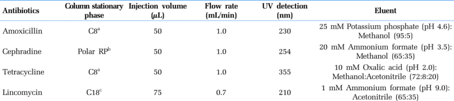
The concentrations of antibiotics in the aqueous samples were determined by HPLC, using an Agilent 1200 Series HPLC system (Agilent Technologies, Santa Clara, CA, USA) equipped with a UV absorbance detector operated at 230, 254, 355, and 210 nm for amoxicillin, cephradine, tetracycline, and lincomycin, respectively. The analytical methods used for each antibiotic are summarized in Table 1. Triplicate subsamples were prepared and analyzed for each sample.
To analyze the mass profile of the cephradine degradation prod-ucts generated by gamma irradiation, the assay was performed using an Agilent 1100 module (Agilent, Palo Alto, CA, USA) equip-ped with a Luna C18 column (150 mm × 2.0 mm, i.d.: 3 μm; Phenomenex, Torrance, CA, USA). The flow rate was set to 0.15 mL/min, and injection volume was 5 μL. A mixture of acetic acid (0.5% v/v) and methanol (42:58, v/v) was used as the mobile phase. All target compounds were eluted out of the column within 15 min. The auto-sampler temperature was operated at 10°C. Mass spectrometric measurements were carried out on a Sciex API 3000 triple-quadrupole tandem mass spectrometry (Applied Biosystems, Foster City, CA, USA) equipped with an electrospray ionization interface in positive mode for cephradine and byproducts. Ions were acquired in multiple reaction monitoring mode with a dwell time of 10 ms. The mass spectrometer conditions were as follows: ion spray voltage: 5.5 kV, curtain gas: 10 L/min, nebulizer gas: 5 L/min, Auxiliary gas: 6.1 L/min, heated capillary temperature: 300°C, interface heater: ON, and collision gas: 5.
2.4. Biological Assay
To measure the minimal inhibitory concentration (MIC) of the antibiotics, we used the antimicrobial susceptibility (AMS) test described by Jorgensen and Hindler [39]. Briefly, 120 μL of serially diluted antibiotic was dispensed into the wells of a 96-well micro-plate, and these dilutions were subsequently inoculated with 60 μL of the bacterial strains in Mueller-Hinton broth. The final number of Staphylococcus aureus or Escherichia coli in the reaction mixtures was approximately 1.0 × 10⁵ CFU/mL. After inoculation, the micro-plates were incubated in a shaking incubator for 18-20 h at 37°C. Bacterial growth was measured after the incubation using a microplate reader (ELx800; BioTek, Winooski, VT, USA) at a wavelength of 595 nm. The minimum concentration of antibiotic, at which over 95% of bacterial growth was inhibited, was considered the MIC.
The concentrations of residual antibiotics in the samples after gamma irradiation were estimated using the AMS test and MIC of the antibiotics. The samples were serially diluted and inoculated with the bacterial strains at a final concentration of 1.0 × 10⁵ CFU/mL. After measuring bacterial growth, antibiotic concentration was calculated by multiplying the MIC of the relevant antibiotic
by the dilution factor at which bacterial growth was inhibited. If the survival rates of two consecutively diluted samples decreased significantly, then the dilution range was subdivided and the growth of bacteria was re-measured to precisely estimate the inhibition range. All experiments were performed in triplicate.
3. Results and Discussion
3.1. Minimal Inhibitory Concentration of Antibiotics for the
Test Strains
The antibiotics used in this study were differentiated into two groups based on their mechanisms of action: β-lactam antibiotics, such as amoxicillin and cephradine, inhibit bacterial cell wall syn-thesis, whereas non β-lactam antibiotics, such as lincomycin and tetracycline, inhibit protein synthesis [40]. The inhibitory effects of the different concentrations of antibiotics on growth of the test strains are shown in Fig. 1. Among the antibiotics examined, tetracy-cline was the most effective at inhibiting the growth of both test organisms, and the MICs for E. coli and S. aureus were ≤ 0.625 Table 1. Details of the High-performance Liquid Chromatography Methods Used to Analyze the Antibiotics
Antibiotics Column stationary phase Injection volume(μL) Flow rate(mL/min) UV detection (nm) Eluent
Amoxicillin C8a 50 1.0 230 25 mM Potassium phosphate (pH 4.6):
Methanol (95:5)
Cephradine Polar RPb 50 1.0 254 20 mM Ammonium formate (pH 3.5):
Methanol (65:35)
Tetracycline C8a 50 1.0 355 10 mM Oxalic acid (pH 2.0):
Methanol:Acetonitrile (72:8:20)
Lincomycin C18c 75 0.7 210 1 mM Ammonium formate (pH 9.0):
Acetonitrile (65:35) aLuna 5 μ C8(2) 100A 150 × 4.6 mm (Phenomenex, Torrance, CA, USA)
bSynergi 4 μ Polar-RP column 150 × 4.6 mm (Phenomenex)
cZorbax SB-C18 250 × 3.0 mm (Agilent Technologies, Santa Clara, CA, USA)
a
b
Fig. 1. Growth curve of Escherichia coli KCTC 1682 and (a) Staphylococcus aureus KCTC 1621 at (b) Various concentrations of amoxicillin, cephradine, lincomycin, and tetracycline. Cell growth (%) was calculated as the OD595 nm value of the antibiotic sample was divided by the OD595 nm value of the blank (distilled water).
and ≤ 0.109 mg/L, respectively. The MICs determined using S. aureus were significantly lower for the other antibiotics (amoxicillin, cephradine, and lincomycin) than those determined using E. coli. The MIC value of amoxicillin, cephradine, and linco-mycin against S. aureus were ≤ 0.125, ≤ 1.75, and ≤ 0.109 mg/L, respectively. E. coli exhibited high resistance to amoxicillin and cephradine with MIC values ≤ 10.00 and ≤ 16.00 mg/L, respectively. The MIC value of lincomycin for E. coli was > 30 mg/L. These results indicate that S. aureus was more sensitive than E. coli to each of the antibiotics examined.
Hydrophilic antibiotics (amoxicillin, cephradine, and tetracy-cline) pass more easily through pore-forming porins compared to hydrophobic antibiotics (lincomycin), which diffuse across the lipid bilayer [41, 42]. Resistance to hydrophobic antibiotics in Gram-neg-ative bacteria may be either due to a decrease in penetration of the antibiotic through the outer membrane or due to specific mecha-nisms, such as a gene mutation or acquisition of resistance genes [41, 43]. Moreover, Gram-negative bacteria are generally more read-ily resistant to antibiotic compounds because their outer membrane protects the peptidoglycans [41, 42]. Most β-lactam antibiotics, including amoxicillin and cephradine, work by inhibiting cell wall
biosynthesis in bacteria and are mainly active against Gram-positive bacteria, such as S. aureus. Therefore, the results suggest that S. aureus is suitable for estimating the residual concentrations of these antibiotics.
3.2. Concentrations of Residual Antibiotics after Treatment
with Gamma Irradiation
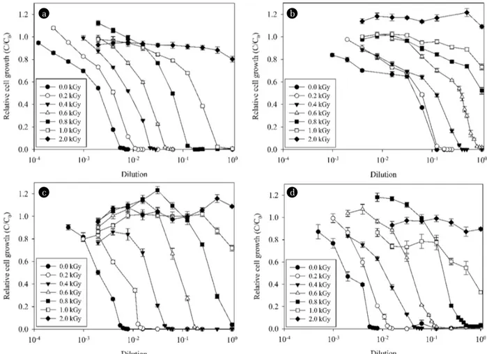
The residual concentrations of the antibiotics in the samples treated with gamma irradiation were measured using both biological and chemical methods. In the biological assay, the samples treated with up to 2.0 kGy of gamma irradiation were serially diluted, and the growth of S. aureus was observed (Fig. 2). Using the dilution factor at which growth of the strain was completely inhibited (≥ 95%) and the MIC of the relevant antibiotic for S. aureus, we calculated the residual bioactive concentrations.
Inactivation of the antibiotics was directly proportional to the strength of gamma irradiation, as observed previously [18, 19, 44]. The four antibiotics used in this study at an initial concentration of 30 mg/L were completely inactivated by the 2.0 kGy gamma irradiation treatment (Fig. S1). With the exception of cephradine,
a b
c d
Fig. 2. Survival rates of Staphylococcus aureus KCTC 1621 in samples of amoxicillin, (a) Cephradine, (b) Lincomycin, and (c) Tetracycline (d) Treated with 0-2 kGy of gamma irradiation. Relative cell growth (C/C0) was calculated as the OD595 nm value of the sample was divided by the OD595 nm value of the blank (distilled water).
the residual concentrations of antibiotics estimated using HPLC and the AMS test were not significantly different (Table 2). In the case of cephradine, the residual concentrations in the samples treated with 0.2-0.6 kGy gamma irradiation were significantly different between the two methods (paired t-test, p < 0.04), whereas the values for the samples treated with irradiation greater than 1.0 kGy were similar. The residual concentrations of cephradine treated with 0.2 kGy gamma irradiation were 23.56 ± 0.00 and 14.27 ± 0.02 mg/L as determined by the AMS test and HPLC assay, respectively (Table 2). The corresponding values for the samples treated with 0.6 kGy were 3.68 ± 0.00 and 1.49 ± 0.03 mg/L. This discrepancy between the two methods may be attributable to the partially decom-posed byproducts of cephradine generated by the gamma irradiation, and these byproducts are capable of inhibiting bacterial growth. These results suggest the potential risk of increased eco-toxicity from cephradine after exposure to gamma irradiation treatment.
The byproducts of cefazolin, cephapirin, cephalexin, and ceph-radine are toxic according to the Microtox test [32]. Hafkemeyer et al. [45] reported that a degradation product of ceftazidim, which is a third-generation cephalosporin antibiotic, exhibits an inhibitory effect against RNase H. The degradation product of cefazolin, a first-generation cephalosporin antibiotic, was identified as a pri-mary toxic byproduct using the zebrafish embryo toxicity test [46]. These cases provide indirect evidence that the degradation products (byproducts) of cephalosporin antibiotics are probably “bioactive” substances.
3.3. Decomposition of Cephradine by the Gamma Irradiation
Treatment
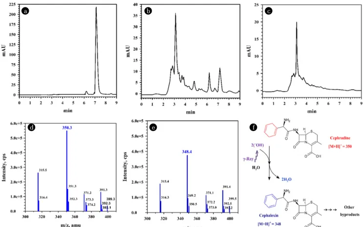
Two distinct peaks with retention times of 3.10 and 6.17 min were detected in the LC chromatograms of cephradine, which accounted for the very lower portion than the main peak (7.16 min) in the
chromatograms of untreated cephradine (Fig. 3). When the irradi-ation dose increased to 0.6 kGy, the peak area of 6.17 min increased to 72.4% of the area of 7.16 min and the peak with a retention time of 6.17 min disappeared above 1 kGy. Other studies have shown that 30 mg/L of antibiotics (e.g., amoxicillin, cefaclor, ceph-radine, tetracycline, lincomycin, and sulfamethazine) were com-pletely degraded and mineralized after 1 kGy gamma radiation [18, 19, 36]. These findings suggest that the peak at 6.17 min was a “bioactive” substance and a microbial activity inhibitor (Fig. 3B and Table 2). In contrast, the area of the peak at 3.10 min continually increased with increasing strength of gamma rays (Fig. 3C). These results indicate that the compound showing a peak at 3.10 min was a degradation product of gamma irradiation and was not an inhibitor of bacterial growth (Table 2). The gamma radiolysis of water generates some active species as given Eq. (1) [47].
H2O → ˙OH(2.7) + eaq-(2.6) + H˙(0.55) +
H2(0.45) + H2O2(0.71) + H3O+(2.6) (1) The values in parenthesis are the average radiation chemical yield (G-value), which was defined as the number of product mole-cules formed per 100 eV absorbed at pH 6.0-8.5 [47]. Hydroxyl radicals (˙OH) and hydrated electrons (eaq-) are two main reactive species. It is known that ˙OH radical is powerful oxidants, non-selective and highly reactive with organic matter, while eaq- is a strong reducing agent. The gamma irradiation may degrade antibiotics through oxidation and reduction pathway [47]. As shown in Fig. 3F and Table 2, absorbed gamma irradiation doses in the range of 0.2-0.6 kGy caused a dose-dependent degradation of the antibiotic and a decrease in cephradine (7.16 min, [M+H]+ = 350) content due to radiation conversion to byproduct (6.17 min, [M+H]+ = 348). Wang and Lin [32] reported that cephradine ([M+H]+ = 350) Table 2. The Residual Concentrations of Antibiotics in the Samples Treated with Gamma Irradiation, Estimated Using HPLC and the AMS Test
Antibiotics (mg/L) Gamma
irradiation (kGy)
Amoxicillin Cephradine Lincomycin Tetracycline
HPLC AMSa HPLC AMS HPLC AMS HPLC AMS
0.0 (± 0.03)31.16 b (± 0.00)27.94 (± 0.02)29.72 (± 0.00)23.56 (± 0.90)31.63 (± 0.00)33.43 (± 0.51)28.10 (± 0.00)31.07 0.2 (± 0.15)15.11 (± 0.00)13.97 (± 0.23)14.27 (± 0.00)23.56 (± 0.03)12.61 (± 0.00)14.63 (± 0.04)11.46 (± 0.00)13.59 0.4 (± 0.04)5.87 (± 0.00)6.98 (± 0.01)4.74 (± 0.00)8.41 (± 0.03)4.02 (± 0.25)4.18 (± 0.04)3.36 (± 0.23)3.65 0.6 (± 0.01)2.71 (± 0.00)3.49 (± 0.03)1.49 (± 0.00)3.68 (± 0.05)1.83 (± 0.03)1.01 (± 0.01)1.12 (± 0.05)1.56 0.8 0.76 (± 0.03) 1.22 (± 0.00) 0.13 (± 0.00) < 2.94 0.74 (± 0.01) 0.18 (± 0.00) 0.21 (± 0.00) 0.35 (± 0.01) 1.0 (± 0.01)0.17 (± 0.00)0.19 ND < 2.94 ND < 0.18 (± 0.00)0.03 < 0.17 2.0 NDc < 0.15 ND < 2.94 ND < 0.18 ND < 0.17
aThe residual concentrations measured by AMS test were estimated by the toxicity of residual parent antibiotics and byproducts. bNumbers in parentheses indicate the standard deviation of three replicates.
reacted immediately with ˙OH radicals and were transformed cepha-lexin ([M+H]+ = 348). Therefore, the gamma radiolysis route results in the formation of cephalexin and later formed byproducts [48, 49]. Moreover, both cephradine and cephalexin are β-lactam anti-biotics and within the class of first-generation cephalosporins. The structurally similar cephradine and cephalexin exhibited similar levels of toxicity after irradiation. López Peñalver et al. [50] evaluated toxicity during the irradiation-induced degradation of tetracycline in an aquatic environment using the Vibrio fischeri inhibition test. Similar to our results, they revealed that toxicity increased at 0.1-0.4 kGy possibly due to the production of more toxic byproducts, which decreased when the dose was increased to 1.0 kGy. Our studies also demonstrate that the four antibiotics can be decomposed by 2.0 kGy gamma irradiation without toxic effect of their byproducts.
Although degradation of the original drug is readily achieved in wastewater treatment systems (e.g., chlorination, ozonation, and AOPs), the byproducts generated can be less biodegradable, have similar biological activity, and/or more be toxic than the parent compound [12, 25, 32]. For example, Dantas et al. [51] observed a slight increase in acute toxicity during the first-stage of ozonation of sulfamethoxazole using the Microtox test. Alsager et al. [12] also tested the biological activity of ozone-treated antibiotics with a well-established E. coli test. The synergistic effect of toxicity
caused by intermediate products or byproducts of antibiotics cannot be ignored and warrants future research [32, 47]. Wang et al. [52] reported that the toxicity of intermediate products or byproducts of antibiotics after ionizing irradiation (gamma ray and electron beam) are significant when evaluating the potential dangers to human and ecological systems. They also suggested that more stud-ies should be conducted to explore bio-toxicity.
4. Conclusions
Although the measurement sensitivities of our two assay methods were almost identical, the AMS biological assay detected toxic antibiotic derivatives that were not detected by HPLC. The results presented in this study suggest that the AMS biological assay may be more useful than the HPLC chemical assay for measuring bio-active residual antibiotics in environmental samples. Therefore, we suggest that the chemical assay should be used in parallel with the bioassay for measuring antibiotics and their byproducts. It is also necessary to use a sufficiently strong irradiation dose to ensure that potentially toxic byproducts are completely degraded and do not affect the ecosystem when using gamma irradiation to treat environmental samples containing antibiotics.
a b c
d e f
Fig. 3. Chromatograms of the liquid chromatography analysis of untreated cephradine (a) and of cephradine treated with 0.6 kGy (b) and 1.0 kGy (c) gamma irradiation. Mass chromatograms of cephradine ((d), Retention time of 7.16 min in (a) and (b)) and byproduct ((e), Retention time of 6.17 min in (b)). Predicted gamma radiolysis products and degradation pathway of cephradine (f). The byproducts were detected in positive mode as [M+H]+.
Acknowledgments
This work was supported by Nuclear Research & Development Program through an NRF grant funded by the Ministry of Science and ICT, Korea.
References
1. Lanzky PF, Halting-Sørensen B. The toxic effect of the antibiotic metronidazole on aquatic organisms. Chemosphere 1997;35: 2553-2561.
2. Glassmeyer ST, Furlong ET, Kolpin DW, et al. Transport of chemical and microbial compounds from known wastewater discharges: Potential for use as indicators of human fecal contamination. Environ. Sci. Technol. 2005;39:5157-5169. 3. Golet EM, Alder AC, Giger W. Environmental exposure and
risk assessment of fluoroquinolone antibacterial agents in waste-water and river waste-water of the Glatt Valley Watershed, Switzerland. Environ. Sci. Technol. 2002;36:3645-3651.
4. Metcalfe CD, Miao XS, Koenig BG, Struger J. Distribution of acidic and neutral drugs in surface waters near sewage treatment plants in the lower Great Lakes, Canada. Environ. Toxicol. Chem. 2003;22:2881-2889.
5. Petrović M, Gonzalez S, Barceló D. Analysis and removal of emerging contaminants in wastewater and drinking water. TrAC Trend. Anal. Chem. 2003;22:685-696.
6. Rizzo L, Fiorentino A, Anselmo A. Advanced treatment of urban wastewater by UV radiation: effect on antibiotics and anti-biotic-resistant E. coli strains. Chemosphere 2013;92:171-176. 7. Schwartz T, Kohnen W, Jansen B, Obst U. Detection of anti-biotic-resistant bacteria and their resistance genes in waste-water, surface waste-water, and drinking water biofilms. FEMS Microbiol. Ecol. 2003;43:325-335.
8. Volkmann H, Schwartz T, Bischoff P, Kirchen S, Obst U. Detection of clinically relevant antibiotic-resistance genes in municipal wastewater using real-time PCR (TaqMan). J. Microbiol. Meth. 2004;56:277-286.
9. Alexy R, Kümpel T, Kümmerer K. Assessment of degradation of 18 antibiotics in the closed bottles test. Chemosphere 2004;57:505-512.
10. Kümmerer K, Al-Ahmad A, Mersch-Sundermann V. Biodegradability of some antibiotics, elimination of the genotox-icity and affection of wastewater bacteria in a simple test. Chemosphere 2000;40:701-710.
11. Zhou P, Su C, Li B, Qian Y. Treatment of high-strength pharma-ceutical wastewater and removal of antibiotics in anaerobic and aerobic biological treatment processes. J. Environ. Eng. 2006;132:129-136.
12. Alsager OA, Alnajrani MN, Abuelizz HA, Aldaghmani IA. Removal of antibiotics from water and waste milk by ozonation: Kinetics, byproducts, and antimicrobial activity. Ecotoxicol. Environ. Saf. 2018;158:114-122.
13. Balcıoğlu IA, Ötker M. Treatment of pharmaceutical wastewater containing antibiotics by O3 and O3/H2O2 processes. Chemosphere 2003;50:85-95.
14. Batt AL, Kim S, Aga DS. Comparison of the occurrence of
anti-biotics in four full-scale wastewater treatment plants with vary-ing designs and operations. Chemosphere 2007;68:428-435. 15. Homen V, Santos L. Degradation and removal methods of
anti-biotics from aqueous matrices-A review. J. Environ. Manage. 2011;92:2304-2347.
16. Liu J, Sun Q, Zhang C, et al. Removal of typical antibiotics in the advanced treatment process of productive drinking water. Desalin. Water Treat. 2016;57:11386-11391.
17. Sayed M, Khan JA, Shah LA, et al. Degradation of quinolone antibiotic, norfloxacin, in aqueous solution using gamma-ray irradiation. Environ. Sci. Pollut. Res. 2016;23:13155-13168. 18. Yu S, Choi D, Lee M. Kinetic and modeling of radiolytic decom-position of antibiotics. WIT Trans. Ecol. Environ. 2008;109:39-47. 19. Yu S, Lee B, Lee M, Cho IH, Chang SW. Decomposition and mineralization of cefaclor by ionizing radiation: Kinetics and effects of the radical scavengers. Chemosphere 2008;71:2106-2112. 20. Andreozzi R, Canterino M, Marotta R, Paxeus N. Antibiotic
removal from wastewaters: the ozonation of amoxicillin. J. Hazard. Mater. 2005;22:243-250.
21. Andreozzi R, Canterino M, Giudice RL, Marotta R, Pinto G, Pollio A. Lincomycin solar photodegradation, algal toxicity and removal from wastewaters by means of ozonation. Water Res. 2006;40:630-638.
22. Deegan AM, Shaik B, Nolan K, et al. Treatment options for wastewater effluents from pharmaceutical companies. Int. J. Environ. Sci. Technol. 2011;8:649-666.
23. Addamo M, Augugliaro V, Di Paola A, et al. Removal of drugs in aqueous systems by photoassisted degradation. J. Appl. Electrochem. 2005;35:765-774.
24. Abellán MN, Bayarri B, Giménez, J, Costa J. Photocatalytic degra-dation of sulfamethoxazole in aqueous suspension of TiO2. Appl. Catal. B: Environ. 2007;74:233-241.
25. Radjenović J, Petrović M, Barceló D. Complementary mass spec-trometry and bioassays for evaluating pharmaceutical-trans-formation products in treatment of drinking water and waste-water TrAC Trend. Anal. Chem. 2009;28:562-580.
26. Ternes TA, Stüber J, Herrmann N, et al. Ozonation: A tool for removal of pharmaceuticals, contrast media and musk fra-grances from wastewater? Water Res. 2003;37:1976-1982. 27. Vogna D, Marotta R, Napolitano A, Andreozzi R, d’Ischia M.
Advanced oxidation of the pharmaceutical drug diclofenac with UV/H2O2 and ozone. Water Res. 2004;38:414-422.
28. Baumgarten S, Schröder HF, Charwath C, Lange M, Beier S, Pinnekamp J. Evaluation of advanced treatment technologies for the elimination of pharmaceutical compounds. Water Sci. Technol. 2007;56:1-8.
29. Ben W, Qiang Z, Adams C, Zhang H, Chen L. Simultaneous determination of sulfonamides, tetracyclines and tiamulin in swine wastewater by solid-phase extraction and liquid chroma-tography-mass spectrometry. J. Chromatogr. A 2008;1202:173-180. 30. Sirtori C, Zapata A, Oller I, Gernjak W, Agüera A, Malato S. Decontamination industrial pharmaceutical wastewater by combining solar photo-Fenton and biological treatment. Water Res. 2009;43:661-668.
31. Joos B, Ledergerber B, Flepp M, Bettex JD, Lüthy R, Siegenthaler W. Comparison of high-pressure liquid chromatography and bioassay for determination of ciprofloxacin in serum and urine.
Antimicrob. Agents Chemother. 1985;27:353-356.
32. Wang XH, Lin AYC. Phototransformation of cephalosporin anti-biotics in an aqueous environment results in higher toxicity. Environ. Sci. Technol. 2012;46:12417-12426.
33. Li K, Yediler A, Yang M, Schulte-Hostede S, Wong MH. Ozonation of oxytetracycline and toxicological assessment of its oxidation by-products. Chemosphere 2008;72:473-478 34. Chanda PK, Ganguly T, Das M, Lee CY, Luong TT, Sau S.
Detection of antistaphylococcal and toxic compounds by bio-logical assay systems developed with a reporter Staphylococcus aureus strain harboring a heat inducible promoter lacZ tran-scriptional fusion. BMB Rep. 2007;40:936-943.
35. Thompson SA, Maani EV, Lindell AH, King CJ, McArthur JV. Novel tetracycline resistance determinant isolated from an envi-ronmental strain of Serratia marcescens. Appl. Environ. Microbiol. 2007;73:2199-2206.
36. Kim HY, Yu S, Lee MJ, Kim TH, Kim SD. Radiolysis of selected antibiotics and their toxic effects on various aquatic organisms. Radiat. Phys. Chem. 2009;78:267-272.
37. Jenkins RD, Stevens SL, Craythorn JM, Thomas TW, Guinan ME, Matsen JM. False susceptibility of enterococci to amino-glycosides with blood-enriched Mueller-Hinton agar for disk susceptibility testing. J. Clin. Microbiol. 1985;22:369-374. 38. ASTM. Standards on dosimetry for radiation processing:
ISO/ASTM 51607:2004. ASTM International. 2004. West Conshohocken, Pennsylvania.
39. J orgensen JH, Hindler JF, Reller LB, Weinstein MP. New con-sensus guidelines from the Clinical and Laboratory Standards Institute for antimicrobial susceptibility testing of infrequently isolated or fastidious bacteria. Clin. Infect. Dis. 2007;44:280-286. 40. Walsh C. Antibiotics: Actions, origins, resistance. Washington
D.C.: ASM Press; 2003.
41. Delcour AH. Outer membrane permeability and antibiotic resistance. Bioch. Biophys. Acta-Proteins Proteom. 2009;1794: 808-816.
42. Shokier HA, EI-Adly AA, Hussein H, Shabon MH,
EI-Shanshoury IH. Effect of gamma rays on antibiotic resistance of Staphylococcus aureus and Pseudomonas aeruginosa isolated from human skin. J. Radiat. Res. Appl. Sci. 2010;3:619-637. 43. Ofek I, Cohen S, Rahmani R, et al. Antibacterial synergism
of polymyxin B nonapeptide and hydrophobic antibiotics in experimental gram-negative infections in mice. Antimicrob. Agents Chemother. 1994;38:374-377.
44. Kang SH, Chang JG, Ka SK, Kim HY, Kim SD, Lee MJ. A study on the aquatic eco-risk assessment of antibiotics treated by radiation. J. Korean Soc. Water Wast. 2012;26:373-381. 45. Hafkemeyer P, Neftel K, Hobi R, et al. HP 0.35, a cephalosporin
degradation product is a specific inhibitor of lentiviral RNAses H. Nucleic Acids Res. 1991;19:4059-4065.
46. Zhang J, Meng J, Li Y, Hu C. Investigation of the toxic functional group of cephalosporins by zebrafish embryo toxicity test. Arch. Pharm. 2010;343:553-560.
47. Wang J, Chu L. Irradiation treatment of pharmaceutical and personal care products (PPCPs) in water and wastewater: an overview. Radiat. Phys. Chem. 2016;125:56-64.
48. Abuirjeie MA, Abdel-Aziz AA, Abdel-Hamid ME. Feasibility studies on radiation sterilization of cephradine. Drug Dev. Ind. Pharm. 1990;16:1661-1673.
49. Signoretti EC, Onori S, Valvo L, et al. Ionizing radiation induced effects on cephradine. Influence of sample moisture content, irradiation dose and storage conditions. Drug Dev. Ind. Pharm. 1993;19:1693-1708.
50. López Peñalver JJ, Gómez Pacheco CV, Sánchez Polo M, Rivera Utrilla J. Degradation of tetracyclines in different water matrices by advanced oxidation/reduction processes based on gamma radiation. J. Chem. Technol. Biot. 2013;88:1096-1108. 51. Dantas RF, Contreras S, Sans C, Esplugas S. Sulfamethoxazole
abatement by means of ozonation. J. Hazard. Mater. 2008;150: 790-794.
52. Wang J, Zhuan R, Chu L. The occurrence, distribution and degradation of antibiotics by ionizing radiation: An overview. Sci. Total Environ. 2019;646:1385-1397.