An Unusual Occurrence of Epidermoid Cyst in the Inner Mucosa of the Upper Lip:
A Case Report
Jihye Lee, Namki Choi, Seonmi Kim
Department of Pediatric Dentistry, School of Dentistry, Chonnam National University
Epidermoid cysts are rare benign lesions in the oral cavity that may be either congenital or acquired. The cysts are usually slow-growing and asymptomatic until becoming secondarily infected or large enough to interfere with mastication and speech. Consequently, diagnosis is often delayed. The condition is also uncommon in newborns and infants. Most of the lesions occur in the floor of the mouth and rarely in the upper lip. This report describes the case of a 29-month-old girl with a palpable mass in the inner mucosa of the upper lip. The lesion was surgically enucleated using an intraoral approach and histopathologically diagnosed as epidermoid cyst.
Key words : Epidermoid cyst, Upper lip, Surgical enucleation
Abstract
Corresponding author : Seonmi Kim
Department of Pediatric Dentistry, School of Dentistry, Chonnam National University, 33 Yongbong-ro, Buk-gu, Gwangju, 61186, Korea Tel: +82-62-530-5660 / Fax: +82-62-530-5669 / E-mail: gracekim@jnu.ac.kr
Received March 3, 2020 / Revised March 31, 2020 / Accepted March 28, 2020
Ⅰ. Introduction
Epidermoid cysts are common benign skin lesions that con-sist of epithelial-lined cavities, which are filled with viscous or semisolid epithelial degradation substances[1]. Such cysts can develop anywhere in the body. The incidence in the head and neck region ranges from 1.6 - 6.9%. Oral forms account for less than 0.01%[2]. The floor of the mouth is the most com-monly involved site in the oral cavity[3]. Rare cases have been reported in the tongue, buccal mucosa, parotid gland, and lips[2,4].
Epidermoid cysts are usually benign, slow-growing, and painless. Most of the lesions are asymptomatic. As the lesions grow beyond, some symptoms such as difficulty in breathing and swallowing can appear[3]. These masses generally occur
in young adults, with pediatric presentation being less com-mon[2].
In this case report, an epidermoid cyst located in the inner mucosa of the upper lip of a pediatric patient was detected and surgically treated. As reported in oral and maxillofacial surgery literature, such epidermoid cysts are not frequently diagnosed by pediatric dentists. Therefore, this is a rare case report of an epidermoid cyst in the upper lip from a pediatric dentistry department.
Ⅱ. Case Report
A 29-month-old girl visited the Department of Pediatric Dentistry, Chonnam National University Dental Hospital, with a mass in the inner mucosa of the upper lip near the labial
fren- ulum. The lesion was first detected at an infant oral examina-tion 2 weeks earlier. There was no history of previous surgery or trauma associated with the site of the lesion.
Intraoral examination revealed a well-demarcated, solitary swelling in the inner mucosa of the upper lip measuring 12.0 X 12.0 mm in its greatest dimensions. The overlying mucosa was normal in color and texture without any signs of inflammation (Fig. 1). The swelling was firm, non-tender, movable, smooth and not associated with discharge on palpation. On physical examination, no other swelling was noted elsewhere in the body. There was no associated lymphadenopathy.
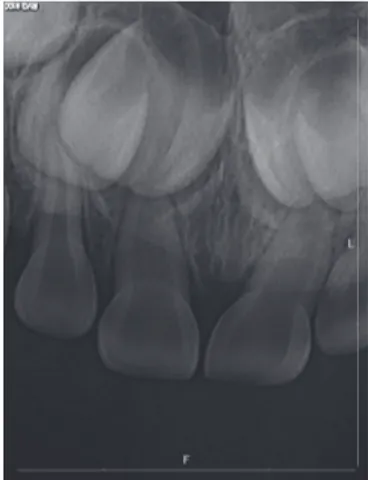
Also, no unusual findings were detected on periapical radio- graphs (Fig. 2). Additional imaging techniques such as com-puted tomography (CT) or magnetic resonance imaging (MRI) were not performed as the patient was young, and the parents
refused to consent. Based on the clinical examination, a provi-sional diagnosis of lipoma or fibroma was made.
An excisional biopsy under oral sedation was planned due to the patient’s cooperation. Under local anesthesia, a care-ful dissection was performed to enucleate the cyst (Fig. 3A). Underlying the inner mucosa of the upper lip, it was a well-encapsulated mass with yellow lesions measuring 12.0 X 12.0 X 18.0 mm. It could be separated from the surrounding tissues. Yellowish material was also present inside the capsule (Fig. 3B). The specimen was sent for histopathological examination. The area was then irrigated and primary closure was followed.
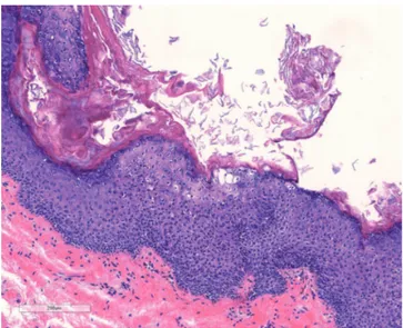
Histopathological examination showed a cystic cavity lined with stratified squamous epithelium and filled with lamellated keratin (Fig. 4). No skin appendages were identified. Histologi-cal analysis confirmed the diagnosis of epidermoid cyst.
Fig. 1. Intraoral view showing the swelling of the inner
mu-cosa of the upper lip.
Fig. 2. Periapical radiography shows
normal characteristics of the structures.
The patient was followed-up for 6 months, with no signs of recurrence. There were no scars or adhesions in the labial frenulum (Fig. 5). The movement of the lip was normal, imply-ing that the function of the orbicularis oris muscle was not impaired.
Ⅲ. Discussion
Histologically, dermoid cysts have been classified into epi-dermoid, Histologically, dermoid cysts have been classified into epi-dermoid, and teratoid cysts. Epidermoid cysts are lined with epithelium and derived from epidermal and
con-nective tissue. Dermoid cysts have cavities lined with a similar epithelium and containing structures such as hair follicles, se-baceous and sweat glands in the underlying connective tissue. Teratoid cysts have cavities lined by epithelium and containing derivatives of the endoderm and mesoderm, such as muscle, intestinal mucosa, respiratory mucosa, bone, blood vessels, and appendages. Overall, epidermoid cysts are the most fre-quently found type[4,5]
The etiology of epidermoid cyst is unclear. A suggested theory is that oral epidermoid cyst can be congenital or ac-quired[6,7]. The congenital type may result from the failure of the surface ectoderm to separate from the underlying neural tube, or the invagination of the surface ectoderm along the embryologic fusion sites[6]. The acquired type is caused by trauma, with implantation of the surface epithelial element into the deeper tissue[6]. There are no clinical or histopathologi-cal differences between the congenital and acquired types[7]. In this case, the lesion developed in the upper lip. Since the lips are vulnerable to traumatic injuries, the lesion could be acquired. On the other hand, the cysts could be congenital as the patient was an infant with no history of trauma or surgery. It was difficult to determine whether the epidermoid cyst was congenital or acquired.
Epidermoid cysts have the typical characteristics of benign lesions. This case also presented with the same clinical find-ings. Depending on the size and location of the mass, the cysts could present with obstructive symptoms such as dys-pnea, dysphonia, dysphagia[3]. Moreover, the cyst wall may rupture[8] or get secondarily infected[9]. Normally, secondary infection is unusual, but it can cause life-threatening situa-tions when it occurs[9]. In rare cases, the formation of a sinus tract from the oral cavity to the skin and muscle fusion defects have been reported[10]. There are studies of malignant trans-formation in long-standing, untreated epidermoid cysts[11,12]. Epidermoid cysts are usually solitary lesions, as in this case. However, multiple masses can be occur in the floor of the mouth[13].
There have been a limited number of cases of epidermoid cysts found in the upper lip. The first case was diagnosed by Kuroyanagi et al.[14] in 1973. Koh[15] reported the case of a cyst with a sinus tract extension to the skin. Kim and Hong[16] described a giant epidermoid cyst in a patient wearing a den-ture. Dogan and Bucak[10], Phukan et al.[17], Mahalakshmi et al.[18], and recently, Chen et al .[19] have also reported epider-moid cysts in the upper lip. All 7 cases showed characteristic Fig. 4. Histopathological section showing a cyst lined by
keratinized squamous epithelium, cavity filled with lamel-lated keratin.
histological findings of typical epidermoid cysts similar to the present case.
It is necessary to make a differential diagnosis with other lesions. In this case, infection was ruled out because of the absence of pain, signs of inflammation, and focus of infection. A neoplastic hypothesis was excluded as the clinical findings were not suggestive of malignancy.
The lip contains adipose tissue, connective tissue, blood vessels, nerves, and salivary glands. Any lesion that originates from these components may occur in the lip. The most com-mon clinical differential diagnosis of a cystic lip mass is muco-cele. Considering characteristics of the lesion based on palpa-tion and appearance, the cyst was different from mucocele[20]. Other clinical differential diagnoses of lip masses include mucous retention cyst, sialolith, fibroma, and lipoma[21-23]. Mucous retention cysts usually occur in older adults. Sialo-liths may also present as masses in the upper lip. However, sialoliths are usually not cystic[21]. Fibromas and lipomas are benign lesions with similar clinical features to the lesion. Fibro-mas are the most frequently found soft tissue lesions in the oral cavity and the lips[22]. Lipomas are benign soft tissue tu-mors composed of adipose tissue with a slightly yellowish, soft and fluctuant swelling[23]. In this case, a provisional diagnosis of fibroma or lipoma was made, as the lesion was soft, mov-able, and fluctuant with a cystic consistency.
Histopathological examination of the cyst is required to con-firm the diagnosis. Histologically, epidermoid cysts are lined with stratified squamous epithelium and usually filled with cheese-like material or keratin[24]. Although a diagnosis can be confirmed by histopathological examination, imaging tech-niques such as CT or MRI are used as important tools. They precisely delineate the location of the lesions and their relation with adjacent structures[24].
In this case, excisional biopsy was performed under the provisional diagnosis that the lesion was benign. However, it was hard to determine the depth of the lesion because there was no 3D image evaluation during the surgical procedure. The upper lip is the boundary between the oral cavity and the skin. As the lesion can also penetrate the tissues, it would be important to understand the depth of the lesion through preoperative image evaluation. In addition, it is significant to predict possible complications through 3D image of the cysts and obtain patient consent in advance. It would be helpful to assess whether the anatomical structure was damaged by the location or growth of the lesion prior to surgery.
The treatment of choice is surgical excision, using an intra-oral or extraintra-oral approach depending on the location of the lesion[4]. In this case, surgical excision was performed using an intraoral approach by placing a horizontal incision over the lesion. Care had to be taken to avoid postoperative inflamma-tion or recurrence.
Prognosis is good, and a recurrence rate of < 3% has been reported[25]. A 5% rate of malignant transformation of the teratoid variety has been reported[11,12]. In this case, the in-traoral wound healed without complications. No signs of infec-tion or recurrence were noted during the 6 months follow up.
Ⅳ. Summary
An exceptionally atypical case of epidermoid cyst occurring in the inner mucosa of the upper lip was treated surgically in this study. Although epidermoid cysts are rarely encountered in the oral cavity, there is a possibility of occurrence of the lesions. It requires that clinicians are aware of the lesion and consider it in their differential diagnosis. Pediatric dentists are often the first to recognize oral pathologies. Therefore, a care-ful evaluation of soft tissue as well as hard tissue should be performed during oral examinations.
Authors’ Information
Jihye Lee http://orcid.org/0000-0003-4607-9992 Namki Choi http://orcid.org/0000-0003-4830-8568 Seonmi Kim http://orcid.org/0000-0001-5103-767XReferences
1. Dutt SN, Hock YL, East DM, et al. : Epidermoid cyst of the submandibular gland. Indian J Otolaryngol, 52:378-379, 2000.
2. New GB, Erich JB : Dermoid cysts of the head and neck. Surg Gynecol Obstet, 65:48-55, 1937.
3. De Ponte FS, Brunelli A, Marchetti E, Bottini DJ : Sublingual epidermoid cyst. J Craniofac Surg, 13:308-310, 2002. 4. Ozan F, Polat HB, Ay S, Goze F : Epidermoid cyst of the
buccal mucosa: a case report. J Contemp Dent Pract , 8:90-96, 2007.
5. Jham BC, Duraes GV, Jham AC, Santos CR : Epidermoid cyst of the floor of the mouth: a case report. J Can Dent Assoc, 73:525-528, 2007.
6. Smirniotopoulos JG, Chiechi MV : Teratomas, dermoids, and epidermoids of the head and neck. Radiographics, 15:1437-1455, 1995.
7. Longo F, Maremonti P, Califano L, et al. : Midline dermoid cysts of the floor of the mouth: report of 16 cases and re-view of surgical techniques. Plast Reconstr Surg , 112:1560-1565, 2003.
8. Suh KS, Kang DY, Jang MS, et al. : Usefulness of dermosco-py in the differential diagnosis of ruptured and unruptured epidermal cyst. Ann Dermatol, 29:33-38, 2017.
9. Cortezzi W, De Albuquerque EB : Secondarily infected epidermoid cyst in the floor of the mouth causing a life-threatening situation: report of a case. J Oral Maxillofac Surg, 52:762-764, 1994.
10. Dogan F, Bucak IH : Congenital epidermoid cyst results in muscle fusion defect in the upperlip. Case Rep Otolaryn-gol, 2014:1-3, 2014.
11. Ikeda I, Ono T : Basal cell carcinoma originating from an epidermoid cyst. J Dermatol, 17:643-646, 1990.
12. Lopez-Rios F, Rodriguez-Peralto JL, Castano E, Benito A : Squamous cell carcinoma arising in a cutaneous epidermal cyst: case report and literature review. Am J Dermatopa-thol, 21:174-177, 1999.
13. Calderon S, Kaplan I : Concomitant sublingual and sub-mental epidermoid cysts: a case report. J Oral Maxillofac Surg, 51:790-792, 1993.
14. Kuroyanagi K, Kawabata T, Tooi M : Epidermoic cyst of up-per lip: report of a case. Bull Tokyo Dent Coll, 14:95-98, 1973.
15. Koh SW : Midline dermoid cyst of the upper lip: case re-port. J Korean Assoc Oral Maxillofac Surg, 37:403-405, 2011.
16. Kim JC, Hong IP : Rare giant upper lip epidermal cyst in a patient wearing a denture. Arch Craniofac Surg, 17:222-224, 2016.
17. Phukan JP, Sinha A, Pal S, Jalan S : Cytodiagnosis of epider-moid cyst of the upper lip: A common lesion in an uncom-mon site. J Lab Physicians, 6:60-62, 2014.
18. Mahalakshmi S, Reddy S, Ramamurthy TK, Shilpa B : Rare locations of epidermoid cyst: case reports and review. Ethiop J Health Sci, 26:595-601, 2016.
19. Chen B, Lu H, Gao Z, et al. : Excision of sebaceous cyst by intraoral approach a case report. Medicine, 96:8803-8806, 2017.
20. Mustapha IZ, Boucree SA : Mucocele of the upper lip: case
report of an uncommon presentation and its differential diagnosis. J Can Dent Assoc, 70:318-321, 2004.
21. Daley TD : The canalicular adenoma: considerations on dif-ferential diagnosis and treatment. J Oral Maxillofac Surg, 42:728-730, 1984.
22. Oda D : Soft tissue lesions in children. Oral Maxillofacial Surg Clin N Am, 17:383-402, 2005.
23. Morais HH, Vajgel A, Vasconcellos RJ, et al. : Congenital li-poma of the lip: a case report. J Oral Sci, 51:489-491, 2009. 24. Mirza S, Fadi S, Napaki S, Abualruz A : Case report of com- plicated epidermoid cyst of the floor of the mouth: Radiol-ogy-hystopathology correlation. Qatar Med J, 2014:12-16, 2014.
25. MacNeil SD, Moxham JP : Review of floor of mouth dys-ontogenic cysts. Ann Otol Rhinol Laryngol, 119:165-173, 2010.
국문초록