저작자표시-비영리-변경금지 2.0 대한민국 이용자는 아래의 조건을 따르는 경우에 한하여 자유롭게 l 이 저작물을 복제, 배포, 전송, 전시, 공연 및 방송할 수 있습니다. 다음과 같은 조건을 따라야 합니다: l 귀하는, 이 저작물의 재이용이나 배포의 경우, 이 저작물에 적용된 이용허락조건 을 명확하게 나타내어야 합니다. l 저작권자로부터 별도의 허가를 받으면 이러한 조건들은 적용되지 않습니다. 저작권법에 따른 이용자의 권리는 위의 내용에 의하여 영향을 받지 않습니다. 이것은 이용허락규약(Legal Code)을 이해하기 쉽게 요약한 것입니다. Disclaimer 저작자표시. 귀하는 원저작자를 표시하여야 합니다. 비영리. 귀하는 이 저작물을 영리 목적으로 이용할 수 없습니다. 변경금지. 귀하는 이 저작물을 개작, 변형 또는 가공할 수 없습니다.
Do balloons larger than 15 mm in size increase the risk of
adverse events in endoscopic large balloon dilation?
by
Gil Ho Lee
Major in Medicine
Department of Medical Sciences
The Graduate School, Ajou University
Do balloons larger than 15 mm in size increase the risk of
adverse events in endoscopic large balloon dilation?
by
Gil Ho Lee
A Dissertation Submitted to The Graduate School of
Ajou University in Partial Fulfillment of the Requirements for
the Degree of
Master of Medicine
Supervised by
Jin Hong Kim, M.D., Ph.D.
Major in Medicine
Department of Medical Sciences
The Graduate School, Ajou University
This certifies that the dissertation
of
Gil Ho Lee
is approved.
SUPERVISORY COMMITTEE
Jin Hong Kim
Byung Moo Yoo
Ki Myung Lee
The Graduate School, Ajou University
June, 20th, 2014
i
- Abstract -
Do balloons larger than 15 mm in size increase the risk of adverse
events in endoscopic large balloon dilation?
Endoscopic large balloon dilation (EPLBD) using large-diameter balloons (12-20 mm) was introduced to facilitate the removal of large bile duct stones and minimize the need for endoscopic mechanical lithotripsy (EML). Much recent literature has suggested EPLBD is safe and effective for extraction of large or difficult stones without any additional risk of life-threatening pancreatitis or perforation. Nevertheless, limited data exist on the maximal balloon size that would minimize fatal adverse events associated with EPLBD. In the current study, we aimed to assess the safety profiles of EPLBD according to balloon size and to identify the proper maximal size of a large balloon for treating large bile duct stones. A total of 279 patients who underwent EPLBD were included in the present study. There were 114 patients in the EPLBD with endoscopic biliary sphincterotomy (EST) group and 165 patients in the EPLBD without EST group. In the EPLBD with EST group, there were 49 patients in the EPLBD with a larger balloon (>15 mm) group and 65 patients in the EPLBD with a smaller balloon (12-15 mm) group. Although no significant difference was found between the larger and smaller balloon groups in terms of adverse events, there was a trend toward the larger balloon group having a higher rate of severe to fatal adverse events. In the EPLBD without EST group, there were 36 patients in the EPLBD with a larger balloon (>15 mm) group and 129 patients in the EPLBD with a smaller balloon (12-15 mm) group. The safety
variables did not differ significantly between the two groups, and no severe to fatal adverse event occurred in either group. In conclusion, our study shows that EPLBD with a larger balloon (>15 mm) tends to have more risk of severe to fatal adverse events compared with a smaller balloon (12-15 mm) for removing large bile duct stones. Large multicenter trials will be needed to reveal the statistical relationships between adverse events and balloon size.
__________________________________________________________________________
iii
TABLE OF CONTENTS
ABSTRACT ··· i
TABLE OF CONTENTS ··· iii
LIST OF FIGURES ··· iv LIST OF TABLES ··· v Ⅰ. INTRODUCTION ··· 1 Ⅱ. METHODS ··· 3 A. Patients ··· 3 B. Endoscopic procedures ··· 3 C. Measurements ··· 4 D. Statistical analyses ··· 5 Ⅲ. RESULTS ··· 6
A. EPLBD with EST group ··· 6
B. EPLBD without EST group··· 13
Ⅳ. DISCUSSION ··· 16
Ⅴ. CONCLUSION ··· 20
REFERENCES ··· 21
List of Figures
Fig. 1. Endoscopic and cholangiographic images from case 1. ··· 11
v
List of Tables
Table 1. Baseline clinical characteristics of the patients – with EST group ··· 7
Table 2. Technical outcomes after EPLBD – with EST group ··· 8
Table 3. Safety profiles of EPLBD – with EST group ··· 9
Table 4. Characteristics in two patients suffering severe to fatal
adverse events following EPLBD ··· 10
Table 5. Baseline clinical characteristics of the patients – without EST group ··· 13
Table 6. Technical outcomes after EPLBD – without EST group ··· 14
Table 7. Safety profiles of EPLBD – without EST group ··· 15
I. Introduction
Since the introduction of endoscopic biliary sphincterotomy (EST) in 1974 (Classen and Demling, 1974; Kawai et al, 1974), it has been the standard endoscopic method for removing common bile duct (CBD) stones. However, it still carries the risk of short-term adverse events, such as bleeding, perforation, pancreatitis, and cholangitis (Cotton et al, 1991; Sherman et al, 1991; Boender et al, 1994; Leung et al, 1995). Endoscopic papillary balloon dilation (EPBD) was first described as an alternative to EST in 1982 (Staritz et al, 1982), with the belief that it was superior to EST because of the low risk of bleeding and perforation and functional preservation of the sphincter of Oddi (Kozarek, 1988; Mathuna et al, 1995; Minami, 1995; Sato et al, 1997; Yasuda et al, 2001). However, EPBD has been less frequently used than EST because of its limited ampullary opening for extraction of large stones and the risk of life-threatening post-ERCP pancreatitis (Bergman et al, 1997; Disario et al, 2004; Weinberg et al, 2006).
Endoscopic papillary large balloon dilation (EPLBD) using large-diameter balloons (12-20 mm) with EST was introduced in 2003 to facilitate the removal of large bile duct stones and minimize the need for endoscopic mechanical lithotripsy (EML) (Ersoz et al, 2003). In contrast to initial presumptions that EPLBD with EST would cause a higher incidence of potentially serious adverse events such as pancreatitis and bile duct perforation (Kim et al, 2007; Lee et al, 2007; Attasaranya et al, 2008; Espinel and Pinedo, 2008; Misra and Dwivedi, 2008), the literature has suggested it is safe and effective for extracting large or difficult
2
-stones without any additional risk of life-threatening pancreatitis or perforation (Itoi et al, 2009; Kim et al, 2009; Ghazanfar et al, 2010; Itoi et al, 2010; Kurita et al, 2010). As a simplified alternative technique, EPLBD without a preceding EST has also been established as a safe and effective treatment of large bile duct stones (Jeong et al, 2009; Kim et al, 2010; Chan et al, 2011; Kim et al, 2011; Stefanidis et al, 2011; Youn et al, 2011; Oh and Kim, 2012; Rebelo et al, 2012; Harada et al, 2013; Hwang et al, 2013; Park et al, 2013; Paspatis et al, 2013; Poincloux et al, 2013; Rosa et al, 2013; Sakai et al, 2013; Yang et al, 2013; Yoon et al, 2013).
Nevertheless, limited data exist on the maximum balloon size that can be used while still minimizing fatal adverse events associated with EPLBD. In the current study, we aimed to assess the safety profiles of EPLBD according to the balloon size and to identify the suitable maximum size of a large balloon to treat large bile duct stones.
II. Methods
A. Patients
From March 2004 to July 2013, we retrospectively reviewed the ERCP database system at our center (Ajou University Hospital, Suwon, South Korea) and included patients with bile duct stones who met the eligible criteria. The eligible criteria were: (1) age >20 years and (2) EPLBD (12-20 mm) regardless of whether a preceding EST was performed. Patients were excluded when they had any of the following: (1) bleeding diathesis (international normalized ratio >1.5 or thrombocytopenia <50,000/μL), (2) acute gallstone pancreatitis, (3) intrahepatic bile duct stones, or (4) obvious distal extrahepatic bile duct stenosis. A total of 279 patients were enrolled in the current study. This study was approved by the Institutional Review Board of our center.
B. Endoscopic procedures
All ERCPs were performed using a side-viewing duodenoscope (JF-260V or TJF-260V; Olympus Corp., Tokyo, Japan) with patients under sedation with a combination of intravenous midazolam and meperidine. Prophylactic antibiotics or protease inhibitors were not routinely administered before the procedure.
4
-rescue method when standard technique after EST had failed. The extent of EST before EPLBD was classified into two groups: (1) “large” if EST was completed anywhere between two-thirds of the total length of the ampulla and up to the major horizontal fold crossing the intramural portion of the bile duct and (2) “limited” if EST was from the orifice of the ampulla proximally up to but not exceeding two-thirds of the ampulla.
An over-the-wire hydrostatic balloon with a maximum size of 20 mm (CRE wire-guided dilator, Boston Scientific Corp., Natick, MA, USA) was used for papillary balloon dilation. After it was positioned across the major papilla, the balloon was gradually inflated with diluted contrast using an inflation device. Once the waist of the balloon disappeared, we maintained the pressure for about 60 seconds. The extent of balloon dilation was determined according to the size of the stones and the diameter of the distal CBD proximal to the tapered segment. Bile duct stones were extracted with a retrieval balloon and/or a basket. EML was used to fragment stones when the standard method using a balloon and basket failed. A second session of ERCP was done within 7 days if the complete removal of stones was unsuccessful in the first attempt.
C. Measurements
Baseline dermographics, stone and bile duct characteristics, and procedure-related adverse events were analyzed. Maximal dimensions of stones and distal bile ducts were measured at their widest points on the cholangiogram and corrected for magnification using the external diameter of the duodenoscope’s distal end as a reference. Stone number was also
documented on cholangiogram and was taken during initial filling of the bile duct and after optimum opacification of the biliary tree. Adverse events were classified and graded according to the consensus criteria proposed by Cotton et al (1991) and Tyoko Guidelines(TG 13) (Takada et al, 2013).
D. Statistical analyses
Statistical analyses were performed using SPSS software for Windows (PASW Statistics 18, SPSS Inc., Chicago, IL, USA). Categorical variables were analyzed by either chi-square test or Fisher’s exact test. The significance of difference for quantitative data was determined using an unpaired t-test and presented as mean ± standard deviation. A p value <0.05 was considered significant.
6
-III. Results
A total of 279 patients who underwent EPLBD were included in the present study. There were 114 patients in the EPLBD with EST group and 165 patients in the EPLBD without EST group.
EPLBD with EST group
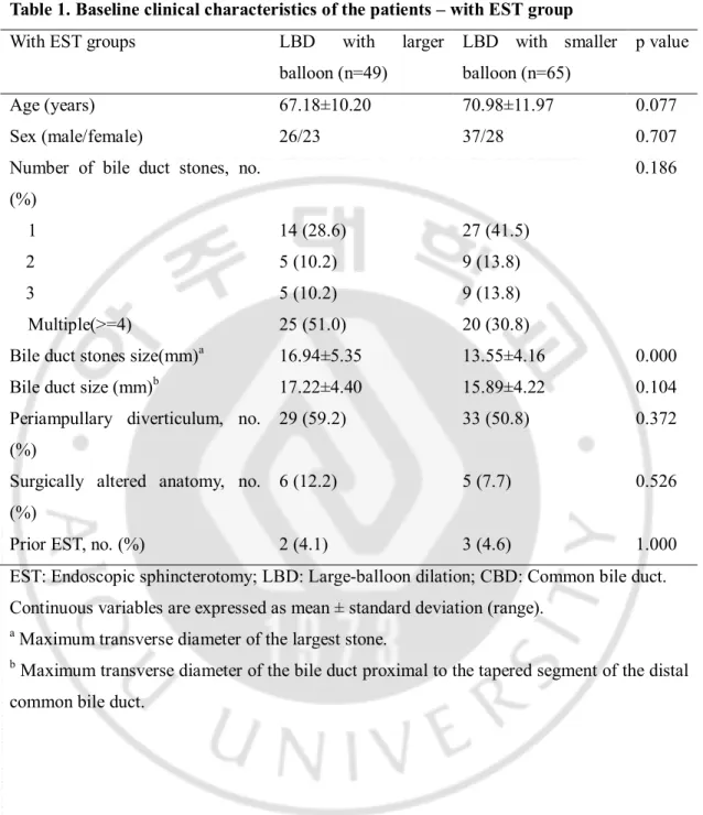
There were 49 patients in the EPLBD with a larger balloon (>15 mm) group and 65 patients in the EPLBD with a smaller balloon (12-15 mm) group. Demographics and clinical data of these groups are summarized in Table 1. Two groups showed homogenous features in terms of age, sex, number of stones, presence of periampullary diverticulum, and underlying surgically altered anatomy. Stone size was significantly larger in the larger balloon group, as expected.
Table 1. Baseline clinical characteristics of the patients – with EST group
With EST groups LBD with larger
balloon (n=49) LBD with smaller balloon (n=65) p value Age (years) 67.18±10.20 70.98±11.97 0.077 Sex (male/female) 26/23 37/28 0.707
Number of bile duct stones, no. (%) 0.186 1 14 (28.6) 27 (41.5) 2 5 (10.2) 9 (13.8) 3 5 (10.2) 9 (13.8) Multiple(>=4) 25 (51.0) 20 (30.8)
Bile duct stones size(mm)a 16.94±5.35 13.55±4.16 0.000
Bile duct size (mm)b 17.22±4.40 15.89±4.22 0.104
Periampullary diverticulum, no. (%)
29 (59.2) 33 (50.8) 0.372
Surgically altered anatomy, no. (%)
6 (12.2) 5 (7.7) 0.526
Prior EST, no. (%) 2 (4.1) 3 (4.6) 1.000
EST: Endoscopic sphincterotomy; LBD: Large-balloon dilation; CBD: Common bile duct. Continuous variables are expressed as mean ± standard deviation (range).
a Maximum transverse diameter of the largest stone.
b Maximum transverse diameter of the bile duct proximal to the tapered segment of the distal
common bile duct.
From the perspective of technical outcomes (Table 2), there was no statistically significant difference in the complete stone removal rate, stone removal rate in the first ERCP session, stone removal rate without EML, and the rate of use of EML in terms of balloon size.
8
-Table 2. Technical outcomes after EPLBD – with EST group
With EST groups LBD with larger
balloon (n=49)
LBD with smaller balloon (n=65)
p value Diameter of inflated balloon
(mm)
18.58±1.18 13.13±1.38 0.000
Stone removal rate in first ERCP session, no. (%)
42 (85.7) 49 (75.4) 0.174
Complete stone removal without ML, no. (%)
26 (73.5) 45 (69.2) 0.621
Use of EML, no. (%) 10 (20.4) 17 (26.2) 0.475
Complete stone removal irrespective of whether ML was used, no. (%)
45 (91.8) 61 (93.8) 0.723
Ballooning time (sec) 62.04±20.91 63.38±21.09 0.736
EST: Endoscopic sphincterotomy; LBD: Large-balloon dilation; ML: Mechanical lithotripsy. Continuous variables are expressed as mean ± standard deviation (range).
Safety profiles are shown in Table 3. Although no significant difference was found between the larger and smaller balloon groups in terms of adverse events, there was a trend toward the larger balloon group having a higher rate of severe to fatal adverse events (4.1% vs. 0.0%).
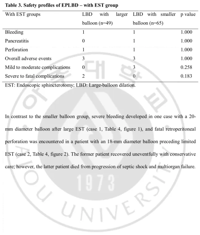
Table 3. Safety profiles of EPLBD – with EST group
With EST groups LBD with larger
balloon (n=49) LBD with smaller balloon (n=65) p value Bleeding 1 1 1.000 Pancreatitis 0 1 1.000 Perforation 1 1 1.000
Overall adverse events 3 3 1.000
Mild to moderate complications 0 3 0.258
Severe to fatal complications 2 0 0.183
EST: Endoscopic sphincterotomy; LBD: Large-balloon dilation.
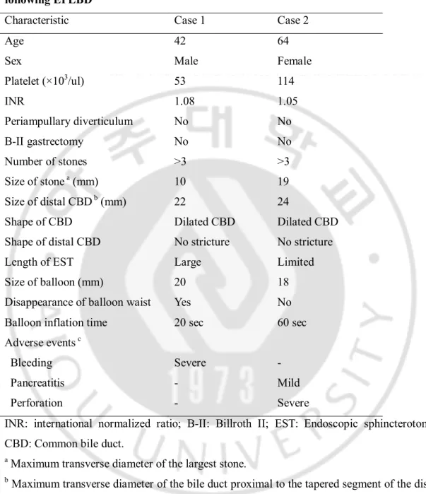
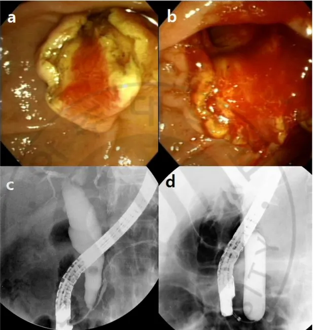
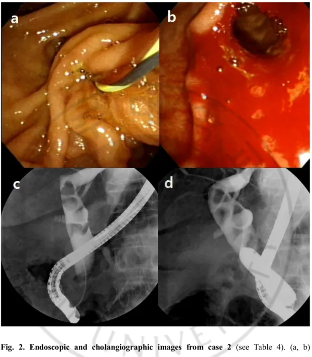
In contrast to the smaller balloon group, severe bleeding developed in one case with a 20-mm diameter balloon after large EST (case 1, Table 4, figure 1), and fatal retroperitoneal perforation was encountered in a patient with an 18-mm diameter balloon preceding limited EST (case 2, Table 4, figure 2). The former patient recovered uneventfully with conservative care; however, the latter patient died from progression of septic shock and multiorgan failure.
- 10 -
Table 4. Characteristics in two patients suffering severe to fatal adverse events following EPLBD
Characteristic Case 1 Case 2
Age 42 64
Sex Male Female
Platelet (×103/ul) 53 114 INR 1.08 1.05 Periampullary diverticulum No No B-II gastrectomy No No Number of stones >3 >3 Size of stone a (mm) 10 19 Size of distal CBD b (mm) 22 24
Shape of CBD Dilated CBD Dilated CBD
Shape of distal CBD No stricture No stricture
Length of EST Large Limited
Size of balloon (mm) 20 18
Disappearance of balloon waist Yes No
Balloon inflation time 20 sec 60 sec
Adverse events c
Bleeding Severe -
Pancreatitis - Mild
Perforation - Severe
INR: international normalized ratio; B-II: Billroth II; EST: Endoscopic sphincterotomy; CBD: Common bile duct.
a Maximum transverse diameter of the largest stone.
b Maximum transverse diameter of the bile duct proximal to the tapered segment of the distal
common bile duct.
Fig. 1. Endoscopic and cholangiographic images from case 1 (see Table 4). (a, b) Endoscopic images before and after the balloon inflation in case 1. (c, d) Cholangiographic images before and after the balloon inflation in case 1.
- 12 -
Fig. 2. Endoscopic and cholangiographic images from case 2 (see Table 4). (a, b) Endoscopic images before and after the balloon inflation in case 2. (c, d) Cholangiographic images before and after the balloon inflation in case 2. Fatal outcomes occurred in these patients after EPLBD, in which no distal common bile duct (CBD) stricture (c) was obvious.
EPLBD without EST group
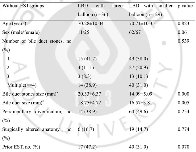
There were 36 patients in the EPLBD with a larger balloon (>15 mm) group and 129 patients in the EPLBD with a smaller balloon (12-15 mm) group. Baseline characteristics were not significantly different, according to balloon size, except stone size and diameter of distal CBD (Table 5).
Table 5. Baseline clinical characteristics of the patients – without EST group
Without EST groups LBD with larger
balloon (n=36) LBD with smaller balloon (n=129) p value Age (years) 70.28±10.04 70.71±10.35 0.823 Sex (male/female) 11/25 62/67 0.061
Number of bile duct stones, no. (%) 0.539 1 15 (41.7) 49 (38.0) 2 4 (11.1) 27 (20.9) 3 3 (8.3) 13 (10.1) Multiple(>=4) 14 (38.9) 40 (31.0)
Bile duct stones size (mm)a 20.33±6.37 14.09±5.09 0.000
Bile duct size (mm)b 18.75±4.72 16.57±3.81 0.005
Periampullary diverticulum, no. (%)
14 (38.9) 64 (49.6) 0.254
Surgically altered anatomy , no. (%)
6 (16.7) 19 (14.7) 0.774
Prior EST, no. (%) 17 (47.2) 40 (31.0) 0.070
EST: Endoscopic sphincterotomy; LBD: Large-balloon dilation; CBD: Common bile duct. Continuous variables are expressed as mean ± standard deviation (range).
a Maximum transverse diameter of the largest stone.
b Maximum transverse diameter of the bile duct proximal to the tapered segment of the distal
common bile duct.
- 14 -
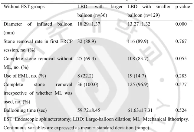
Two groups showed similar technical outcomes (Table 6).
Table 6. Technical outcomes after EPLBD – without EST group
Without EST groups LBD with larger
balloon (n=36)
LBD with smaller balloon (n=129)
p value Diameter of inflated balloon
(mm)
18.29±1.37 13.27±1.32 0.000
Stone removal rate in first ERCP session, no. (%)
32 (88.9) 116 (89.9) 0.767
Complete stone removal without ML, no. (%)
25 (69.4) 108 (83.7) 0.055
Use of EML, no. (%) 8 (22.2) 19 (14.7) 0.283
Complete stone removal irrespective of whether ML was used, no. (%)
36 (100.0) 125 (96.9) 0.577
Ballooning time (sec) 59.72±8.45 61.63±17.31 0.524
EST: Endoscopic sphincterotomy; LBD: Large-balloon dilation; ML: Mechanical lithotripsy. Continuous variables are expressed as mean ± standard deviation (range).
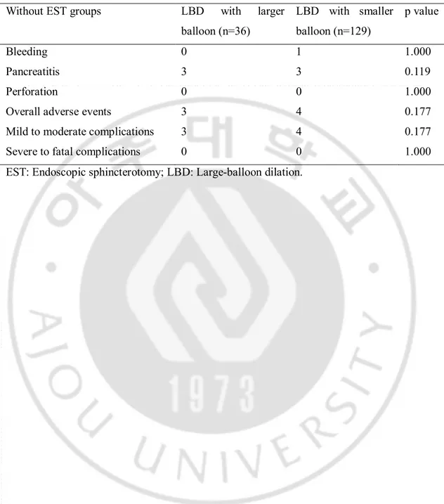
The safety variables did not differ significantly between the two groups, and no severe to fatal adverse event occurred in either group (Table 7).
Table 7. Safety profiles of EPLBD – without EST group
Without EST groups LBD with larger
balloon (n=36) LBD with smaller balloon (n=129) p value Bleeding 0 1 1.000 Pancreatitis 3 3 0.119 Perforation 0 0 1.000
Overall adverse events 3 4 0.177
Mild to moderate complications 3 4 0.177
Severe to fatal complications 0 0 1.000
- 16 -
IV. Discussion
Approximately 10% of bile duct stones are difficult to remove using conventional basket and balloon techniques preceding EST because of the large size of CBD stone compared with the diameter of distal CBD or the ampullary opening (Binmoeller et al, 1993). In such cases, EML has generally been performed as a rescue procedure for successful stone extraction (Cipolletta et al, 1997; Binmoeller and Schafer, 2001). Although EML provides a considerable success rate of stone clearance, it carries a potential risk of bile duct injury or impaction of the stone-capturing basket as well (Hwang et al, 2013). Also, small, fragmented stones could act as a nidus for stone recurrence if they are not completely removed (Ando et al, 2003).
As an alternative to conventional large EST with EML, EPLBD was introduced for successful extraction of large or difficult CBD stones to minimize the need of EML and short-term adverse events associated with large EST or EPBD (Ersoz et al, 2003). EPLBD is now performed as a primary therapeutic tool for difficult stones, after limited EST or without EST; it also could be adopted in patients who underwent a failed standard technique using large EST (Stefanidis et al, 2011). Recent literature has reported that EPLBD with or without EST enabled considerable initial success and overall success of stone clearance that is similar to conventional large EST and did not increase the risk of procedure-related adverse events, especially pancreatitis, which is the main concern when performing EPBD (Youn et al, 2011). Although EPLBD did not show statistically significant superiority to conventional
large EST in terms of needing EML, a further well-designed study is warranted. Most recent studies about the need for EML after EPLBD or conventional large EST used the number of patients requiring EML to calculate the rate of EML use instead of EML frequency on each patient; the latter could be a more accurate variable for estimating the need for EML.
Despite of its various technical advantages and considerable safety profiles, EPLBD still has an important critical issue to resolve with regard to reasonable maximal balloon size: severe perforation or bleeding may lead to mortality and needs to be minimized. Until recently, no definitive study has been conducted on this issue. Therefore, in the current study, we classified the patients who underwent EPLBD into a larger balloon group (>15 mm) and a smaller balloon group (12-15 mm) and compared the incidence of procedure-related adverse events between the two groups, focusing preferentially on severe to fatal adverse events.
Among the patients who underwent EPLBD with EST, two patients suffered severe to fatal adverse events in the larger balloon group, and one of them died from septic shock associated with severe retroperitoneal perforation (Table 3). Although no statistically significant difference was identified between the larger and smaller balloon groups (4.1% vs. 0.0%, p = 0.183), it is worth noting that a larger balloon showed a tendency toward a higher incidence of severe to fatal adverse events, because mortality associated with EPLBD is mainly attributed to severe perforation or bleeding (Park et al, 2013). Theoretically, the over inflation of a transpapillary balloon could increase the shear stress of the distal CBD and lead to retroperitoneal perforation and cause significant bleeding through a large vessel injury at the proximal part of the ampullary orifice (Oh and Kim, 2012; Park et al, 2013).
- 18 -
Until now, there was no clear evidence that balloon size >15 mm is a risk factor for severe to fatal adverse events after EPLBD because stretching of the distal CBD resulted from large stones and intraluminal pressure increased the compliance of the distal CBD (Oh and Kim, 2012; Park et al, 2013). However, distal biliary stricture, which is confirmed as major risk factor for post-EPLBD perforation in the recent multicenter study, may be unclear in the initial cholangiography and be apparent only during the balloon inflation process. In such cases, an overinflated balloon could result in unexpected fatal outcomes, even though expert recommendations are followed (i.e., that the size of balloon should not exceed the distal CBD diameter and balloon dilation should be halted if the central waist of the balloon persists after reaching 75% of targeted inflation pressure (Lee and Han, 2012).
During EPLBD, if immediate bleeding occurs, it can be controlled by subsequent balloon inflation. Delayed bleeding may occur if the balloon compresses the injured vessel for an insufficient time (Lee and Han, 2012). However, severe bleeding is found only in the large balloon group in the present study; therefore, we presume that severe delayed bleeding can arise if a major vessel injury occurs in the papillary roof during balloon inflation. A large balloon can cause additional major vessel injury, leading to severe or fatal delayed bleeding.
As a result, a larger scale multicenter trial is needed to confirm whether a larger than 15 mm balloon increases severe to fatal adverse events in patients with EPLBD and EST.
According to Park et al (2013), perforation tended to be a more severe or fatal adverse event compared with bleeding or pancreatitis, and most fatal perforations were observed in the group of patients who had undergone EPLBD with EST. In the present study, the patient who
suffered from fatal perforation had also undergone EPLBD with EST. When standard EST is performed, a full or large incision may be made, possibly leading to perforation (Yang and Hu, 2013). EPLBD also leads to perforation in patients who had distal CBD stricture or received an overinflated balloon. Although EST size tends to be smaller in EPLBD with EST compared with standard EST, a synergetic effect between the mechanism of perforation in EST and EPLBD leads to more occurrences of perforation in patients who underwent EPLBD with EST using a larger balloon, which has a greater risk of over inflation compared with a smaller one.
This is the first study in which severe to fatal adverse events after EPLBD were analyzed according to balloon size. The current study had certain limitations because our data were analyzed retrospectively and all the patients were enrolled from a single center.
- 20 -
V. Conclusion
Our study shows that EPLBD with a larger balloon (>15 mm) tends to have more risk of severe to fatal adverse events compared with a smaller balloon (12-15 mm) for the removal of large bile duct stones. A large-scale multicenter trial will be needed to reveal statistical relationships between adverse events and balloon size.
References
1. Ando T, Tsuyuguchi T, Okugawa T, Saito M, Ishihara T, Yamaguchi T, Saisho H: Risk factors for recurrent bile duct stones after endoscopicpapillotomy. Gut 52: 116–121, 2003
2. Attasaranya S, Cheon YK, Vittal H, Howell DA, Wakelin DE, Cunningham JT, Ajmere N, Ste Marie Jr RW, Bhattacharya K, Gupta K, Freeman ML, Sherman S, McHenry L, Watkins JL, Fogel EL, Schmidt S, Lehman GA: Large-diameter biliary orifice balloon dilation to aid in endoscopic bile duct stone removal: a multicenter series. Gastrointestinal Endoscopy 67: 1046-1052, 2008
3. Bergman JJ, Rauws EA, Fockens P, van Berkel AM, Bossuyt PM, Tijssen JG, Tytgat GN, Huibregtse K: Randomised trial of endoscopic balloon dilation versus endoscopic sphincterotomy for removal of bileduct stones. Lancet 349: 1124-1129, 1997
4. Binmoeller KF, Bruckner M, Thonke F, Soehendra N: Treatment of difficultbile duct stones using mechanical, electrohydraulic and extracorporealshock wave lithotripsy. Endoscopy 25: 201–206, 1993
5. Binmoeller KF, Schafer TW: Endoscopic management of bile duct stones. J Clin Gastroenterol 32: 106–118, 2001
6. Boender J, Nix GA, de Ridder MA, van Blankenstein M, Schutte HE, Dees J, Wilson JH: Endoscopic papillotomy for common bile duct stones: factors influencing the complication rate. Endoscopy 26: 209-216, 1994
7. Chan HH, Lai KH, Lin CK, Tsai WL, Wang EM, Hsu PI, Chen WC, Yu HC, Wang HM, Tsay FW, Tsai CC, Chen IS, Chen YC, Liang HL, Pan HB: Endoscopic papillary large balloon dilation alone without sphincterotomy for the treatment of large common bile duct stones. BMC Gastroenterol 11: 69, 2011
- 22 -
Marmo R: Endoscopic mechanical lithotripsy of difficult common bileduct stones. Br J Surg 84: 1407–1409, 1997
9. Classen M, Demling L: Endoscopic sphincterotomy of the papilla of vater and extraction of stones from the choledochal duct (author's transl). Dtsch Med Wochenschr 99: 496-497, 1974
10. Cotton PB, Lehman G, Vennes J, Geenen JE, Russell RC, Meyers WC, Liguory C, Nickl N: Endoscopic sphincterotomy complications and their management: an attempt at consensus. Gastrointest Endosc 37: 383-393, 1991
11. Disario JA, Freeman ML, Bjorkman DJ, Macmathuna P, Petersen BT, Jaffe PE, Morales TG, Hixson LJ, Sherman S, Lehman GA, Jamal MM, Al-Kawas FH, Khandelwal M, Moore JP, Derfus GA, Jamidar PA, Ramirez FC, Ryan ME, Woods KL, Carr-Locke DL, Alder SC: Endoscopic balloon dilation compared with sphincterotomy for extraction of bile duct stones. Gastroenterology 127: 1291-1299, 2004
12. Ersoz G, Tekesin O, Ozutemiz AO, Gunsar F: Biliary sphincterotomy plus dilation with a large balloon for bile duct stones that are difficult to extract. Gastrointest Endosc 57: 156-159, 2003
13. Espinel J, Pinedo E: Large balloon dilation for removal of bile duct stones [In Spanish with English abstract]. Rev Esp Enferm Dig 100: 632-636, 2008
14. Ghazanfar S, Qureshi S, Leghari A, Taj MA, Niaz SK, Quraishy MS: Endoscopic balloon sphincteroplasty as an adjunct to endoscopic sphincterotomy in removing large and difficult bile duct stones. J Pak Med Assoc 60: 1039-1042, 2010
15. Harada R, Maguchi H, Takahashi K, Katanuma A, Osanai M, Yane K, Hashigo S, Kaneko M, Katoh R, Katoh S: Large balloon dilation for the treatment of recurrent bile duct stones prevents short-term recurrence in patients with previous endoscopic sphincterotomy. J Hepatobiliary Pancreat Sci 20: 498-503, 2013
16. Hwang JC, Kim JH, Lim SG, Kim SS, Shin SJ, Lee KM, Yoo BM: Endoscopic large-balloon dilation alone versus endoscopic sphincterotomy plus large-large-balloon dilation for the treatment of large bile duct stones. BMC Gastroenterol 13: 15-20, 2013
17. Itoi T, Itokawa F, Sofuni A, Kurihara T, Tsuchiya T, Ishii K, Tsuji S, Ikeuchi N, Moriyasu F: Endoscopic sphincterotomy combined with large balloon dilation can reduce the procedure time and fluoroscopy time for removal of large bile duct stones. Am J Gastroenterol 104: 560-565, 2009
18. Itoi T, Sofuni A, Itokawa F, Kurihara T, Tsuchiya T, Ishii K, Tsuji S, Ikeuchi N, Umeda J, Moriyasu F: New large-diameter balloon-equipped sphincterotome for removal of large bile duct stones (with videos). Gastrointest Endosc 72: 825-830, 2010
19. Jeong S, Ki SH, Lee DH, Lee JI, Lee JW, Kwon KS, Kim HG, Shin YW, Kim YS: Endoscopic large-balloon sphincteroplasty without preceding sphincterotomy for the removal of large bile duct stones: a preliminary study. Gastrointest Endosc 70: 915-922, 2009
20. Kawai K, Akasaka Y, Murakami K, Tada M, Koli Y: Endoscopic sphincterotomy of the ampulla of Vater. Gastrointest Endosc 20: 148-151, 1974
21. Kim HG, Cheon YK, Cho YD, Moon JH, Park DH, Lee TH, Choi HJ, Park SH, Lee JS, Lee MS: Small sphincterotomy combined with endoscopic papillary large balloon dilation versus sphincterotomy. World J Gastroenterol 15: 4298-4304, 2009
22. Kim KO, Kim TN, Lee SH: Endoscopic papillary large balloon dilation for the treatment of recurrent bile duct stones in patients with prior sphincterotomy. J Gastroenterol 45: 1283-1288, 2010
23. Kim MK, Kim MH, Lee TY, Oh HC, Kwon SH, Han JH, Choi HO, Park SJ, Kim TH, Lee SS, Seo DW, Lee SK: Combined endoscopic sphincterotomy and large balloon sphincteroplasty for bile duct stones [In Korean with English abstract]. Korean J Med 73: 474-480, 2007
24. Kim TH, Oh HJ, Lee JY, Sohn YW: Can a small endoscopic sphincterotomy plus a large-balloon dilation reduce the use of mechanical lithotripsy in patients with large bile duct stones? Surg Endosc 25: 3330-3337, 2011
25. Kozarek RA: Balloon dilation of the sphincter of Oddi. Endoscopy 20 Suppl 1: 207-210, 1988
- 24 -
26. Kurita A, Maguchi H, Takahashi K, Katanuma A, Osanai M: Large balloon dilation for the treatment of recurrent bile duct stones in patients with previous endoscopic sphincterotomy: preliminary results. Scand J Gastroenterol 45: 1242-1247, 2010
27. Lee DK, Han JW: Endoscopic papillary large balloon dilation: guidelines for pursuing zero mortality. Clin Endosc 45(3): 299-304, 2012
28. Lee SH, Hong SW, Cho YD, Cheon YK, Kim SG, Jang JY, Kim YS, Moon JH, Lee JS, Lee MS, Shim CS, Kim BS: The safety and effectiveness of medium endoscopic sphincterotomy with endoscopic papillary large balloon dilation for removing difficult common bile duct stones [In Korean with English abstract]. Korean J Gastroint Endosc 35: 80-86, 2007
29. Leung JW, Chan FK, Sung JJ, Chung S: Endoscopic sphincterotomy-induced hemorrhage: a study of risk factors and the role of epinephrine injection. Gastrointest Endosc 42: 550-554, 1995
30. Mathuna PM, White P, Clarke E, Merriman R, Lennon JR, Crowe J: Endoscopic balloon sphincteroplasty (papillary dilation) for bile duct stones: efficacy, safety, and follow-up in 100 patients. Gastrointest Endosc 42: 468-474, 1995
31. Minami A, Nakatsu T, Uchida N, Hirabayashi S, Fukuma H, Morshed SA, Nishioka M: Papillary dilation vs sphincterotomy in endoscopic removal of bile duct stones. A randomized trial with manometric function. Dig Dis Sci 40: 2550-2554, 1995
32. Misra SP, Dwivedi M: Large-diameter balloon dilation after endoscopic sphincterotomy for removal of difficult bile duct stones. Endoscopy 40: 209-213, 2008
33. Oh MJ, Kim TN: Prospective comparative study of endoscopic papillary large balloon dilation and endoscopic sphincterotomy for removal of large bile duct stones in patients above 45 years of age. Scand J Gastroenterol 47: 1071-1077, 2012
34. Park SJ, Kim JH, Hwang JC, Kim HG, Lee DH, Jeong S, Cha SW, Cho YD, Kim HJ, Kim JH, Moon JH, Park SH, Itoi T, Isayama H, Kogure H, Lee SJ, Jung KT, Lee HS, Baron TH, Lee DK: Factors predictive of adverse events following endoscopic papillary large balloon dilation: results from a multicenter series. Dig Dis Sci 58: 1100-1109, 2013
35. Paspatis GA, Konstantinidis K, Tribonias G, Voudoukis E, Tavernaraki A, Theodoropoulou A, Chainaki I, Manolaraki M, Chlouverakis G, Vardas E, Paraskeva K: Sixty- versus thirty-seconds papillary balloon dilation after sphincterotomy for the treatment of large bile duct stones: a randomized controlled trial. Dig Liver Dis 45: 301-304, 2013
36. Poincloux L, Rouquette O, Privat J, Gorce D, Abergel A, Dapoigny M, Bommelaer G: Large-balloon dilation of the sphincter of Oddi after sphincterotomy or infundibulotomy to extract large calculi or multiple common bile duct stones without using mechanical lithotripsy. Scand J Gastroenterol 48: 246-251, 2013
37. Rebelo A, Ribeiro PM, Correia AP, Cotter J: Endoscopic papillary large balloon dilation after limited sphincterotomy for difficult biliary stones. World J Gastrointest Endosc 4: 180-184, 2012
38. Rosa B, Moutinho Ribeiro P, Rebelo A, Pinto Correia A, Cotter J: Endoscopic papillary balloon dilation after sphincterotomy for difficult choledocholithiasis: A case-controlled study. World J Gastrointest Endosc 5: 211-218, 2013
39. Sakai Y, Tsuyuguchi T, Sugiyama H, Nishikawa T, Kurosawa J, Saito M, Tawada K, Mikata R, Tada M, Ishihara T, Yokosuka O: Endoscopic sphincterotomy combined with large balloon dilation for removal of large bile duct stones. Hepatogastroenterology 60: 58-64, 2013
40. Sato H, Kodama T, Takaaki J, Tatsumi Y, Maeda T, Fujita S, Fukui Y, Ogasawara H, Mitsufuji S: Endoscopic papillary balloon dilatation may preserve sphincter of Oddi function after common bile duct stone management: evaluation from the viewpoint of endoscopic manometry. Gut 41: 541-544, 1997
41. Sherman S, Ruffolo TA, Hawes RH, Lehman GA: Complications of endoscopic sphincterotomy. A prospective series with emphasis on the increased risk associated with sphincter of Oddi dysfunction and nondilated bile ducts. Gastroenterology 101: 1068-1075, 1991
- 26 -
alternative to papillotomy? (author's transl). Dtsch Med Wochenschr 107: 895-897, 1982 43. Stefanidis G, Viazis N, Pleskow D, Manolakopoulos S, Theocharis L, Christodoulou C,
Kotsikoros N, Giannousis J, Sgouros S, Rodias M, Katsikani A, Chuttani R: Large balloon dilation vs. mechanical lithotripsy for the management of large bile duct stones: a prospective randomized study. Am J Gastroenterol 106: 278-285, 2011
44. Takada T, Strasberg SM, Solomkin JS, Pitt HA, Gomi H, Yoshida M, Mayumi T, Miura F, Gouma DJ, Garden OJ, Büchler MW, Kiriyama S, Yokoe M, Kimura Y, Tsuyuguchi T, Itoi T, Gabata T, Higuchi R, Okamoto K, Hata J, Murata A, Kusachi S, Windsor JA, Supe AN, Lee S, Chen XP, Yamashita Y, Hirata K, Inui K, Sumiyama Y; Tokyo Guidelines Revision Committee: TG13: Updated Tokyo Guidelines for the management of acute cholangitis and cholecystitis. J Hepatobiliary Pancreat Sci 20(1):1-7, 2013 45. Weinberg BM, Shindy W, Lo S: Endoscopic balloon sphincter dilation (sphincteroplasty)
versus sphincterotomy for common bile duct stones. Cochrane Database Syst Rev 18, 2006
46. Yang XM, Hu B, Pan YM, Gao DJ, Wang TT, Wu J, Ye X: Endoscopic papillary large-balloon dilation following limited sphincterotomy for the removal of refractory bile duct stones: experience of 169 cases in a single Chinese center. J Dig Dis 14: 125-131, 2013 47. Yang XM, Hu B: Endoscopic sphincterotomy plus large-balloon dilation vsendoscopic
sphincterotomy for choledocholithiasis: A meta-analysis World J Gastroenterol 28; 19(48):9453-60, 2013
48. Yasuda I, Tomita E, Enya M, Kato T, Moriwaki H: Can endoscopic papillary balloon dilation really preserve sphincter of Oddi function? Gut 49: 686-691, 2001
49. Yoon HG, Moon JH, Choi HJ, Kim DC, Kang MS, Lee TH, Cha SW, Cho YD, Park SH, Kim SJ: Endoscopic papillary large balloon dilation for the management of recurrent difficult bile duct stones after previous endoscopic sphincterotomy. Dig Endosc 26(2):259-63, 2014
50. Youn YH, Lim HC, Jahng JH, Jang SI, You JH, Park JS, Lee SJ, Lee DK: The increase in balloon size to over 15 mm does not affect the development of pancreatitis after
endoscopic papillary large balloon dilatation for bile duct stone removal. Dig Dis Sci 56: 1572-1577, 2011
- 28 - - 국문요약 -
내시경적 유두부 큰 풍선 확장술에서 15mm이상의 풍선을
사용하였을 때 부작용이 증가하는가?
12~20mm크기의 풍선을 이용하는 내시경적 유두부 큰 풍선 확장술은 크기가 큰 담도결석의 제거를 용이하게 하고 내시경적 기계적 쇄석술의 필요성을 줄이기 위한 목적으로 소개되었다. 최근의 많은 연구에서는 내시경적 유두부 큰 풍선 확장술이 큰 담도 결적의 제거에 효과적이며 치명적인 췌장염이나 담도 천공의 위험을 높이지 않는 안전한 시술이라는 사실이 보고되고 있다. 그럼에도 불구하고 내시경적 유두부 큰 풍선 확장술에서 치명적인 부작용을 줄이기 위한 최대 풍선 크기에 대한 지침에 대한 자료가 부족한 상황이다. 본 연구는 풍선 크기에 따른 내시경적 유두부 큰 풍선 확장술의 안전성에 초점을 맞추어 적절한 최대 풍선 크기를 확인하기 위해 설계되었다. 총 279명의 환자가 본 연구에 등록되었으며 내시경적 유두부 큰 풍선 확장술과 내시경적 괄약근 절개술을 같이 시행받은 환자가 114명, 내시경적 괄약근 절개술 없이 내시경적 큰 풍선 확장술만을 시행받은 환자가 165명 이었다. 내시경적 유두부 큰 풍선 확장술과 내시경적 괄약근 절개술을 같이 시행받은 환자군에서 49명의 환자는 15mm초과의 풍선을 사용하여 시술받았고 65명의 환자는 12-15mm의 풍선을 사용하여 시술받았다. 비록 15mm초과의 풍선을 사용한 환자군과 12-15mm의 풍선을 사용한 환자군에서 통계적으로 유의한 차이를 보이지는 못했지만12-15mm의 풍선을 사용한 환자군에 비해 15mm초과의 풍선을 사용한 환자군에서 중증도 및 치명도의 부작용이 증가하는 경향성을 보였다. 내시경적 괄약근 절개술 없이 내시경적 큰 풍선 확장술만을 시행받은 환자군에서 36명의 환자는 15mm초과의 풍선을 사용하여 시술받았고 129명의 환자는 12-15mm의 풍선을 사용하여 시술받았다. 두 집단 사이에 안전성과 관련한 부작용의 차이는 관찰되지 않았으며 중증도 및 치명도의 부작용은 관찰되지 않았다. 결론적으로 내시경적 유두부 큰 풍선 확장술에서 12-15mm의 풍선을 사용하는 것에 비해 15mm초과의 풍선을 사용할 경우 중증도 및 치명도의 부작용이 증가하는 경향성을 확인할 수 있었으며 추후 다기관 연구를 통해 이 연관관계의 통계학적 유의성을 증명할 필요가 있다. _________________________________________________________________________ 핵심어: 내시경적 유두부 큰 풍선 확장술, 부작용, 풍선 크기