165
A Study on the Distribution of Microorganisms in Department of
Radiography
Jeong-Hyun Chang1, Eun-Ju Yang1, Young-Jae Kim2,* 1Department of Clinical Laboratory Science, Daegu Haany University
2Department of Radiologic Technology, Daegu Health College
Received: April 22, 2021. Revised: April 28, 2021. Accepted: April 30, 2021.
ABSTRACT
To prevent the secondary hospital-acquired infection (cross-infection) from occurring in the general radiographic room in the department of radiology, the microbial measurement was conducted at the points making direct close contact with radiologists and patients. For the case of radiologists, the microbial measurement and incubation were focused on the x-ray tube handle of the radiation generating device, and, for the case of patients, the microbial measurement and incubation were focused on the chin supporting device, chest-contact point, and handle. Once disinfected with Aniosurf, the sterilized media were gathered and identified, and the microorganisms were confirmed. Based on the identification results, it was confirmed that the points making direct close contact with radiologists showed a value of 103CFU for Proteus mirabilis, Staphylococcus epidermidis, Bacillus spp. and
Candida spp., and that the points making direct close contact with patients showed a value of 103~5 CFU for Proteus mirabilis, Enterococcu faecium, Pseudomonas aeruginosa, NTM(Non-Tuberculosis Mycobacteria) and Candida spp.. It was also confirmed that the types and number of microorganisms gathered from the points making direct close contact with patients were greater. Fortunately, most of the involved microorganisms were observed to be on the skin surface and are known to become extinct when disinfected in accordance with the hospital-acquired infection control rules. However, since even minor exposure to such microorganisms may be lethal for patients with reduced immunity, caution must be taken. In particular, since the points making contact with patients showed a high level of microbial measurement, it was thought that it would be necessary for radiologists and personnel having frequent access to strictly disinfect the parts, such as instruments and handles, making frequent contact with patients. The purpose of this study was to announce the importance of safe microbial control in the radiographic inspection room in hospital, and this study is expected to be used as the baseline data for preventing hospital-acquired secondary infection and Nth infectious diseases.
Keywords: Department of Radiography, Infections, Microorganism
I. INTRODUCTION
Hospital-acquired infection not only leads to secondary diseases[1], but also may serve as a cause that results in diseases fatal to patients with reduced immunity[2].
The increased medical expenses and delayed patient recovery resulting from hospital-acquired cross-infection
may result in a number of social costs, and, this may lead to degrading the medical quality, it is necessary to provide periodic control to guarantee patient safety and quality medical service[2].
The department of radiology in hospital is an essential factor for providing patients with imaging examination. In particular, radiologists in charge of radiography serve as an important point where a
number of bacterial cross infections, including microbial infections, may occur due to direct contact made with patients or indirect contact made through radiation generating devices. Ministry of Food and Drug Safety reported that chest x-ray imaging is an essential inspection that accounts for approximately 27.5% of all the radiological imaging examinations[3,4].
In the preceding studies, microbial observation studies were conducted in the radiology department, centered on general hospitals, and studies were conducted to observe microbes in imaging IPs and cassettes. All of them were characterized by being a cassette part of the space and close proximity of patients, and this paper attempts to proceed with microbial collection centered on the equipment.
The purpose of this study was to classify the points that make frequent contact with patients and with radiologists during such x-ray imaging, to analyze the types of microorganisms existing at those points, and to collect and analyze the data for preventing hospital-acquired cross-infection in the future.
Ⅱ. MATERIAL AND METHODS
To conduct this study, Hospitals A and B located in Daegu were selected the experiment subjects, and the microbial sampling was conducted from September 2020 to October 2020.
1. Study Subjects
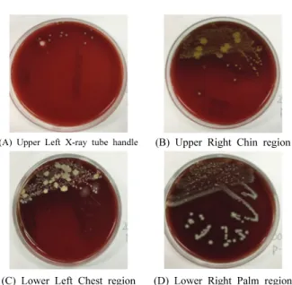
General imaging equipment (DRGEM, E7239X) was used to sample microorganisms as shown in Fig. 1. In detail, the sampling was focused mainly on the tube handle serving as the main point making contact with radiologists, and on the chin supporting device, chest-contact point and patient handle serving as the main points making contact with patients during x-ray imaging.
As far as the sampling process is concerned, once disinfected with Aniosurf, the patient imaging took place, and, after the imaging, sterile cotton-tipped
swabs were used to sample the microorganisms from the above points each limited to an area of 10×10.
According to a thesis by Jung Woo Chan et al.[5], it
was confirmed that most of the bacteria became extinct after they were sterilized with Aniosurf-diluted solution. therefore, the disinfection process was conducted in advance using Aniosurf.
Fig. 1. (A) X-ray tube handle, (B) Chin region, (C) Chest region, (D) Palm region.[5]
2. Study Methods
2.1 Disinfection Methods
The original Aniosurf (ANIOS, ANIOSURF Premium NPC) solution was diluted by a factor of 400, the disinfection took place, the disinfected objects were allowed to stand for 15 minutes, and the patient imaging took place.
2.2 Sampling Methods and Microbial Incubation After the sampling process, to examine to microbial status, TrransystemTM(TransystemTM, Copan Diagnostics Inc., Murrieta, CA, USA) shown in Fig. 2 was used to store and transport the samples.
To accurately confirm the sealed samples, it was necessary to move the samples to the microbial test room as soon as possible. Therefore, the microbial analysis was initiated within 24 hours.
The test was conducted within the biosafety cabinet, the innoculation was conducted based on the
horizontal stroke method using blood agar (synergy innovation), and the incubation was conducted for 48 hours within an incubator under the following conditions: 37℃ and 5% CO2.
Fig. 2. Transystem TM.[5]
2.3 Identification
The cultured strain parts forming a single colony were used in the identification test, and, for each single colony, the bacterial solution was manufactured by using the turbidity meter to adjust the turbidity to No. 0.5(McFarland).
The identification process was conducted using the MICROSCAN (Dade Behring, West Sacramento, CA, USA) automated device, and the results were acquired as shown in Fig. 3.
(A) Upper Left X-ray tube handle (B) Upper Right Chin region
(C) Lower Left Chest region (D) Lower Right Palm region
Fig. 3 Microbial identification result.
Ⅲ. RESULT
The microbial identification was conducted at the points making frequent contact with radiologists and with patients during radiological imaging examination (x-ray imaging), and the results were as follows. The collected samples were classified through the dust spot method, and the bacteria of the number of microorganisms were proposed using the colony forming unit(colony forming unit: CFU/plate).
1. Points Making Frequent Contact with Radiologists Radiologists make direct contact with the tube handle of the radiation generating device during patient positioning and radiography. Table 1 shows the microbial identification results acquired from the handle of the radiation generating device. A total of 4 types were detected as follows and showed a value above 103CFU: Bacillus spp., Candida spp., Proteus mirabilis, and Staphylococcus epidermidis.
2. Patients
Table 2 shows the microbial identification results acquired from each point making frequent contact with patients. From the chin supporting device, proteus mirabilis, enterococcu faecium, pseudomonas aeruginosa, and NTM were detected, and, from the chest-contact point, proteus mirabilis, enterococcu faecium, and pseudomonas aeruginosa were detected, and all of them showed a value above 103CFU
(Colony Forming Unit). From the palm-contact point, NTM and candida spp. were detected and showed a value of 105 CFU (Colony Forming Unit). In total, 5
types were measured.
Table 1. Microorganism detection by site
Site Classification Before Disinfection Tube
handle
Type of microorgan
ism
Proteus mirabilis, Staphylococcus epidermidis, Bacillus spp., Candida spp. CFU/plate 103 CFU
Table 2. Microorganism detection by site(Pt)
Site Classification Before Disinfection Chin
region
Type of microorganism
Proteus mirabilis, Enterococcu faecium, Pseudomonas aeruginosa,
NTM CFU/plate 103 CFU Chest
region
Type of
microorganism faecium, Pseudomonas aeruginosaProteus mirabilis, Enterococcu CFU/plate 103 CFU Palm region Type of microorganism NTM, Candida spp. CFU/plate 105
IV. DISCUSSION
Hospital-acquired infection refers to the infection occurring in patients within 72 hours (within 30 days for surgical patients) after admission at the hospital that was neither present nor incubating at the time of admission, and such infection includes the infection occurring in not only patients, but also hospital employees[7,8]. To prevent such infection. it seems
necessary to avoid making contact with contaminated medical devices or contaminated patients to the utmost extent possible by classifying infections into endogenous infections and exogenous infections. A pre-existing study reported that approximately 50% of the hospital-acquired infections are caused by the tools used in hospital[9,10]. To prevent hospital-acquired
infections, disinfecting and sterilizing medical instruments can be said to be the most important factor.
As far as radiography is concerned, Ministry of Food and Drug Safety announced that chest x-ray imaging is the most frequently executed imaging method from among various imaging methods[3.4].
Since it is the most basic examination, it is a fact that such imaging can be frequently exposed to microorganisms or bacteria. Accordingly, to prevent radiologists, related operation participants and personnel having frequent access from causing
cross-contamination, close-contact points are required to be disinfected to a substantial extent[3,4].
According to a research by Jeong Woo Chan et al.[5], points having frequent contact with radiologists
and with patients were selected as the research subjects, and Aniosurf (ANIOS, ANIOSURF Premium NPC) was used to conduct the pre-/post-disinfection microbial analysis. Based on the results, it was confirmed that the disinfection effect was approximately 90%, and, in some cases involving Bacillus spp., it was revealed that the microorganisms were not perfectly removed and that it was necessary to conduct an additional disinfection,
Of the detected microorganisms, Staphylococcus epidermidis is a bacterium that exists on human skin and is the most frequent hospital-acquired infectious bacterium. In the case where such bacterium is injected into veins through a prosthetic device, it is known to lead to virulence, causing septicaemia at a common frequency[12].
Pseudomonas aeruginosa detected from the patients was reported to cause pneumonia and various types of septicaemia, and was reported to develop a genetic tolerance to a number of antibiotics[13]. Enterococcu
faecium is a type of enterococci that exists as commensal bacteria, and the presence of it means that the environment can be unsanitary due to actions such as not washing hands after using toilet[14]. In addition,
since candida spp. injected into patients with reduced immunity through the veins or blood flow may cause abscess, thrombophlebitis, endocarditis, eye infection or other organ-related infections, it is necessary to take caution[14,15].
According to Bae et al[16], 7 kinds of bacteria were
detected in the microbial measurement using the cassette and IP used for radiographic imaging. Two (ABA) sites, Bacillus sp, Coagulase-negative Staphylococci (CNS), and 1 Enterococcus sp (ENT) were detected. All except ABA were Gram positive bacilli, and only 3% reported that Gram
positive bacilli were detected. In this study, micro-organisms were observed centering on the imaging device, but Bae et al. used the cassette and IP used for radiographic imaging, so it seems that more types of micro-organisms were observed as the degree of direct contact increased.
HS Shin et al[17] analyzed radiology and infection
control in a general hospital, and Staphylococcus, Micrococcus, Pseudomonas stutzeri, and Pseudomonas oryzihabitans were observed. The above paper identified microbial strains in five general hospitals, suggesting that the pathogens of nosocomial infection may be different for each hospital, and at the same time, it shows that pathogens exist in all hospitals.
based on the results acquired in this study, it was confirmed that opportunistic bacteria exist at points making frequent contact with radiologists and with patients, and it was determined that it would be necessary to comply with the hand sanitation and in-hospital disinfection rules to prevent hospital-acquired infections.
The limitation of this paper is that the number of microbial collections is insufficient. In future studies, it is thought that more times of microbial collection and the method according to the collection of microorganisms should be studied differently.
V. CONCLUSION
Based on the results acquired from the microbial measurement focused on points making frequent contact with radiologists and with patients during radiological imaging, it was confirmed that proteus mirabilis, staphylococcus epidermidis, bacillus spp., and candida spp. were detected from the points making contact with radiologists, and that proteus mirabilis, enterococcu faecium, pseudomonas aeruginosa, NTM, and candida spp. were detected from the points making contact with patients. Although most of them were revealed as microorganisms that become extinct as disinfection
takes place, it is necessary to be aware that the secondary infection may occur in hospital at all times. In particular, since the types and number of microorganisms detected from the points making contact with patients were greater than those detected from the points making contact with radiologists, it is thought that it would be necessary to make stricter efforts to control infections resulting from the equipment with which patients make more frequent contact.
Reference
[1] H. S. Kim, "Effects of Hand Washing Enhancement
on Improving Hand Hygiene Performance and Reducing Hospital Infection", Korean Society of Clinical Pathologists Association, 53rd Annual Meeting Abstract, 2015.
[2] M. S. Song, "A Study on the Awareness and
Practice of the Nurce’s on the Nosocomical Infection", Masters of Nurse, Dan Kook University, Korea, 2001.
[3] J. S. Lee, K. H. Jeong, G. H. Kim, I. C. Im, D. C.
Kweon, E. H. Goo, K. R. Dong, W. K. Chung, "Radiology Department Infection Control According to Radiography Frequency and Disinfection Period", Korean Radiation Society, Vol. 5, No. 2, pp. 73-80, 2011. http://dx.doi.org/10.7742/jksr.2011.5.2.073
[4] G. P. Kim, "Radiation exposure of Korean population
from medical diagostic examinations", Ministry of Food and Drug Safety, Vol. 11, No. 579, pp. 213-217, 2013.
https://doi.org/10.23000/TRKO201400011827
[5] W. C. Jung, K. S. Ahn, E. J. Yang, Y. J. Kim,
"Research on ANIOS Disinfection Efficiency to Prevent Infection in Radiography Room", Journal of Korean Society Radiology, Vol. 15, No. 1, pp. 55-61, 2021. https://doi.org/10.7742/jksr.2021.15.1.55
[6] Ministry of Health and Welfare, "Guidelines for
disinfecting instruments and articles used by medical institutions", Health and Welfare Department Notification, No. 2017-101, 2017.
[7] M. Hasan, H. Tuckman, R. Patrick D. Kountz, J.
Topics, Vol. 8, No. 3, pp. 82-89, 2010. http://dx.doi.org/10.1080/00185868.2010.507124
[8] B. U. Wu, R. S. Johanes, S. Kurtz, P. A. Banks,
"The impact of hospital-acquired infection on outcome in acute pancreatis", Gastroenterology, Vol. 135, No. 3 pp. 816-820, 2008.
http://dx.doi.org/10.1053/j.gastro.2008.05.053
[9] J. H. Woo, M. S. Lee, M. H. Jeong, S. O. Lee, D. R. Jeong, E. O. Kim, "National Survey on the Current Status of Nosocomial Infection Control in Korea", Korean Society for Healthcare-associated Control and Prevention, Vol. 2, No. 2, pp. 177-202, 1997.
[10] H. J. Jeon, D. S. Jeon, J. R Kim, J. S. Kim, J. M.
Kim, "Use of Antibiotics and Environmental Survey in Nosocomial Infection", Korean Journal of Laboratory Medicine, Vol. 5, No. 2, pp. 451-462, 1985.
[11] S. G. Shin, H. Y. Lee, "The Pathology of Infection in the Department of Radiology", Journal of radiological science and technology, Vol. 35, No. 3, pp. 211-218, 2012.
[12] J. W. Lee, K. J. Oh, S. C Park, J. S. Rim, "The
Clinical Features of Complicated Urinary Tract Infections by Pseudomonas aeruginosa", The Korean Journal of Urology. Vol. 49, No. 12, pp. 1149-1154, 2008. http://dx.doi.org/10.4111/kju.2008.49.12.1149
[13] Ahu Karaa, Ilker Devrima, Nuri Bayrama, Nagehan
Katipo glub, Ezgi Kıranb, Yeliz Oruc¸ Nevbahar Demirayc, Hurs¸ Apab, Gamze Gülfidand, "Risk of vancomycin-resistant enterococci bloodstream infection among patients colonized with
vancomycin-resistant enterococci ", The Brazilian Journal of Infectious diseases, Vol.19, No.1 pp.58-61, 2015
https://doi.org/10.1016/j.bjid.2014.09.010
[14] C. d' Enfert, B. Hube, "Candida: Comparative and
Functional Genomics", Caister Academic Press, Vol. 7, No. 6, pp. 616, 2007.
[15] F. H. Meyers, E. Jawetz, A. Goldfien, "Review of Medical Pharmacology", Lange Medical Publications. 1978.
[16] SH Bae, MS Lee, CS Lim, GJ Kim, “A Study on
the Measurement of the Pollution Level of Bacteria
and Disinfection of Table and IP Cassette”, Journal of radiological science and technology Vol. 31 No, 3, pp.229-237. 2008
[17] J Shin, C Park, BK Jeon, “Analysis on infection
control of general hospital radiology”, Journal of the Korean Society of Radiology, Vol. 6, No. 5, pp..335-342, 2012
성명 소속 직위 (제1저자) 장정현 대구한의대학교 임상병리학과 교수 (공동저자) 양은주 대구한의대학교 임상병리학과 교수 (교신저자) 김영재 대구보건대학교 방사선학과 교수 연구자 정보 이력
영상의학과 촬영실의 미생물 분포에 관한 연구
장정현1, 양은주1, 김영재2,* 1대구한의대학교 임상병리학과 2대구보건대학교 방사선학과 요 약 영상의학과의 일반방사선 촬영실의 2차 원내감염(교차감염)의 방지를 위해 방사선사, 환자와 직접적인 밀접접촉부분의 미생물을 측정하였다. 방사선사 측은 방사선 발생장치의 엑스선튜브 손잡이, 환자측은 턱 받침대, 가슴닿는 곳, 손잡이를 중심으로 미생물을 측정하여 배양하였다. 에니오설프로 소독한 후 멸균배지 를 이용해 수합하고 동정하여 미생물을 확인하였다. 동정결과 방사선사 측은 Proteus mirabilis, Staphylococc us epidermidis, Bacillus spp., Candida spp., 103CFU, 환자측은 Proteus mirabilis, Enterococcu faecium, Pseudomonas aeruginosa, NTM, Candida spp. 가 103~5 CFU 검출 되었으며 환자측에서 수합된 미생물의 종류와 수가
더 많이 관찰되었다. 다행히 해당되는 미생물은 대부분 피부표면에 있는 것들로 관찰되었으며 원내감염관 리 규칙에 의거하여 소독한다면 대부분 사멸하는 것으로 알려져 있지만 면역력이 떨어진 환자의 경우 소 량의 노출로도 치명적일 수 있으므로 주의해야 한다. 특히, 환자와 접촉한 부분의 미생물이 높게 측정된 것 을 보아 방사선사 및 수시출입자 등은 환자와 접촉이 잦은 기구 및 손잡이 등의 부분에 대해 소독을 철저 히 해야 할 것으로 사료된다. 기존 에니오설프 연구를 기반으로 현재연구를 확대하여 원내 방사선 검사실 의 미생물 안전관리의 중요성을 알리는 목적으로 조사하였고 병원 2차 및 N차 감염병 예방을 위한 기본적 인 자료가 될 수 있을 것이다. 중심단어: 영상의학과촬영실, 감염, 미생물