Volume 2013, Article ID 758763,5pages http://dx.doi.org/10.1155/2013/758763
Research Article
Novel Threadlike Structures May Be Present on the Large
Animal Organ Surface: Evidence in Swine Model
Kyoung-Hee Bae,
1Sang Hyun Park,
2Byung-Cheon Lee,
2Min-Ho Nam,
3Ji Woong Yoon,
4Hee-Min Kwon,
5and Seung Zhoo Yoon
61Nano Primo Research Center, Advanced Institute of Convergence Technology, Seoul National University,
Suwon 443-270, Republic of Korea
2Ki Primo Research Laboratory, Division of Electrical Engineering, KAIST Institute for Information Technology Convergence,
Korea Advanced Institute of Science and Technology (KAIST), Daejeon 305-701, Republic of Korea
3Department of Pathology, College of Korean Medicine, Kyung Hee University, Seoul 130-701, Republic of Korea 4Impedance Imaging Research Center and Department of Public Administration, Kyung Hee University,
Seoul 130-701, Republic of Korea
5Yonsei Institute of Convergence Technology, Yonsei University, 162-1 Sondo-dong, Yeonsu-gu, Incheon 406-840, Republic of Korea 6Department of Anesthesiology and Pain Medicine, College of Medicine, Korea University, No. 5 Anam-dong Sungbuk-gu,
Seoul 136-705, Republic of Korea
Correspondence should be addressed to Seung Zhoo Yoon; yoonsz70@gmail.com Received 11 February 2013; Accepted 2 May 2013
Academic Editor: Yeonhee Ryu
Copyright © 2013 Kyoung-Hee Bae et al. This is an open access article distributed under the Creative Commons Attribution License, which permits unrestricted use, distribution, and reproduction in any medium, provided the original work is properly cited.
Background. The types of embryonic development probably provoke different paths of novel threadlike structure (NTS) develop-ment. The authors hypothesized that NTS may be easily observed on the surface of swine intestines by using trypan blue staining method and visualization under an optical microscope. Methods. General anesthesia was administered to 2 Yorkshire pigs. The abdominal walls of the pigs were carefully dissected along the medial alba. NTSs were identified on organ surfaces under a stereo-scopic microscope after trypan blue staining. Isolated NTS specimens obtained from the large intestine were subjected to 4 ,6-diamidino-2-phenylindole (DAPI) staining and observed using the polarized light microscopy to confirm whether the obtained structure fits the definition of NTS. Results. We found elastic, semitransparent threadlike structures (forming a network structure) that had a milky-white color in situ and in vivo in swine large intestines. The samples showed distinct extinction of polarized light at every 90 degrees, and nucleus was shown to be rod shaped by DAPI staining, indicating that they meet the criteria of NTS. Conclusion. We used a swine model to demonstrate that NTS may be present on large animal organ surfaces. Our results may permit similar studies by using human specimens.
1. Introduction
In 2003, Jiang et al. [1] authored a paper that was instrumental
in establishing the existence of intravascular novel threadlike structures (NTSs). They started an intensive reinvestigation into Kim theory, which was first introduced by Kim in 1963
[2], using modernized techniques, such as fluorescent
micro-scopy, confocal micromicro-scopy, and electron microscopy. There-fore, NTSs have been called Bonghan ducts (BDs) or primo vessels (the name used by Jiang et al.).
Since the first discovery of NTSs, investigators have adopted various methods for confirming the existence of NTSs. These methods include confocal laser scanning micro-scopy; various types of electron microscopy, such as scanning electron microscopy (SEM), cryoSEM, focused-ion-beam SEM, and high voltage transmission electron microscopy (TEM); X-ray microtomography; atomic force microscopy; fluorescent nanoparticles; and immunohistochemistry. As these methods developed, NTSs were found in a gradually increasing number of organs. These organs include blood
of animals could be similarly classified, with the most devel-oped BD existing in humans. Therefore, if NTSs and BDs are the same structure, NTSs may be present on the surfaces of organs in highly developed animals. To our disappointment, NTSs have so far been observed primarily in small animals,
such as rabbits [3], rats [4], and mice [10]. Bovine heart is the
only large animal organ in which NTSs have been observed
[5].
Therefore, we hypothesized that NTS might be easily observable on the surface of swine intestines using trypan blue staining and an optical microscope. 4,6-diamidino-2-phenylindole (DAPI) staining and polarized light microscopy were used to confirm whether any structures obtained fit the definition of NTS.
2. Methods
All experiments were performed in accordance with the Prin-ciples of Laboratory Animal Care, prepared by the National Society of Medical Research, and with the Guide for the Care and Use of Laboratory Animals, prepared by the Institute of Laboratory Animal Resources and published by the National Institute of Health (NIH Publication 85-23, Rev. 1985). The study was approved by the Korea University Institutional Animal Care and Use Committee.
Two Yorkshire pigs, weighing 30–40 kg, were used in this study. Preoperatively, the pigs were fasted for 12 h. After intramuscular preanesthetic medication with Rumpun (0.5– 1 mg/kg) and Zoletil (7–10 mg/kg), the pigs were placed on an operating table in the supine position with the neck extended. Standard monitoring included electrocardiogram, pulse
oximetry, end-tidal CO2, and inspiratory and expiratory gas
concentration. The auricular vein and femoral artery were cannulated to infuse fluids and for invasive blood pressure monitoring, respectively. On confirming the adequacy of the depth of anesthesia, a laryngeal mask airway (LMA, size 3 for pigs up to 30 kg, size 4 for pigs up to 43 kg) was inserted to the pig and the cuff was inflated with 15 mL of air. Correct LMA placement was confirmed by chest expansion and without leakage of air. Anesthesia was maintained with 50% nitrous oxide in oxygen and an end-tidal concentration of 2-3% enflurane. Intravenous rocuronium bromide was used to faci-litate tracheal intubation. After achieving complete mus-cular relaxation, the LMA was removed and the pig was intubated with a standard cuffed endotracheal tube (Hi-Lo, Mallinckrodt Inc.). The size of the ETT was determined by visual inspection of the larynx and confirmed by the ability to pass the tube without resistance. The pigs received
microforceps, and needles, for manipulation. These manipu-lations were performed carefully because they are the primary step for discriminating NTSs from similar-looking artifacts, such as long, thin pure fibrin coagulations or pieces of peri-tonea or serosa. In general, NTSs had more regular mor-phology, elasticity, and color/transparency than artifacts. For NTSs that were difficult to distinguish from artifacts, we used 0.4% trypan blue solution (Sigma, St. Louis, MO, USA) to stain NTSs on the internal organs of swine. After exposure, several milliliters of trypan blue solution were dropped onto the large intestine. About 1 min later, we washed the large intestine several times with phosphate-buffered saline, pH 7.4 (PBS).
The isolated NTS specimens from the large intestine were fixed with either 4% PFA solution or 10% NBF solution for one or two days prior to further analysis and were stored
at 4∘C. The fixed samples were washed two or three times
with PBS solution. To characterize the nuclei in the NTSs, the
specimens were stained with 4,6-diamidino-2-phenylindole
(DAPI; Invitrogen, Eugene, OR, USA). A fluorescence micro-scope (Olympus BX51, Olympus) was used to observe the rod-shaped nuclei. We also confirmed whether phase-con-trast images of the NTS samples contained striations using a phase contrast microscope (Olympus BX51, Olympus).
Finally, we examined the polarizability of the NTS sam-ples using a polarized light microscope (Samwon Optics,
Korea) [11]. For this imaging, the NTS sample was placed
between two polarizers which are in crossed nicols. Two
polarizers were simultaneously rotated for 360∘ while the
position of the sample is fixed, and it was checked for the extinction of polarized light with regard to a constant polar-ized angle.
3. Results
From both pigs, we observed NTSs on the surface of the large
intestine, as shown in Figure 1. The elasticity, milky-white
color, and semitransparency of the structures were consistent
with those reported for small animals, such as rabbits [3], rats
[4], and mice [10]. The threadlike structures were uniform
in thickness, with a diameter of approximately 30𝜇m. This
diameter was similar to that of NTSs from small animals. The phase-contrast microscopy image of the NTSs we
obtained had a striped appearance, as shown in Figure2. This
was in agreement with previous primo vessel images. The most striking feature of the NTS samples was the rod shape
and linear alignment of the nuclei, as depicted in Figure3.
Figure 1: Novel threadlike structures found on the surface of the large intestine of a pig. The circled area indicates the location of the semitransparent threadlike structures that formed a network. Scale bar is 10 mm.
Figure 2: Phase contrast image of a novel threadlike structure from the large intestine of a pig. Three arrows indicate distinctive striations perpendicular to the axis of the vessel.
necessary condition for the NTSs to be identified as primo
vessels, according to Kim [9]. In addition, we obtained
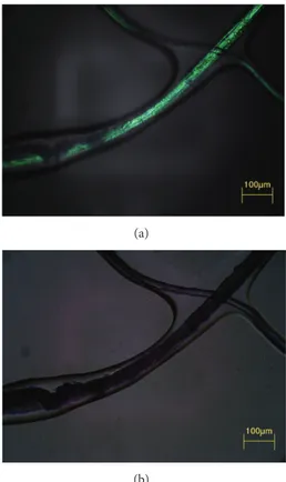
polar-ized light microscopic images, as shown in Figure4. Using
polarized light microscopy, it was confirmed that the intensity
of the polarized light was cyclically changed with 90∘period.
Its maximum intensity and minimum intensity (i.e., extinc-tion) were shown by turns, which is the consistent result with
extravascular primo vessel in the brain [11].
4. Discussion
This is the first report of NTS on the surface of a large animal organ. We found elastic, milky-white, and semitransparent threadlike structures (that formed network structures) in
situ and in vivo on the large intestine of swine. Polarized
light microscopy DAPI staining revealed that the observed structures fit the definition of NTS.
Anatomical observations alone indicated that the NTSs were distinctively different from well-known systems like
nerves, blood vessels, capillaries, and lymph vessels [8]. The
NTSs could be moved freely and could be lifted above the organ surface. There have been no reports of mammalian anatomy in which nerves, blood, or lymph vessels were separated from and raised above the surfaces of organs in normal animals. The NTSs were distinctively different from
Figure 3: Fluorescence image of DAPI-stained nuclei in a novel threadlike structure (NTS). Rod-shaped nuclei are aligned in a broken-line fashion (dotted-oval circle). This characteristic feature of NTS is commonly seen in other animals and organs.
(a)
(b)
Figure 4: Polarized light microscopic images of a novel threadlike structure obtained from the large intestine of a pig. The NTS appeared to be bright (a), while it becomes dark when the polarized angle is rotated 45∘(b). This distinct polarization property means that the NTS contains a significant amount of collagen fibers which are oriented in a single direction. Scale bar is 100𝜇m.
nerves because liquid flowed out from corpuscles that were sparsely located along the NTS. The NTSs were also easily distinguished from blood capillaries or venules due to ery-throcytes because the NTSs were semitransparent and milky white. In addition, the NTSs have been observed inside large
arteries and veins [1]. However, the NTSs could be mistaken
for lymphatic vessels. It is this possibility of confusion with the lymph system that caused medical and veterinary experts
lymph vessels carry lymphocytes that are 5𝜇m or larger. The alignment of rod-shaped nuclei along the major axis of NTSs is accepted as one of the characteristics that can be used to discriminate NTSs from other similar-looking tissues or
artifacts [13]. Our fluorescence images of DAPI-stained
struc-tures (Figure3) showed that the rod shape and linear align
ment of the nuclei aligned the major axis of the NTS. In addi-tion, the NTS samples showed the same polarization property
as the primo vessels in the cranial dura mater (Figure4). This
property is meaningful because it indicates that dense colla-gen fibers which are oriented in a particular direction exists
in the NTS [11]. It also makes the NTSs differentiats from
the blood and lymphatic vessels which do not show distinct change of polarized light with regard to the polarization angle. Therefore, we considered the observed network struc-tures on the swine large intestine surface to be NTSs.
The most striking figure of our result is that thickness of the threadlike structure was uniform, with a diameter of
approximately 30𝜇m. This diameter was similar to that
reported for NTS from small animals. Kim primarily used rabbits as laboratory animals. Although Kim did not present data for other animals, he mentioned that NTSs had been observed in various animals, including humans, other mam-mals, avians, amphibians, fish, and invertebrates, without
suggesting any specific animal species [9]. Until now,
investi-gators have tried to confirm the existence of NTSs using the organs of various animals, such as mice, rats, and rabbits. A
few case studies have been reported for cows [5] and dogs [14].
As we mentioned previously, as the animals are divided from lower to higher, the NTS of animals can be divided in a similar way. The development of the NTS may be progressive, with the most developed one being in humans. Therefore, it is con-ceivable that NTSs in large animals might have a larger diam-eter than the NTSs in small animals because the diamdiam-eter of blood vessels is relatively large in large animals. According to
Lee et al. [5], however, the diameter of NTSs in the bovine
heart is18.38 ± 17.8 𝜇m. Although exact diameter was not
presented, NTSs in dogs are estimated to be smaller than
20𝜇m in diameter [14]. The average NTS diameter in small
animals is about 10𝜇m and ranges from 1 𝜇m to 50 𝜇m [15].
Our results suggest that the diameter of human NTSs may be similar to those of small animals. Such small structures would be difficult to detect with the naked eye.
There are some limitations to the present study. Histologi-cal results of examined specimens should have been present-ed. However, we were not sure as to whether we would find NTSs in the swine large intestine. Therefore, this study was planned to confirm the presence of NTSs on the intestinal surface of this large animal. The results of the present study
Conflict of Interests
The authors listed above declare no conflict of interests.
Acknowledgment
This work was supported by the Industrial Strategic Tech-nology Program of the Ministry of Knowledge Economy (10041120). K.-H. Bae and S. H. Park have equally contributed to this project. The authors give special thanks to Professor K. S. Soh for support and advice. Professor S. Z. Yoon pays especially his homage to Professor K. S. Soh for his great achievements since 2002.
References
[1] X. Jiang, H. K. Kim, H. S. Shin, B. C. Lee, C. Choi, K. S. Soh et al., “Method for observation intravascular Bonghan ducts,” Korean Journal of Oriental Preventive Medical Society, vol. 6, pp. 162– 166, 2002.
[2] B. H. Kim, “On the Kyungrak system,” Journal of the Academy of Medical Sciences of the Democratic People’s Republic of Korea, vol. 90, pp. 1–35, 1963.
[3] B. C. Lee, J. S. Yoo, K. Y. Baik, K. W. Kim, and K. S. Soh, “Novel threadlike structures (Bonghan ducts) inside lymphatic vessels of rabbits visualized with a Janus Green B staining method,” Anatomical Record B, vol. 286, no. 1, pp. 1–7, 2005.
[4] H. M. Johng, J. S. Yoo, T. J. Yoon et al., “Use of magnetic nano-particles to visualize threadlike structures inside lymphatic ves-sels of rats,” Evidence-Based Complementary and Alternative Medicine, vol. 4, no. 1, pp. 77–82, 2007.
[5] B. C. Lee, H. B. Kim, B. Sung et al., “Network of endocardial vessels,” Cardiology, vol. 118, pp. 1–7, 2011.
[6] J. Lim, J. H. Jung, S. Lee et al., “Estimating the density of fluo-rescent nanoparticles in the primo vessels in the fourth ventricle and the spinal cord of a rat,” Journal of Biomedical Optics, vol. 6, no. 11, Article ID 116010, 2011.
[7] H. S. Shin, H. M. Johng, B. C. Lee et al., “Feulgen reaction study of novel threadlike structures (Bonghan ducts) on the surfaces of mammalian organs,” Anatomical Record B, vol. 284, no. 1, pp. 35–40, 2005.
[8] B. C. Lee, J. S. Yoo, V. Ogay et al., “Electron microscopic study of novel threadlike structures on the surfaces of mammalian organs,” Microscopy Research and Technique, vol. 70, no. 1, pp. 34–43, 2007.
[9] B. H. Kim, “The Kyungrak System,” Journal of Jo Sun Medicine, vol. 108, pp. 1–38, 1965.
[10] J. S. Yoo, M. S. Kim, V. Ogay, and K. S. Soh, “In vivo visualization of Bonghan ducts inside blood vessels of mice by using
an Alcian blue staining method,” Indian Journal of Experimental Biology, vol. 46, no. 5, pp. 336–339, 2008.
[11] M. H. Nam, J. Lim, J. H. Yoon et al., “Polarized light microscopy as an additional tool foridentifying primo vascular system in brain” in press.
[12] H. S. Shin, H. M. Johng, B. C. Lee et al., “Feulgen reaction study of novel threadlike structures (Bonghan ducts) on the surfaces of mammalian organs,” Anatomical Record B, vol. 284, no. 1, pp. 35–40, 2005.
[13] B. C. Lee, B. Sung, K. H. Eom et al., “Novel threadlike structures on the surfaces of mammalian abdominal organs are loose bundles of fibrous stroma with microchannels embedded with fibroblasts and inflammatory cells,” Connective Tissue Research, vol. 54, no. 2, pp. 94–100, 2013.
[14] Z. F. Jia, K. S. Soh, Q. Zhou, B. Dong, and W. H. Yu, “Study of novel threadlike structures on the intestinal fascia of dogs,” Jour-nal of Acupuncture and Meridian Studies, vol. 4, no. 2, pp. 98– 101, 2011.
[15] S. Lee, Y. Ryu, Y. Yun et al., “Anatomical discrimination of the differences between torn mesentery tissue and internal organ-surface primo-vessels,” Journal of Acupuncture and Meridian Studies, vol. 3, no. 1, pp. 10–15, 2010.
Submit your manuscripts at
http://www.hindawi.com
Hindawi Publishing Corporation
http://www.hindawi.com Volume 2013
Oxidative Medicine and Cellular Longevity
International Journal of
Endocrinology
Hindawi Publishing Corporationhttp://www.hindawi.com Volume 2013
ISRN
Anesthesiology Hindawi Publishing Corporation
http://www.hindawi.com Volume 2013
PPAR
R e s e a r c h
Hindawi Publishing Corporation
http://www.hindawi.com Volume 2013
Ophthalmology
Journal of Hindawi Publishing Corporationhttp://www.hindawi.com Volume 2013
ISRN
Allergy Hindawi Publishing Corporation
http://www.hindawi.com Volume 2013
BioMed Research International
Hindawi Publishing Corporation
http://www.hindawi.com Volume 2013
ISRN
Addiction Hindawi Publishing Corporation
http://www.hindawi.com Volume 2013
Hindawi Publishing Corporation
http://www.hindawi.com Volume 2013 Computational and Mathematical Methods in Medicine ISRN AIDS Hindawi Publishing Corporation
http://www.hindawi.com Volume 2013
Clinical & Developmental Immunology
Hindawi Publishing Corporation
http://www.hindawi.com Volume 2013
Evidence-Based Complementary and Alternative Medicine
Volume 2013 Hindawi Publishing Corporation
http://www.hindawi.com
Hindawi Publishing Corporation
http://www.hindawi.com Volume 2013
ISRN
Biomarkers
Hindawi Publishing Corporation
http://www.hindawi.com Volume 2013