0099-2240/11/$12.00 doi:10.1128/AEM.02527-10
Copyright © 2011, American Society for Microbiology. All Rights Reserved.
Development of a Streptomyces venezuelae-Based Combinatorial
Biosynthetic System for the Production of Glycosylated Derivatives
of Doxorubicin and Its Biosynthetic Intermediates
䌤
†
Ah Reum Han,
1Je Won Park,
2,4Mi Kyeong Lee,
3Yeon Hee Ban,
2Young Ji Yoo,
2Eun Ji Kim,
2Eunji Kim,
2Byung-Gee Kim,
1Jae Kyung Sohng,
4and Yeo Joon Yoon
2*
Interdisciplinary Programs of Bioengineering, Seoul National University, Seoul 151-742, Republic of Korea1; Department of
Chemistry and Nano Sciences, Ewha Womans University, Seoul 120-750, Republic of Korea2; College of Pharmacy,
Chungbuk National University, Cheongju 361-763, Republic of Korea3; and Department of
Pharmaceutical Engineering, Institute of Biomolecule Reconstruction,
Sun Moon University, Asan 336-708, Republic of Korea4
Received 26 October 2010/Accepted 11 May 2011
Doxorubicin, one of the most widely used anticancer drugs, is composed of a tetracyclic polyketide aglycone andL-daunosamine as a deoxysugar moiety, which acts as an important determinant of its biological activity. This is exemplified by the fewer side effects of semisynthetic epirubicin (4ⴕ-epi-doxorubicin). An efficient combinatorial biosynthetic system that can convert the exogenous aglycone -rhodomycinone into diverse glycosylated derivatives of doxorubicin or its biosynthetic intermediates, rhodomycin D and daunorubicin, was developed through the use of Streptomyces venezuelae mutants carrying plasmids that direct the biosynthesis of different nucleotide deoxysugars and their transfer onto aglycone, as well as the postglycosylation modifica-tions. This system improved epirubicin production from -rhodomycinone by selecting a substrate flexible glycosyltransferase, AknS, which was able to transfer the unnatural sugar donors and a TDP-4-ketohexose reductase, AvrE, which efficiently supported the biosynthesis of TDP-4-epi-L-daunosamine. Furthermore, a range of doxorubicin analogs containing diverse deoxysugar moieties, seven of which are novel rhodomycin D derivatives, were generated. This provides new insights into the functions of deoxysugar biosynthetic enzymes and demonstrates the potential of the S. venezuelae-based combinatorial biosynthetic system as a simple biological tool for modifying structurally complex sugar moieties attached to anthracyclines as an alternative to chemical syntheses for improving anticancer agents.
Anthracyclines, effective anticancer drugs, have a basic 7,8,9,10-tetrahydro-5,12-naphthacenequinone structure that is glycosylated with one or more sugar residues. Doxorubicin and daunorubicin, produced by Streptomyces peucetius, are repre-sentative anthracyclines and are approved for clinical use in, for example, breast and small lung cancers, acute myeloid leukemia, and different sarcomas (1, 2). The anticancer activ-ities of anthracyclines generally result from DNA damage via the inhibition of DNA topoisomerase II and the generation of free radicals, DNA binding and alkylation, and DNA interca-lation (28). These activities are closely related to the sugar components, which usually participate in molecular recogni-tion of cellular targets. Their presence and structure are there-fore important, or even essential, to the biological activity of anthracyclines (45). The severe side effects of anthracycline, such as dose-dependent cardiotoxicity, have been the main problems associated with the therapeutic use of these drugs. The clinically important epirubicin (4⬘-epi-doxorubicin) has successfully overcome these side effects. It is a semisynthetic analogue of doxorubicin with an opposite configuration of the
hydroxyl group at C-4 of the deoxysugar. It displays reduced cardiotoxicity while maintaining similar antitumor properties compared to doxorubicin (12). Therefore, alteration of the sugar portion of anthracyclines is hoped to produce the next generation of anticancer agents with improved activities and reduced toxicities. Great efforts have already been made over the last decade to modify the glycosylation patterns of natural products by combinatorial biosynthesis as an alternative to chemical modification of the structurally complex sugar moiety (31). 4⬘-epi-Daunorubicin had been produced directly by fer-mentation from an S. peucetius mutant in which the dnmV gene involved in the determination of the stereochemistry of the C-4⬘ hydroxyl group was replaced by Streptomyces avermitilis avrE or Saccharopolyspora erythraea eryBIV. However, the pro-duction level of epirubicin was too low to be quantified (24). Little progress to improve the biological production of epiru-bicin has been made. Consequently, it is still manufactured semisynthetically from doxorubicin aglycone or daunorubicin by several chemical steps. Moreover, combinatorial biosynthe-sis of the doxorubicin analogs containing diverse sugar moi-eties has not been reported.
The pikromycin-producing Streptomyces venezuelae ATCC 15439 has been developed as a favorable host for combinato-rial biosynthesis and heterologous expression of natural prod-uct biosynthetic pathways originating in other organisms due to its fast growth and relative ease of genetic manipulation (10, 13–15, 33–35). In the present study, an S. venezuelae-based
* Corresponding author. Mailing address: Department of Chemistry and Nano Sciences, Ewha Womans University, Seoul 120-750, Repub-lic of Korea. Phone: 82-2-3277-4082. Fax: 82-2-3277-3419. E-mail: joonyoon@ewha.ac.kr.
† Supplemental material for this article may be found at http://aem .asm.org/.
䌤Published ahead of print on 20 May 2011.
4912
on June 26, 2016 by Ewha Womans Univ
http://aem.asm.org/
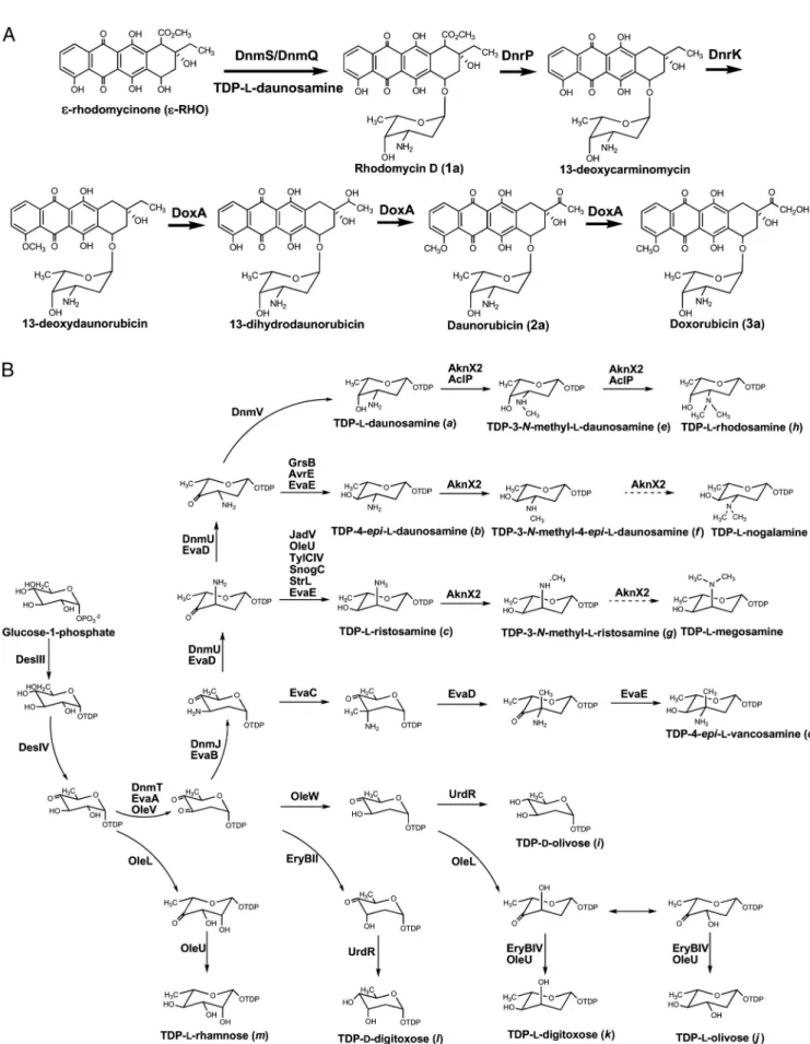
combinatorial biosynthetic system was constructed to convert exogenously fed doxorubicin aglycone ε-rhodomycinone (ε-RHO) into glycosylated doxorubicin analogs. This was achieved through the heterologous expression of different combinations of genes for the synthesis of various nucleotide-activated deoxysugars and their transfer toε-RHO, as well as genes for further elaboration of rhodomycin D (a glycosylated form ofε-RHO) to doxorubicin in a mutant strain of doxoru-bicin-resistant S. venezuelae in which the entire pikromycin biosynthetic gene cluster was deleted (Fig. 1). This genetically amenable and fast-growing S. venezuelae system allowed the exploration of substrate flexibility of anthracycline glycosyl-transferases to transfer desired sugars to aglycone as well as the ability of thymidine-5⬘-diphospho (TDP)-4-ketohexose re-ductases to synthesize TDP-4-epi-L-daunosamine to enhance
the in vivo production of epirubicin from ε-RHO. Further-more, 17 unnatural glycosylated derivatives of doxorubicin or its biosynthetic intermediates were generated (Fig. 2), demon-strating the potential of this S. venezuelae system for engi-neered generation of doxorubicin analogs with modified sugar moieties as well as other polyketides glycosylated with unnat-ural sugars (10, 13–15).
MATERIALS AND METHODS
Bacterial strains, culture conditions, and genetic manipulation.Escherichia coli DH5␣ was used for routine subcloning. S. venezuelae YJ028 lacking the pikromycin polyketide synthase genes and genes for the biosynthesis of TDP-D -desosamine (14) was used as a heterologous host and was maintained on SPA medium (1 g of yeast extract, 1 g of beef extract, 2 g of tryptose, 10 g of glucose, a trace amount of ferrous sulfate, and 15 g of agar/liter). Protoplast formation and transformation procedures of S. venezuelae were performed as described previously (16), and transformants of S. venezuelae were selected in R2YE agar plates (16) supplemented with appropriate antibiotics. The genomic DNAs were prepared from a variety of actinomycetes: S. peucetius ATCC 29050, S.
venezu-elae ATCC 15439, S. venezuvenezu-elae ISP 5230, Streptomyces avermitilis K139, Strep-tomyces galilaeus ATCC 31615, StrepStrep-tomyces fradiae ATCC 19609, StrepStrep-tomyces griseus ATCC 10137, Amycolaptosis orientalis NRRL 18098, Streptomyces antibi-oticus ATCC 11891, Streptomyces griseus subsp. griseus NBRC 13350, and Strep-tomyces nogalater ATCC 27451. The sources of these strains were described in
Table S1 in the supplemental material.
Construction of expression plasmids.The DNA fragments used for the con-struction of expression plasmids in the present study were amplified by PCR with the corresponding primers and template DNAs (see Table S1 in the supplemen-tal material). PCR was performed by using Pfu polymerase (Fermentas) accord-ing to the manufacturer’s recommended conditions. All PCR products were cloned into plasmid Litmus28 (New England Biolabs) and sequenced to confirm their authenticity. Doxorubicin resistance genes were cloned into the integrative plasmid pSET152 (4) derivative containing ermE* promoter (40) and an apra-mycin resistance marker. To construct the plasmids expressing doxorubicin re-sistance genes, a BglII/HindIII fragment containing drrA-drrB and a HindIII/ XbaI fragment containing drrC were PCR amplified from the genomic DNA of
S. peucetius ATCC 29050, ligated into the BglII/XbaI sites of Litmus28, and
moved into BamHI/XbaI sites of pSET152 downstream of the ermE* promoter generating the plasmid pABC.
The genes involved in the conversion ofε-RHO to rhodomycin D or doxoru-bicin derivatives were cloned into the replicative plasmid pSE34 (49), E.
coli-Streptomyces shuttle vector containing ermE* promoter, and thiostrepton
resis-tance marker. To construct plasmids harboring several genes, the following general method was used. The PacI/XbaI fragment containing one gene was cloned to PacI/SpeI-digested Litmus28 carrying other genes. This process was repeated until all of the genes involved in the conversion ofε-RHO to rhodo-mycin D (compound 1a), daunorubicin (compound 2a), doxorubicin (compound 3a), or their derivatives were combined in Litmus28 and then transferred into pSE34 digested with PacI/XbaI. Genes encoding glycosyltransferase/auxiliary protein pairs, TDP-4-ketohexose reductases, and other sugar biosynthetic en-zyme sets were respectively substituted by using EcoRV/NdeI, SnaBI/AvrII, and ClaI/XbaI sites in Litmus28. The catalytic functions of the genes used in the
present study are listed in Table 1. The constructed gene combinations are summarized in Table 2 (plasmids for the production of rhodomycin D, doxoru-bicin, 4⬘-epi-rhodomycin D, and epirubicin), and Table 3 (plasmids for the pro-duction of glycosylated derivatives of rhodomycin D and doxorubicin).
In order to generate pDNS1, the genes encoding the AknS/AknT glycosyl-transferase/auxiliary protein pair and TDP-L-daunosamine biosynthetic enzymes were sequentially cloned in Litmus28 to produce pDNS1_lit28. The insert DNA of pDNS1_lit28 was digested with PacI/XbaI, and ligated to pSE34 to give pDNS1. To construct a pDNS1 derivative carrying dnmQ-dnmS instead of
aknT-aknS, the EcoRV/NdeI fragment carrying dnmQ-dnmS was ligated into EcoRV/
NdeI-digested pDNS1_lit28, generating pDNS2_lit28. The insert DNA of pDNS2_lit28 was cloned into pSE34 to produce pDNS2. Plasmids pDXR1 and pDXR2, containing postglycosylation modification genes were constructed by adding PacI/XbaI fragments released from pDNS1 into PacI/SpeI sites of pSE34 carrying dnrK-dnrP-doxA and dnrK-dnrP-dnrV-doxA, respectively. To generate plasmids expressing the genes encoding enzymes that convertε-RHO to 4⬘-epi-rhodomycin D (compound 1b), SnaBI/AvrII fragment carrying dnmV in pDNS1_lit28 was replaced by the SnaBI/AvrII fragment containing avrE, pro-ducing pEDNS1_lit28. The PacI/XbaI fragment from pEDNS1_lit28 was ligated to pSE34, yielding pEDNS1. To construct pEDNS1 derivatives, in which
aknT-aknS was replaced with the genes encoding other glycosyltransferase/auxiliary
protein pairs, EcoRV/NdeI fragment carrying dnmQ-dnmS, stfPII-stfG, or
snogN-snogE was ligated to pEDNS1_lit28 lacking aknT-aknS, and then the
insert DNAs of pEDNS1_lit28 derivatives were independently cloned to pSE34 producing pEDNS2, pEDNS3, or pEDNS4, respectively. pEDNS1 derivatives carrying genes coding different TDP-4-ketohexose reductases were constructed by replacing avrE in pEDNS1 using SnaBI/AvrII sites with grsB, evaE, jadV, oleU,
tylCIV, strL, or snogC, generating pEDNS5, pEDNS6, pRST1, pRST2, pRST3,
pRST4, or pRST5, respectively. To construct several plasmids (pEVCS, pMDNS1, pMDNS2, pRDS, pMEDNS, pMRST, pDOLV, pDDGT, pLOLV, pLDGT, and pLRHM) that direct the biosynthesis of different deoxysugars and their attachment, ClaI/XbaI fragments carrying different sugar gene cassettes were separately cloned into ClaI/XbaI-digested pEDNS1_lit28. The PacI/XbaI fragments of the resulting plasmids were independently cloned into pSE34. The PacI/XbaI fragment released from pEDNS1, pRST1, pMDNS1, or pEVCS was cloned into the PacI/SpeI sites of pSE34 harboring dnrK-dnrP-dnrV-doxA to generate pEDXR, pRSDXR, pMDXR, or pEVDXR, respectively.
Production and analysis of anthracycline glycosides. S. venezuelae strains
expressing different combinations of sugar biosynthetic genes, genes encoding glycosyltransferase/auxiliary protein pairs, and genes participating in postglyco-sylation modifications were cultivated at 30°C for 5 days on R2YE agar supple-mented with 23.3M (10 mg/liter) ε-RHO. The extraction of the anthracyclines produced in the S. venezuelae cultures was carried out as follows. The grown culture was diced, extracted with 50% methanol, and then centrifuged at 5,000⫻
g for 10 min. The crude extract was passed through an Oasis HLB (Waters)
solid-phase extraction column, preconditioned with 3 ml of methanol, followed by 3 ml of 50% (vol/vol) methanol. The column was washed with 3 ml of 50% (vol/vol) methanol and then air dried for ca. 30 s. The attached anthracyclines were eluted three times with 1 ml of 5% (vol/vol) methanolic formic acid, evaporated to dryness at room temperature by vacuum centrifugation. Rhodo-mycin D (compound 1a), daunorubicin (compound 2a), doxorubicin (compound 3a), and their glycosylated derivatives produced by S. venezuelae mutant strains were analyzed by high-performance liquid chromatography-electrospray ioniza-tion-tandem mass spectrometry (MS/MS). Analytical HPLC-ESI-MS/MS was performed in a Waters/Micromass Quattro micro/MS interface using a Phenomenex Synergi Polar-RP column (150 mm by 4.6 mm, 4m) in positive-ion mode. The analytes were eluted at a flow rate of 250l/min using a gradient of 5 mM (wt/vol) ammonium acetate and 0.05% (vol/vol) acetic acid in water (solution A) and 80% (vol/vol) acetonitrile with the same additive con-centrations (solution B) at 20 to 70% solution B for 25 min, increasing to 80% solution B for 15 min, maintained at 80% solution B for 9 min, and then reduced to 20% solution B for 11 min for column re-equilibration. Quantification was conducted in multiple-reaction-monitoring (MRM) mode using ESI-MS/MS by selecting the two mass ions set to detect a transition of the parent ion to the product ion specific to the selected analytes. The productivities of all of the anthracyclines produced in the S. venezuelae-based bioconversion system are summarized in Tables 2 and 3. Independent experiments were carried out in triplicate. Preparative HPLC was performed with the same analytical column and mobile phase as the analytical HPLC system for isolating and purifying rhodo-mycin D (compound 1a) and its derivatives. Samples were prepared for nuclear magnetic resonance (NMR) spectroscopy, by each compound being dissolved in 250l of CD3OD and then placed in a 5-mm Shigemi advanced NMR microtube
(Sigma) matched to the solvent. NMR data for each purified compound were
on June 26, 2016 by Ewha Womans Univ
http://aem.asm.org/
FIG. 1. Proposed pathways fromε-rhodomycinone into rhodomycin D, daunorubicin, and doxorubicin (A) and biosynthesis of the different deoxysugars directed by the plasmids described in the present study (B).
on June 26, 2016 by Ewha Womans Univ
http://aem.asm.org/
acquired at 298 K on a Bruker Avance 500-MHz spectrometer. Proton and carbon chemical shifts were referenced to the tetramethylsilane signal. The structure of each compound was assigned based on its1
H-NMR spectrum com-pared to literature values.13C analysis was also carried out if necessary.ε-RHO
was purchased from Galilaeus (Kaarina, Finland). Standard doxorubicin (com-pound 3a), daunorubicin (com(com-pound 2a), and epirubicin (com(com-pound 3b) were obtained from GeneChem, Inc. (Daejeon, South Korea).
RESULTS
Construction of S. venezuelae mutant strains for bioconver-sion of-rhodomycinone to doxorubicin. To confer
self-resis-tance to doxorubicin (compound 3a) in the anthracycline-non-producing strain S. venezuelae, pABC carrying the three doxorubicin resistance genes drrA, drrB (9), and drrC (19) was introduced into the chromosome of S. venezuelae mutant
YJ028 bearing a deletion of the entire pikromycin biosynthetic gene cluster encoding the polyketide synthases and TDP-D
-desosamine biosynthetic enzymes, generating S. venezuelae YJ183. The mutant strain YJ183 was resistant to 25.9 M commercially available doxorubicin hydrochloride, compared to a resistance to⬍1.7 M in S. venezuelae YJ028. S. venezu-elae YJ183 was used as heterologous host in subsequent ex-periments.
In order to biotransform ε-RHO to rhodomycin D (com-pound 1a), the first glycosylated intermediate of doxorubicin (compound 3a) biosynthesis in S. peucetius ATCC 29050 (Fig. 1A), two plasmids were constructed: pDNS1 and pDNS2, which contained sets of genes encoding the glycosyltransferase/ auxiliary protein pair, together with TDP-L-daunosamine
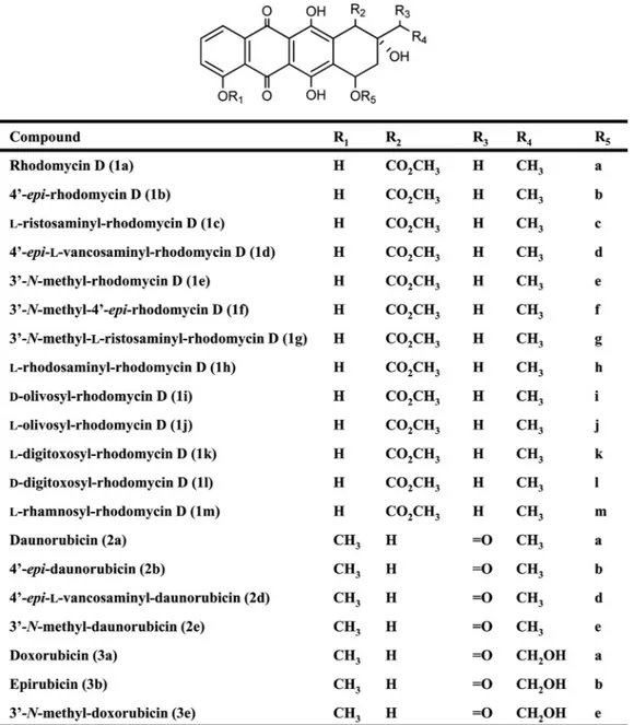
bio-FIG. 2. Proposed structures for rhodomycin D, daunorubicin, doxorubicin and their derivatives. Column R5: a,L-daunosamine; b, 4-epi-L
-daunosamine; c,L-ristosamine; d, 4-epi-L-vancosamine; e, 3-N-methyl-L-daunosamine; f, 3-N-methyl-4-epi-L-daunosamine; g, 3-N-methyl-L -ris-tosamine; h,L-rhodosamine; i,D-olivose; j,L-olivose; k,L-digitoxose; l,D-digitoxose; m,L-rhamnose.
on June 26, 2016 by Ewha Womans Univ
http://aem.asm.org/
synthetic genes (29) (Table 2). For the biosynthesis of the common deoxysugar biosynthetic intermediate TDP-4-keto-6-deoxy-D-glucose, the glucose-1-phosphate thymidylyl
trans-ferase gene desIII and glucose-4,6-dehydratase gene desIV of S. venezuelae ATCC 15439 (48) were used instead of the corre-sponding genes in the TDP-L-danunosamine biosynthetic gene
cluster of S. peucetius ATCC 29050. The AknS/AknT in pDNS1 encodes the rhodosaminyltransferase/auxiliary protein
pair involved in the biosynthesis of aclacinomycin A from S. galilaeus ATCC 31615 (18), whereas pDNS2 contains dnmS/ dnmQ, which encodes the glycosyltransferase/auxiliary protein pair participating in glycosylation of ε-RHO with TDP-L
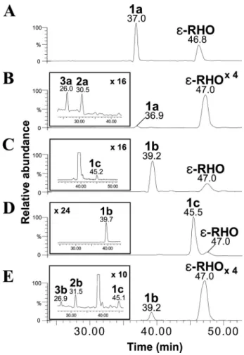
-daunosamine from S. peucetius ATCC 29050 (32). The auxil-iary helper proteins such as AknT and DmnQ are required for efficient in vivo glycosylation (11, 21). Both pDNS1 and pDNS2 were independently introduced into S. venezuelae YJ183 to yield the mutant strains YJ183/pDNS1 and YJ183/pDNS2, re-spectively. The transformants were cultured on R2YE agar supplemented with ε-RHO, extracted by solid-phase extrac-tion, and then analyzed by HPLC-ESI-MS/MS. The peak corresponding to rhodomycin D (compound 1a; 7-O-L -daunosaminyl-ε-RHO), selected at a mass transition from m/z 558 to 130, was detected by HPLC-ESI-MS/MS with a reten-tion time (Rt) of 37.0 min (Fig. 3A and see Fig. S1-1B in the supplemental material for its MS/MS fragmentation pattern). The production of rhodomycin D (compound 1a) in YJ183/ pDNS1 was 2.5-fold higher than in YJ183/pDNS2 (Table 2), indicating that aknS/aknT can more efficiently transfer
TDP-L-daunosamine to the exogenousε-RHO in the heterologous
host S. venezuelae. Therefore, further study of modifications of rhodomycin D (compound 1a) to doxorubicin (compound 3a) was carried out using AknS as glycosyltransferase. Upon HPLC-ESI-MS/MS analysis, YJ183/pDXR1, carrying dnrP, dnrK, and doxA, which encode the enzymes responsible for the postglycosylation modifications of rhodomycin D (compound 1a) in the doxorubicin biosynthetic pathway (20, 22, 23), in addition to the genes contained in pDNS1, produced only
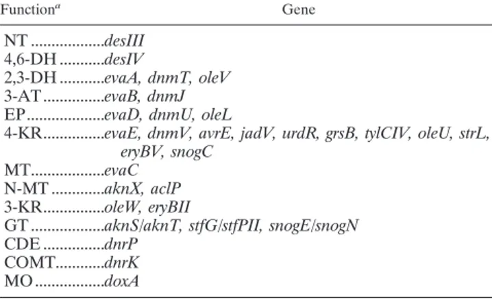
TABLE 1. Genes used in this study and their catalytic functions Functiona Gene NT ...desIII 4,6-DH ...desIV 2,3-DH ...evaA, dnmT, oleV 3-AT ...evaB, dnmJ EP...evaD, dnmU, oleL
4-KR...evaE, dnmV, avrE, jadV, urdR, grsB, tylCIV, oleU, strL, eryBV, snogC
MT...evaC N-MT ...aknX, aclP 3-KR...oleW, eryBII
GT ...aknS/aknT, stfG/stfPII, snogE/snogN CDE ...dnrP
COMT...dnrK MO ...doxA
aAbbreviations: NT, nucleotidyltransferase; 4,6-DH, 4,6-dehydratase;
2,3-DH, 2,3-dehydratase; 3-AT; 3-aminotransferase; EP, epimerase; 4-KR, 4-ke-toreductase; MT, methyltransferase; N-MT, N-methyltransferase; 3-KR, 3-ketoreductase; GT, glycosyltransferase/auxiliary protein pair; CDE, 10-carbomethoxy-13-deoxycarminomycin esterase; COMT, carminomycin
o-methyltransferase; MO, cytochrome P450 monooxygenase.
TABLE 2. Biosynthetic gene sets expressed in the engineered strain of S. venezuelae for bioconversion ofε-RHO to doxorubicin or epirubicin and productivities of doxorubicin, its intermediates, and epirubicin, by S. venezuelae mutant strains
Plasmid Deoxysugar biosynthetic gene set GTa Post-GTb Compound
(productivity)c
DNS1 desIII-desIV-dnmT-dnmJ-dnmU-dnmV aknS/aknT 1a (14.3⫾ 0.21) DNS2 desIII-desIV-dnmT-dnmJ-dnmU-dnmV dnmS/dnmQ 1a (5.7⫾ 0.65) DXR1 desIII-desIV-dnmT-dnmJ-dnmU-dnmV aknS/aknT dnrK-dnrP-doxA 1a (10.8⫾ 1.36) DXR2 desIII-desIV-dnmT-dnmJ-dnmU-dnmV aknS/aknT dnrK-dnrP-dnrV-doxA 1a (2.8⫾ 0.5)
2a (0.9⫾ 0.04) 3a (1.1⫾ 0.13)
EDNS1 desIII-desIV-dnmT-dnmJ-dnmU-avrE aknS/aknT 1b (13.4⫾ 2.51)
1c (0.2⫾ 0.05)
EDNS2 desIII-desIV-dnmT-dnmJ-dnmU-avrE dnmS/dnmQ 1b (0.1⫾ 0.03)
EDNS3 desIII-desIV-dnmT-dnmJ-dnmU-avrE stfG/stfPII 1b (10.9⫾ 0.92)
EDNS4 desIII-desIV-dnmT-dnmJ-dnmU-avrE snogE/snogN 1b (5.1⫾ 1.16)
EDNS5 desIII-desIV-dnmT-dnmJ-dnmU- grsB aknS/aknT 1b (0.2⫾ 0.04)
EDNS6 desIII-desIV-dnmT-dnmJ-dnmU- evaE aknS/aknT 1b (10.0⫾ 1.02)
1c (4.8⫾ 1.21) RST1 desIII-desIV-dnmT-dnmJ-dnmU-jadV aknS/aknT 1b (0.4⫾ 0.06) 1c (15.2⫾ 1.94) RST2 desIII-desIV-dnmT-dnmJ-dnmU-oleU aknS/aknT 1b (0.2⫾ 0.06) 1c (15.1⫾ 1.43) RST3 desIII-desIV-dnmT-dnmJ-dnmU-tylCIV aknS/aknT 1c (0.5⫾ 0.14) RST4 desIII-desIV-dnmT-dnmJ-dnmU-strL aknS/aknT 1c (0.9⫾ 0.07) RST5 desIII-desIV-dnmT-dnmJ-dnmU-snogC aknS/aknT 1c (0.7⫾ 0.05) EDXR desIII-desIV-dnmT-dnmJ-dnmU-avrE aknS/aknT dnrK-dnrP-dnrV-doxA 1b (6.0⫾ 1.22) 1c (0.38⫾ 0.1) 2b (3.7⫾ 0.23) 3b (0.9⫾ 0.11)
a
GT, glycosyltransferase/auxiliary protein-encoding gene pair.
b
Post-GT, postglycosylation enzyme-encoding genes.
cCompounds: 1a, rhodomycin D; 2a, daunorubicin; 3a, doxorubicin; 1b, 4⬘-epi-rhodomycin D; 1c,
L-ristosaminyl-rhodomycin D; 2b, 4⬘-epi-daunorubicin; 3b,
epirubicin. The productivity is expressed as the mean concentration (M) ⫾ the standard error of the mean.
on June 26, 2016 by Ewha Womans Univ
http://aem.asm.org/
rhodomycin D (compound 1a), whereas YJ183/pDXR2, which harbors the additional gene dnrV, produced rhodomycin D (compound 1a), daunorubicin (compound 2a; m/z 528⬎ 130 in MRM mode), and doxorubicin (compound 3a; m/z 544⬎ 130 in MRM mode) at Rts of 36.9, 30.5, and 26.0 min, respectively (Fig. 3B and Table 2; see also Fig. S1-2B and Fig. S1-3B in the supplemental material for the MS/MS fragmentation patterns of compounds 2a and 3a, respectively). These results are con-sistent with previous results implicating dnrV in enzymatic re-actions catalyzed by DoxA, although its exact function was not established (20).
Bioconversion of-rhodomycinone to epirubicin. Epirubicin
(compound 3b) production in the S. venezuelae system re-quired the TDP-4-ketohexose reduction step which confers an equatorial C-4 hydroxyl group to TDP-4-keto-L-daunosamine,
the glycosylation step attaching TDP-4-epi-L-daunosamine to ε-RHO, producing 4⬘-epi-rhodomycin D (compound 1b), and the postglycosylation steps leading to epirubicin (compound 3b). Glycosyltransferase/auxiliary protein pairs capable of con-verting ε-RHO to 4⬘-epi-rhodomycin D (compound 1b) effi-ciently were first screened using a TDP-4-epi-L-daunosamine
biosynthetic gene cassette containing avrE, a gene encoding TDP-4-ketohexose reductase in theL-oleandrose biosynthetic
pathway from S. avermitilis (46), in place of dmnV since it has
already been reported that avrE can support 4 ⬘-epi-daunoru-bicin (compound 2b) production in S. peucetius (24). Four plasmids were constructed: pEDNS1, pEDNS2, pEDNS3, and pEDNS4 carrying aknS/aknT, dnmS/dnmQ, stfG/stfPII, and snogE/snogN, respectively (Table 2). StfG from S. steffisburgen-sis NRRL 3193 and SnogE from S. nogalater ATCC 27451 are glycosyltransferases involved in the biosynthesis of steffimycin and nogalamycin, respectively (30, 42). HPLC-ESI-MS/MS analyses showed that 4⬘-epi-rhodomycin D (compound 1b; 7-O-4⬘-epi-L-daunosaminyl-ε-RHO; m/z 558 ⬎ 130 in MRM mode) was detected at an Rt of 39.2 min from all of the YJ183 mutants carrying each of the constructed plasmids, with pEDNS1 supporting the highest production (Fig. 3C; see also Fig. S1-4B in the supplemental material for its MS/MS frag-mentation pattern), confirming that AknS is the most efficient glycosyltransferase toward the unnatural sugar donor TDP-4-epi-L-daunosamine among the tested glycosyltransferases
(Ta-ble 2). Interestingly, a new small peak with the same mass transition of the parent ion to the product ion and MS/MS fragmentation pattern as those of 4⬘-epi-rhodomycin D (com-pound 1b) was detected at an Rt of 45.2 min from the culture of YJ183/pEDNS1 and determined as a novelL -ristosaminyl-rhodomycin D (compound 1c; 7-O-L-ristosaminyl-ε-RHO)
(Fig. 3C; see also Fig. S1-5B in the supplemental material for
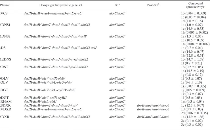
TABLE 3. Biosynthetic gene sets expressed in the engineered strain of S. venezuelae for bioconversion ofε-RHO to derivatives of rhodomycin D and doxorubicin and productivities of derivatives of rhodomycin D and doxorubicin by S. venezuelae mutant strains
Plasmid Deoxysugar biosynthetic gene set GTa Post-GTb Compound
(productivity)c
EVCS desIII-desIV-evaA-evaB-evaD-evaE-evaC aknS/aknT 1b (0.04⫾ 0.009) 1c (0.03⫾ 0.004) 1d (1.0⫾ 0.16) MDNS1 desIII-desIV-dnmT-dnmJ-dnmU-dnmV-aknX2 aknS/aknT 1a (1.0⫾ 0.07) 1e (14.9⫾ 0.53) 1h (0.005⫾ 0.002) MDNS2 desIII-desIV-dnmT-dnmJ-dnmU-dnmV-aclP aknS/aknT 1a (1.3⫾ 0.05) 1e (10.5⫾ 0.09) 1h (0.004⫾ 0.0007) RDS desIII-desIV-dnmT-dnmJ-dnmU-dnmV-aknX2-aclP aknS/aknT 1a (0.7⫾ 0.04) 1e (14.0⫾ 0.07) 1h (12.8⫾ 0.51)
MEDNS desIII-desIV-dnmT-dnmJ-dnmU-avrE-aknX2 aknS/aknT 1b (14.7⫾ 1.78)
1f (0.7⫾ 0.21) MRST desIII-desIV-dnmT-dnmJ-dnmU-jadV-aknX2 aknS/aknT 1b (0.2⫾ 0.05) 1c (14.5⫾ 2.15) 1g (0.8⫾ 0.12) DOLV desIII-desIV-oleV-urdR-oleW aknS/aknT 1i (0.3⫾ 0.07) LOLV desIII-desIV-oleV-oleL-oleU-oleW aknS/aknT 1j (0.6⫾ 0.10)
1k (0.02⫾ 0.005) LDGT desIII-desIV-oleV-oleL-eryBIV-oleW aknS/aknT 1j (0.05⫾ 0.009)
1k (0.3⫾ 0.07) DDGT desIII-desIV-oleV-urdR-eryBII aknS/aknT 1l (0.5⫾ 0.05)
LRHAM desIII-desIV-oleL-oleU aknS/aknT 1m (0.3⫾ 0.04)
RSDXR desIII-desIV-dnmT-dnmJ-dnmU-jadV aknS/aknT dnrK-dnrP-dnrV-doxA 1c (12.5⫾ 0.07) EVDXR desIII-desIV-evaA-evaB-evaD-evaE-evaC aknS/aknT dnrK-dnrP-dnrV-doxA 1d (0.7⫾ 0.03)
2d (0.06⫾ 0.0035) MDXR desIII-desIV-dnmT-dnmJ-dnmU-dnmV-aknX2 aknS/aknT dnrK-dnrP-dnrV-doxA 1e (13.9⫾ 1.86)
2e (0.1⫾ 0.02) 3e (0.3⫾ 0.02)
aGT, glycosyltransferase/auxiliary protein-encoding gene pair. bPost-GT, postglycosylation enzyme-encoding genes.
cCompounds: 1a, rhodomycin D; 1b, 4⬘-epi-rhodomycin D; 1c,L-ristosaminyl-rhodomycin D; 1d, 4⬘-epi-L-vancosaminyl-rhodomycin D; 1e, 3⬘-N-methyl-rhodomycin
D; 1f, 3⬘-N-methyl-4⬘-epi-L-daunosaminyl-rhodomycin D; 1g, 3⬘-N-methyl-L-ristosaminyl-rhodomycin D; 1h,L-rhodosaminyl-rhodomycin D; 1i,D-olivosyl-rhodomycin D; 1j,L-olivosyl-rhodomycin D; 1k,L-digitoxosyl-rhodomycin D; 1l,D-digitoxosyl-rhodomycin D; 1m,L-rhamnosyl-rhodomycin D; 2d, 4⬘-epi-L -vancosaminyl-daunoru-bicin; 2e, 3⬘-N-methyl-daunorubicin; 3e, 3⬘-N-methyl-doxorubicin. The productivity is expressed as the mean concentration (M) ⫾ the standard error of the mean.
on June 26, 2016 by Ewha Womans Univ
http://aem.asm.org/
its MS/MS fragmentation pattern), which might be generated by unexpected 5-epimerase activity of DmnU (Fig. 1B).
Next, several TDP-4-ketohexose reductases were tested for efficient biosynthesis of TDP-4-epi-L-daunosamine by con-structing the pEDNS1 derivatives: pEDNS5, pEDNS6, pRST1, pRST2, pRST3, pRST4, and pRST5, which include grsB (GenBank accession no. AF128273; http://www.ncbi.nlm.nih .gov/GenBank/index.html), evaE (5), jadV (44), oleU (46), tylCIV (3), strL (37), and snogC (42), respectively, that encode TDP-4-ketohexose reductases in other deoxysugar biosynthetic
pathways but act on different sugar intermediates (Table 2). Only small amounts of 4⬘-epi-rhodomycin D (compound 1b) were produced in the YJ183/pEDNS5 strain containing grsB (Table 2). Replacement of avrE by evaE (pEDNS6) led to the formation of twice as much 4⬘-epi-rhodomycin D (compound 1b) than L-ristosaminyl-rhodomycin D (compound 1c). jadV (pRST1; Fig. 3D) and oleU (pRST2) producedL
-ristosaminyl-rhodomycin D (compound 1c) as major product and only trace amounts of 4⬘-epi-rhodomycin D (compound 1b) (Table 2). Strains YJ183/pRST3, YJ183/pRST4, and YJ183/pRST5 pro-duced only small amounts of L-ristosaminyl-rhodomycin D
(compound 1c), but no 4⬘-epi-rhodomycin D (compound 1b) (Table 2). No TDP-4-ketohexose reductase tested supported higher production of 4⬘-epi-rhodomycin D (compound 1b) than AvrE (Table 2). Based on the selection of AknS and AvrE as an efficient glycosyltransferase and 4-ketohexose reductase to support 4⬘-epi-rhodomycin D (compound 1b) production in a S. venezuelae system, pEDXR was constructed by adding a dnrK-dnrP-dnrV-doxA gene cassette into pEDNS1 to produce epirubicin (compound 3b) fromε-RHO (Table 2). HPLC-ESI-MS/MS analysis showed that YJ183/pEDXR produced epiru-bicin (compound 3b; 4⬘-epi-L-daunosaminyl-doxorubicinone; Rt, 26.9 min; m/z 544 ⬎ 130 in MRM mode), along with 4⬘-epi-rhodomycin D (compound 1b; Rt, 39.2 min), L -ris-tosaminyl-rhodomycin D (compound 1c; Rt, 45.1 min), and 4⬘-epi-daunorubicin (compound 2b; 4⬘-epi-L -daunosaminyl-daunorubicinone; Rt, 31.5 min; m/z 528⬎ 130 in MRM mode) (Fig. 3E and see Fig. S1-6B and Fig. S1-7B in the supplemental material for the MS/MS fragmentation patterns of compounds 2b and 3b, respectively).
Generation of glycosylated derivatives of rhodomycin D.
The substrate flexibility of AknS for the unnatural sugar donor TDP-4-epi-L-daunosamine suggests the possibility of a
combi-natorial biosynthesis of unnatural rhodomycin D analogs con-taining diverse sugar moieties. The plasmids pEVCS, pDOLV, pDDGT, pLOLV, pLDGT, and pLRHM containing combina-tions of genes for the biosynthesis of TDP-4-epi-L-vancosamine
(5), TDP-D-olivose (39), TDP-D-digitoxose (36), TDP-L -olivose (8), TDP-L-digitoxose (8), and TDP-L-rhamnose (39)
(Table 3), respectively, as well as aknS/aknT, were constructed based on previous studies. In addition, three further plasmids were constructed: pMDNS1, pMEDNS, and pMRST, which contained the newly designed biosynthetic pathways directing the biosynthesis of TDP-L-rhodosamine (38), TDP-L -nogala-mine (42), and TDP-L-megosamine (43), respectively.
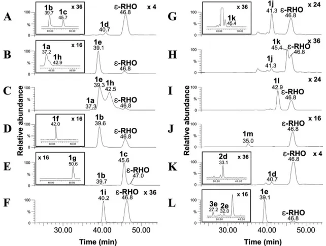
YJ183/pEVCS produced 4⬘-epi-L-vancosaminyl-rhodomycin D (compound 1d; 7-O-4⬘-epi-L-vancosaminyl-ε-RHO; Rt, 40.7
min; m/z 572⬎ 144 in MRM mode) as expected, 4⬘-epi-rho-domycin D (compound 1b; Rt, 39.7 min), andL
-ristosaminyl-rhodomycin D (compound 1c; Rt, 45.7 min), probably due to the skipping of the C-3 methylation step catalyzed by EvaC (Fig. 4A and Table 3; see also Fig. S1-8B in the supplemental material for the MS/MS fragmentation pattern of compound 1d). YJ183/pMDNS1 produced 3⬘-N-methyl-rhodomycin D (compound 1e; 7-O-3⬘-N-methyl-L-daunosaminyl-ε-RHO; Rt,
39.1 min; m/z 572⬎ 144 in MRM mode), an N-monomethyl-ated biosynthetic intermediate ofL-rhodosaminyl-rhodomycin
D, as a major product and its biosynthetic intermediate rho-domycin D (compound 1a) (Fig. 4B and Table 3; see also Fig. S1-9B in the supplemental material for the MS/MS
fragmen-FIG. 3. HPLC-ESI-MS/MS chromatograms of cultures of S. ven-ezuelae strains. (A) rhodomycin D (compound 1a; m/z 558⬎ 130) and ε-rhodomycinone (ε-RHO; m/z 429 ⬎ 322) detected from culture of YJ183/pDNS1. (B) Rhodomycin D (compound 1a; m/z 558⬎ 130) and ε-RHO (m/z 429 ⬎ 322) detected from culture of YJ183/pDXR2. The peaks of doxorubicin (compound 3a; m/z 544⬎ 130) and daunorubicin (compound 2a; m/z 528⬎ 130) are illustrated as an inset to chromato-gram B. (C) 4⬘-epi-Rhodomycin D (compound 1b; m/z 558 ⬎ 130) and ε-RHO (m/z 429 ⬎ 322) detected from culture of YJ183/pEDNS1. The peak ofL-ristosaminyl-rhodomycin D (compound 1c; m/z 558⬎ 130) is illustrated as an inset to chromatogram C. (D)L -Ristosaminyl-rhodo-mycin D (compound 1c; m/z 558⬎ 130) and ε-RHO (m/z 429 ⬎ 322) produced from culture of YJ183/pRST1. The 4⬘-epi-rhodomycin D (compound 1b; m/z 558⬎ 130) is illustrated as an inset to chromato-gram D. (E) 4⬘-epi-Rhodomycin D (compound 1b; m/z 558 ⬎ 130) and ε-RHO (m/z 429 ⬎ 322) observed from culture of YJ183/pEDXR. The peaks of epirubicin (compound 3b; m/z 544⬎ 130), 4⬘-epi-daunorubi-cin (compound 2b; m/z 528⬎ 130), andL-ristosaminyl-rhodomycin D (1c; m/z 558⬎ 130) are illustrated as an inset to chromatogram E. The magnification factors of peak intensities of several compounds were displayed at the top of chromatograms.
on June 26, 2016 by Ewha Womans Univ
http://aem.asm.org/
tation pattern of compound 1e). A small peak attributable to
L-rhodosaminyl-rhodomycin D (compound 1h; 7-O-L -rhodos-aminyl-ε-RHO; Rt, 42.9 min; m/z 586 ⬎ 158 in MRM mode) was also detected from YJ183/pMDNS1 (Fig. 4B and Table 3; see also Fig. S1-10B in the supplemental material for the MS/MS fragmentation pattern of compound 1h). pMDNS2 was constructed in which the N-methyltransferase gene aknX2 of the aclacinomycin A-producing S. galilaeus ATCC 31615 was replaced with aclP from another aclacinomycin A pro-ducer, S. galilaeus ATCC 31133 (Table 3). YJ183/pMDNS2
showed a similar production pattern to YJ183/pMDNS1 (Ta-ble 3). However, YJ183/pRDS, containing both aknX2 and aclP, produced significant amounts of a compound predicted to beL-rhodosaminyl-rhodomycin D (compound 1h) (Fig. 4C and Table 3). YJ183/pMEDNS produced a compound pre-dicted to be 3⬘-N-methyl-4⬘-epi-rhodomycin D (compound 1f; 7-O-3⬘-N-methyl-4⬘-epi-L-daunosaminyl-ε-RHO; Rt, 42.0 min;
m/z 572⬎ 144 in MRM mode) and its biosynthetic
interme-diate 4⬘-epi-rhodomycin D (compound 1b), but no -L
-noga-losaminyl-rhodomycin D (Fig. 4D and Table 3; see also Fig.
FIG. 4. HPLC-ESI-MS/MS chromatograms of cultures of S. venezuelae strains. (A) 4⬘-epi-L-Vancosaminyl-rhodomycin D (compound 1d; m/z 572⬎ 144) and ε-RHO (m/z 429 ⬎ 322) detected from culture of YJ183/pEVCS. The peaks of 4⬘-epi-rhodomycin D (compound 1b; m/z 558 ⬎ 130) and l-ristosaminyl-rhodomycin D (compound 1c; m/z 558⬎ 130) are illustrated as an inset to chromatogram A. (B) 3⬘-N-Methyl-rhodomycin D (compound 1e; m/z 572⬎ 144) and ε-RHO (m/z 429 ⬎ 322) detected from culture of YJ183/pMDNS1. The peaks of rhodomycin D (compound 1a; 558⬎ 130) andL-rhodosaminyl-rhodomycin D (compound 1h; m/z 586⬎ 158) are illustrated as an inset to chromatogram B. (C) Rhodomycin D (compound 1a; m/z 558⬎ 130), 3⬘-N-methyl-rhodomycin D (compound 1e; m/z 572 ⬎ 144), andL-rhodosaminyl-rhodomycin D (compound 1h; m/z 586⬎ 158) detected from culture of YJ183/pRDS. (D) 4⬘-epi-Rhodomycin D (compound 1b; m/z 558 ⬎ 130) and ε-RHO (m/z 429 ⬎ 322) detected from culture of YJ183/pMEDNS. The peak of 3⬘-N-methyl-4⬘-epi-rhodomycin D (compound 1f; m/z 572 ⬎ 144) is illustrated as an inset to chromatogram D. (E) 4⬘-epi-Rhodomycin D (1b; m/z 558 ⬎ 130) andL-ristosaminyl-rhodomycin D (compound 1c; m/z 558⬎ 130) detected from culture of YJ183/pMRST. The peak of 3⬘-N-methyl-L-ristosaminyl-rhodomycin D (compound 1g; m/z 572⬎ 144) is illustrated as an inset to chromato-gram E. (F)D-Olivosyl-rhodomycin D (compound 1i; m/z 559⬎ 393) and ε-RHO (m/z 429 ⬎ 322) observed from culture of YJ183/pDOLV. (G)L-Olivosyl-rhodomycin D (compound 1j; m/z 559⬎ 393) and ε-RHO (m/z 429 ⬎ 322) observed from culture of YJ183/pLOLV. The peak of
L-digitoxosyl-rhodomycin D (compound 1k; m/z 559⬎ 393) is illustrated as an inset to chromatogram G. (H)L-Olivosyl-rhodomycin D (compound 1j; m/z 559⬎ 393),L-digitoxosyl-rhodomycin D (compound 1k; m/z 559⬎ 393), and ε-RHO (m/z 429 ⬎ 322) observed from culture of YJ183/pLDGT. (I)D-Digitoxosyl-rhodomycin D (compound 1l; m/z 559⬎ 393) and ε-RHO (m/z 429 ⬎ 322) observed from culture of YJ183/pDDGT. (J)L -Rhamnosyl-rhodomycin D (compound 1m; m/z 575⬎ 393) and ε-RHO (m/z 429 ⬎ 322) observed from culture of YJ183/pLRHM. (K) 4⬘-epi-L -Vancosaminyl-rhodomycin D (compound 1d; 572⬎ 144) and ε-RHO (m/z 429 ⬎ 322) observed from culture of YJ183/pEVDXR. The peak of 4⬘-epi-L -vancosaminyl-daunorubicin (compound 2d; m/z 542⬎ 144) is illustrated as an inset to chromatogram K. (L) 3⬘-N-Methyl-rhodomycin D (compound 1e; 572 ⬎ 144) andε-RHO (m/z 429 ⬎ 322) observed from culture of YJ183/pMDXR. The peaks of 3⬘-N-methyl-doxorubicin (compound 3e; m/z 558 ⬎ 144) and 3⬘-N-methyl-daunorubicin (compound 2e; m/z 542 ⬎ 144) are illustrated as an inset to chromatogram L. The magnification factors of peak intensities of several compounds were displayed at the top of chromatograms.
on June 26, 2016 by Ewha Womans Univ
http://aem.asm.org/
S1-11B in the supplemental material for the MS/MS fragmen-tation pattern of compound 1f), suggesting that the N-methyl-transferase AknX2 acted as a monomethylN-methyl-transferase on TDP-4⬘-epi-L-daunosamine. YJ183/pMRST also generated a compound consistent with the mass of monomethylated
L-ristosaminyl-rhodomycin D (compound 1g; 7-O-3
⬘-N-methyl-L-ristosaminyl-ε-RHO; Rt, 50.6 min; m/z 572 ⬎ 144 in MRM
mode), its biosynthetic intermediateL -ristosaminyl-rhodomy-cin D (compound 1c), and a trace amount of 4 ⬘-epi-rhodomy-cin D (compound 1b), but noL-megosaminyl-rhodomycin D (Fig. 4E and Table 3; see also Fig. S1-12B in the supplemental material for the MS/MS fragmentation pattern of compound 1g). This result again indicates that AknX2 functioned as a monomethyltransferase toward TDP-L-ristosamine. As antici-pated, HPLC-ESI-MS/MS analysis of the culture extract of YJ183/pDOLV produced a peak consistent with the mass of
D-olivosyl-rhodomycin D (compound 1i; 7-O-D-olivosyl-
ε-RHO; Rt, 40.2 min; m/z 559⬎ 393 in MRM mode) (Fig. 4F and Table 3; see also Fig. S1-13B in the supplemental material for the MS/MS fragmentation pattern of compound 1i). YJ183/ pLOLV and YJ183/pLDGT, respectively, produced com-pounds consistent with the mass ofL-olivosyl-rhodomycin D (compound 1j; 7-O-L-olivosyl-ε-RHO; Rt, 41.3 min; m/z 559 ⬎
393 in MRM mode) and L-digitoxosyl-rhodomycin D (com-pound 1k; 7-O-L-digitoxosyl-ε-RHO; Rt, 45.4 min; m/z 559 ⬎
393 in MRM mode) as major products (Fig. 4G and H and Table 3; see also Fig. S1-14B and Fig. S1-15B in the supple-mental material for the MS/MS fragmentation patterns of compounds 1j and 1k, respectively), with small amounts of the other compound, probably due to tautomerization between TDP-4-keto-L-olivose and TDP-4-keto-L-digitoxose (8). Peaks
consistent with the masses of D-digitoxosyl-rhodomycin D (compound 1l; 7-O-D-digitoxosyl-ε-RHO; Rt, 42.9 min; m/z 559
⬎ 393 in MRM mode) andL-rhamnosyl-rhodomycin D (com-pound 1m; 7-O-L-rhamnosyl-ε-RHO; Rt, 35.0 min; m/z 575 ⬎
393 in MRM mode) were observed in the culture extracts of YJ183/pDDGT and YJ183/pLRHM, respectively (Fig. 4I and J and Table 3; see also Fig. S1-16B and Fig. S1-17B in the supplemental material for the MS/MS fragmentation patterns of compounds 1l and 1m, respectively).
Generation of glycosylated derivatives of doxorubicin. In order to modify further the relatively abundant glycosylated rhodomycin D derivatives 4⬘-epi-L-vancosaminyl-rhodomycin
D (compound 1d), 3⬘-N-methyl-rhodomycin D (compound 1e), and L-ristosaminyl-rhodomycin D (compound 1c) into their
glycosylated analogs of doxorubicin, the plasmids pEVDXR, pMDXR, and pRSDXR were constructed by adding the four genes dnrP, dnrK, doxA, and dnrV into pEVCS, pMDNS1, and pRST1, respectively. When pEVDXR was expressed in S.
ven-ezuelae mutant YJ183, metabolites expected to 4⬘-epi-L
-vanco-saminyl-daunorubicin (compound 2d; 7-O-4⬘-epi-L
-vancosami-nyl-daunorubicinone; Rt, 33.1 min; m/z 542 ⬎ 144 in MRM mode) and 4⬘-epi-L-vancosaminyl-rhodomycin D (compound
1d) were produced (Fig. 4K and Table 3; see also Fig. S1-18B in the supplemental material for the MS/MS fragmentation pattern of compound 1d). From the organic extract of YJ183/ pMDXR supplemented with ε-RHO, three peaks were ob-served at Rts of 27.2, 32.0, and 39.1 min, predicted to be 3⬘-N-methyl-doxorubicin (compound 3e; 7-O-3⬘-N-methyl-L
-daunosaminyl-doxorubicinone; m/z 558⬎ 144 in MRM mode),
3⬘-N-methyl-daunorubicin (compound 2e; 7-O-3⬘-N-methyl-L
-daunosaminyl-daunorubicinone; m/z 542 ⬎ 144 in MRM mode), and 3⬘-N-methyl-rhodomycin D (compound 1e), re-spectively (Fig. 4L and Table 3; see also Fig. S1-19B and S1-20B in the supplemental material for the MS/MS fragmen-tation patterns of compounds 2e and 3e, respectively). YJ183/ pRSDXR led to the production of onlyL
-ristosaminyl-rhodo-mycin D (compound 1c) but not any glycosylated derivatives of doxorubicin and daunorubicin (data not shown).
Structural elucidation of glycosylated derivatives of rhodo-mycin D.The structures of the anthracycline derivatives gen-erated in small amounts from S. venezuelae YJ183 mutant strains were predicted based on the ESI-MS/MS fragmentation patterns of the glycosylated anthracyclines (see Fig. S1 in the supplemental material). Although the configurations of sugar moieties cannot be determined by ESI-MS/MS, they were pre-dicted from the deduced catalytic specificities of the sugar biosynthetic enzymes. However, the structures of rhodomycin D (compound 1a), 4⬘-epi-rhodomycin D (compound 1b), L -ristosaminyl-rhodomycin D (compound 1c), 4⬘-epi-L
-vanco-saminyl-rhodomycin D (compound 1d), and 3 ⬘-N-methyl-rho-domycin D (compound 1e) were deduced mainly by the chemical shifts of particular protons in NMR (see the supple-mental material). The limited amounts of purified rhodomycin D and its derivatives did not allow the precise determination of coupling constants from 2D NMR.
The structure of rhodomycin D (compound 1a) was deduced from the spectroscopic data by comparison with literature val-ues (6, 17; see also the supplemental material). The1H-NMR
chemical shifts of 4⬘-epi-rhodomycin D (compound 1b) were similar to those of rhodomycin D (compound 1a) except for the upfield shifts of H-4⬘ and H-5⬘, suggesting the presence of 4⬘-epi-L-daunosamine as a sugar moiety (7; see also the sup-plemental material). The ESI-MS/MS spectrum ofL
-ristosami-nyl-rhodomycin D (compound 1c) was similar to those of rhodomycin D (compound 1a) and 4⬘-epi-rhodomycin D (com-pound 1b) (see the supplemental material). The1H-NMR data
of L-ristosaminyl-rhodomycin D (compound 1c) were also largely similar to those of rhodomycin D (compound 1a) ex-cept for the upfield shifts of H-3⬘ and H-4⬘ (see the supple-mental material). In addition, characteristic signals for the
L-ristosaminyl moiety were observed at␦C49.27 (C-3⬘), ␦C71.18
(C-4⬘), and ␦C64.07 (C-5⬘) in the13C-NMR spectrum (26; see
also the supplemental material). Therefore, the structure ofL
-ristosaminyl-rhodomycin D (compound 1c), which has not been reported yet, could be deduced from the1H- and13
C-NMR data. The 1H-NMR chemical shifts of 4⬘-epi-L
-vanco-saminyl-rhodomycin D (compound 1d) were similar to those of 4⬘-epi-rhodomycin D (compound 1b) (see the supplemental material). However, an additional methyl signal at ␦H1.45 (3⬘-CH3) was shown instead of the H-3⬘ signal from
4⬘-epi-rhodomycin D (compound 1b), which is characteristic of the 4⬘-epi-L-vancosamine moiety (26, 41). The 1H-NMR data of
3⬘-N-methyl-rhodomycin D (compound 1e) was similar to that of rhodomycin D, but for differences in the sugar moiety sig-nals (see supplementary text). In the 13C-NMR data,
addi-tional methyl signals appeared at␦C30.45, suggesting the pres-ence of a 7⬘-CH3moiety (see the supplemental material). The structure of 3⬘-N-methyl-rhodomycin D (compound 1e), which
on June 26, 2016 by Ewha Womans Univ
http://aem.asm.org/
has not yet been reported, was predicted by the1H- and13
C-NMR data.
DISCUSSION
The simple S. venezuelae-based combinatorial biosynthetic system developed here facilitated improved biological produc-tion of epirubicin by selecting an efficient TDP-4-ketohexose reductase, AvrE, and glycosyltransferase, AknS, which were able to synthesize and transfer unnatural TDP-4-epi-L -daunosamine toε-RHO. Although an S. peucetius mutant in which dnmV was substituted with avrE has been reported to produce 4⬘-epi-daunorubicin with trace amounts of epirubicin, the present study is, to the best of our knowledge, the first report of a considerable amount of epirubicin (0.9M ⬇ 0.5 mg/liter) being produced by a biological method. The accumu-lation of large amounts of 4⬘-epi-rhodomycin D (compound 1b) in the YJ183/pEDXR strain (Fig. 3E) and the low conver-sion yields of rhodomycin D derivatives into doxorubicin de-rivatives in YJ183 strains expressing pEVDXR, pMDXR, and pRSDXR (Fig. 4K and L and Table 3) suggest that the post-glycosylation modification steps catalyzed by DnrP, DnrK, and DoxA are rate-limiting and that engineering of more efficient postglycosylational modification enzymes could further en-hance epirubicin production. Furthermore, two doxorubicin derivatives (compounds 3b and 3e), three daunorubicin deriv-atives (compounds 2b, 2d, and 2e), and 12 rhodomycin D derivatives (compounds 1b to 1m) were generated, as well as doxorubicin (compound 3a) and its intermediates (compounds 1a and 2a). Among the 20 anthracyclines produced in the present study, seven glycosylated derivatives of rhodomycin D (compounds 1c, 1e to 1g, 1i, 1k, and 1l) are novel anthracy-clines that have not been previously described, demonstrating the potential of this combinatorial biosynthetic method for the engineered generation of doxorubicin analogs with modified sugar moieties and the creation of improved anticancer agents. Analysis of the metabolites produced by various combina-tions of genes encoding glycosylaransferases and sugar biosyn-thesis provided valuable information on their substrate flexi-bility and functions, although concurrent biochemical experiments were not carried out here. Introduction of the respective plasmids into S. venezuelae YJ183 did not cause any phenotypic difference, implying that expression plasmids did not affect the expression or activities of other host genes in-volved in growth, differentiation, and the production of other secondary metabolites. The glycosyltransferase AknS has been shown to accept aklavinone andε-RHO as sugar acceptors and
L-rhodosamine, L-rhamnose, L-daunosamine, and 2-deoxy-L -fucose as sugar donors (18, 21). Production of structurally diverse rhodomycin D derivatives in the present study showed that AknS has relaxed substrate specificity toward variousL
-aminosugars andL- andD-neutral sugars. The flexibility of the glycosyltransferase StfG in recognizing seven neutral sugars with different degrees of deoxygenation has been established in other work (30). Generation of 4⬘-epi-rhodomycin D (com-pound 1b) indicated that ε-RHO and 4-epi-L-daunosamine could be recognized and accepted by StfG. The glycosyltrans-ferase SnogE showed the highest amino acid identity (59%) when the amino acid sequences of the glycosyltransferases SnogE, SnogD, and SnogZ present in the nogalamycin
biosyn-thetic gene cluster from S. nogalager (42) were compared to that of AknS. The sequence comparison suggests that SnogE might transfer TDP-L-nogalose at the C-7 hydroxyl group of
nogalamycin aglycone, and the production of 4 ⬘-epi-rhodomy-cin D by SnogE also supports this hypothesis. In addition, the auxiliary protein SnogN, which is essential for the glycosyl transfer activity of SnogE, was found. When the pEDNS4 derivative lacking snogN was expressed in YJ183, 4 ⬘-epi-rho-domycin D (compound 1b) was not detected (data not shown). Many previous studies have reported that the glycosylferase/auxiliary protein pair is necessary for the efficient trans-fer of deoxysugar in the biosynthesis of macrolide and anthra-cycline compounds (11, 21, 50), and the gene encoding the auxiliary protein was generally located immediately upstream of the glycosyltransferase gene in anthracycline and macrolide biosynthetic gene clusters. However, snogN has been found to be located 7 kb away from snogE, making it an unusual ar-rangement of genes.
The formation of 4⬘-epi-rhodomycin D (compound 1b),L
-ristosaminyl-rhodomycin D (compound 1c), or both during the test of TDP-4-ketohexose reductases is an interesting outcome of the present study. It can be explained through DnmU being able to act not only as a 3,5-epimerase (in the formation of 4⬘-epi-rhodomycin D [compound 1b]) but also as a 5-epimerase (in the formation ofL-ristosaminyl-rhodomycin D [compound
1c)] to cause an equilibrium of C-3,C-5 and C-5 epimers of TDP-3-amino-2,3,6-trideoxy-4-keto-D-glucose, one of them
be-ing a better substrate for each of the TDP-4-ketohexose re-ductases tested (Fig. 1B). It has been shown that GDP-man-nose-3⬘,5⬘-epimerase (GME), an enzyme involved in the conversion of GDP-D-mannose to GDP-L-galactose, catalyzes
two different epimerization reactions resulting in the produc-tion of two discrete products: GDP-L-glucose (by
5-epimeriza-tion) and GDP-L-galactose (by 3,5-epimerization). (47). These two distinct epimerization reactions of GME support the hypoth-esis that DnmU could catalyze both 3,5 and 5 epimerizations, producing TDP-4-keto-L-daunosamine and TDP-4-keto-L
-ris-tosamine, respectively. Production of 4⬘-epi-rhodomycin D (1b) andL-ristosaminyl-rhodomycin D (compound 1c) by YJ183
mu-tants harboring pEDNS1 and its derivatives (pEDNS5, pEDNS6, pRST1, pRST2, pRST3, pRST4, and pRST5) indicates that the TDP-4-ketohexose reductases examined could accept 4-keto-L -daunosamine, 4-keto-L-ristosamine, or both as substrates. AvrE,
which supported the highest epirubicin production, accepted TDP-4-keto-L-daunosamine (the C-3,C-5 epimer of
TDP-3-ami-no-2,3,6-trideoxy-4-keto-D-glucose) as a glycosyl donor more ef-ficiently than it did TDP-4-keto-L-ristosamine (the C-5 epimer of
TDP-3-amino-2,3,6-trideoxy-4-keto-D-glucose). GrsB, which is in-volved in the biosynthesis of streptomycin, was also capable of reducing TDP-4-keto-L-daunosamine to produce TDP-4-epi-L -daunosamine. JadV, OleU, TylCIV, StrL, and SnogC, which par-ticipate in the biosynthesis of jadomycin B, oleandomycin, tylosin, streptomycin, and nogalamycin, respectively, were able to reduce TDP-4-keto-L-ristosamine mostly. EvaE, which encodes a TDP-4-ketohexose reductase involved in the biosynthesis of chloroer-emomycin, can act on both intermediates generating both TDP-4-epi-L-daunosamine and TDP-L-ristosamine.
Production of 4⬘-epi-rhodomycin D (compound 1b) and L -ristosaminyl-rhodomycin D (compound 1c) in YJ183/pEVCS showed relaxed substrate specificity of both 5-epimerase EvaD
on June 26, 2016 by Ewha Womans Univ
http://aem.asm.org/
and 4-ketoreductase EvaE from the 4-epi-L-vancosamine
biosyn-thetic pathway. It has previously been reported that 5-epimerase EvaD can accept
TDP-3-amino-2,3,6-trideoxy-3-methyl-4-keto-D-glucose and epimerize at C-5 to produce TDP-4-keto-L -van-cosamine, which is reduced at C-4 by 4-ketoreductase EvaE to generate TDP-4-epi-L-vancosamine (5). In addition, it had been demonstrated that EvaD also possesses the activity of 3,5-epimerase, catalyzing double epimerization of TDP-4-ke-to-6-deoxy-D-glucose at C-3 and C-5 (27). In the present study,
4⬘-epi-rhodomycin D (compound 1b),L -ristosaminyl-rhodomy-cin D (compound 1c), and 4⬘-epi-L-vancosaminyl-rhodomycin
D (compound 1d) produced in YJ183/pEVCS indicated EvaD epimerizes both at C-5 and at C-3 and C-5 in TDP-3-amino-2,3,6-trideoxy-4-keto-D-glucose, and EvaE reduces both prod-ucts of EvaD to synthesize both TDP-L-ristosamine and
TDP-4-epi-L-daunosamine, which showed that EvaD and EvaE can recognize the unnatural substrates lacking a methyl group at C-3 in their natural substrates.
3⬘-N-Methyl-rhodomycin D (compound 1e),
3⬘-N-methyl-4⬘-epi-rhodomycin D (compound 1f), and 3⬘-N-methyl-L
-ristosami-nyl-rhodomycin D (compound 1g) were found from the cultures of YJ183 mutants expressing the plasmids pMDNS1, pMEDNS, and pMRST, respectively, which contain aknX2 encoding
N-methyltransferase from the biosynthetic pathway of TDP-L
-rhodosamine (TDP-3-dimethyl-L-daunosamine). This showed
that AknX2 was able to transfer a methyl group at the C-3 amine in TDP-L-daunosamine, TDP-4-epi-L-daunosamine, and TDP-L
-ristosamine, indicating that AknX2 possesses the activity of N-methyltransferase and a certain relaxation of specificity. AknX2 had been thought to be a N,N-dimethyltransferase catalyzing the transfer of two methyl groups at the C-3 amine in TDP-L
-daunosamine based on sequence comparison with other N,N-dimethyltransferases that have been experimentally characterized (38). However, YJ183 mutant carrying L-daunosamine biosyn-thetic genes and aknX2 (YJ183/pMDNS1) produced significant amounts of 3⬘-N-methyl-rhodomycin D (compound 1e) and a trace amount ofL-rhodosaminyl-rhodomycin D (compound 1h),
suggesting that another N-methyltransferase might act on TDP-3-N-methyl-L-daunosamine for synthesizing TDP-L-rhodosamine.
Interestingly, S. galilaeus 3AR-33 (25), an aklavinone-accumulat-ing mutant derived from another aclacinomycin A producer, S. galilaeus ATCC 31133, has the aclacinomycin A biosynthetic gene cluster which has a similar genetic organization to that of S. galilaeus ATCC 31615 but harbors another N-methyltransferase gene, aclP, at a different location from that of aknX2 in S. galilaeus ATCC 31615. When TDP-L-daunosamine biosynthesis genes and aclP were expressed in YJ183 (YJ183/pMDNS2), a large amount of 3⬘-N-methyl-rhodomycin D (compound 1e) and a trace amount ofL-rhodosaminyl-rhodomycin D (compound 1h) were
observed. YJ183 mutant containing the TDP-L -daunosamine-syn-thesizing gene cassette, aknX2, and aclP (YJ183/pRDS) produced significant amounts of 3⬘-N-methyl-rhodomycin D (compound 1e) andL-rhodosaminyl-rhodomycin D (compound 1h).
There-fore, it is likely that two N-methyltransferases, AknX2 and AclP, are required for the efficient biosynthesis of TDP-L-rhodosamine.
ACKNOWLEDGMENTS
This study was supported by National Research Laboratory program 20100018430 through the Nation Research Foundation of Korea (NRF); NRF grants 2010K000890 and 20100001487 funded by the
Ministry of Education, Science, and Technology; the National R&D Program for Cancer Control (grant 0620029); and the Marine and Extreme Genome Research Center Program of the Ministry of Land, Transportation and Maritime Affairs, Republic of Korea.
REFERENCES
1. Acramone, F., et al. 1997. New developments in antitumor anthracyclines. Pharmacol. Ther. 76:117–124.
2. Acramone, F., and G. Cassinelli. 1998. Biosynthetic anthracyclines. Curr. Med. Chem. 5:391–419.
3. Bate, N., A. R. Butler, I. P. Smith, and E. Cundliffe. 2000. The mycarose-biosynthetic genes of Streptomyces fradiae, producer of tylosin. Microbiology
146:139–146.
4. Bierman, M., et al. 1992. Plasmid cloning vectors for the conjugal transfer of DNA from Escherichia coli to Streptomyces spp. Gene 116:43–49. 5. Chen, H., et al. 2000. Deoxysugars in glycopeptide antibiotics: enzymatic
synthesis of TDP-L-epivancosamine in chloroeremomycin biosynthesis. Proc. Natl. Acad. Sci. U. S. A. 97:11942–11947.
6. Essery, J. M., and T. W. Doyle. 1980. The synthesis of daunosaminyl ε-rho-donycinone, daunosaminyl 10-epi-ε-rhodomycinone, daunosaminyl ε-pyr-romycinone, and 10-descarbomethoxy-ε-pyrromycin. Can. J. Chem. 58:1869– 1874.
7. Fan, E., W. Shi, and T. L. Lowary. 2007. Synthesis of daunorubicin analogues containing truncated aromatic cores and unnatural monosaccharide resi-dues. J. Org. Chem. 72:2917–2928.
8. Fischer, C., et al. 2002. Digitoxosyltetracenomycin C and glucosyltetraceno-mycin C, two novel elloraglucosyltetraceno-mycin analogues obtained by exploring the sugar donor substrate specificity of glycosyltransferase ElmGT. J. Nat. Prod. 65: 1685–1689.
9. Guilfoile, P. G., and C. R. Hutchinson. 1991. A bacterial analog of the mdr gene of mammalian tumor cells is present in Streptomyces peucetius, the producer of daunorubicin and doxorubicin. Proc. Natl. Acad. Sci. U. S. A.
88:8553–8557.
10. Hong, J. S. J., S. H. Park, C. Y. Choi, J. K. Sohng, and Y. J. Yoon. 2004. New olivosyl derivatives of methymycin/pikromycin from an engineered strain of
Streptomyces venezuelae. FEMS Microbiol. Lett. 238:291–299.
11. Hong, J. S. J., et al. 2007. Functional analysis of desVIII homologues involved in glycosylation of macrolide antibiotics by interspecies complementation. Gene 386:123–130.
12. Hurteloup, P., and F. Ganzina. 1986. Clinical studies with new anthracy-clines: epirubicin, idarubicin, esorubicin. Drugs Exp. Clin. Res. 12:233–246. 13. Jung, W. S., et al. 2006. Heterologous expression of tylosin polyketide syn-thase and production of a hybrid macrolide in Streptomyces venezuelae. Appl. Microbiol. Biotechnol. 72:763–769.
14. Jung, W. S., et al. 2007. Bioconversion of 12-, 14-, and 16-membered ring aglycones to glycosylated macrolides in an engineered strain of Streptomyces
venezuelae. Appl. Microbiol. Biotechnol. 76:1373–1381.
15. Jung, W. S., et al. 2008. Enhanced heterologous production of desosaminyl macrolides and their hydroxylated derivatives by overexpression of the pikD regulatory gene in Streptomyces venezuelae. Appl. Environ. Microbiol. 74: 1972–1979.
16. Kieser, T., M. J. Bibb, M. J. Buttner, K. F. Chater, and D. A. Hopwood. 2000. Practical Streptomyces genetics. John Innes Centre, Norwich, United King-dom.
17. Kim, B. S., S. S. Moon, and B. K. Hwang. 2000. Structure elucidation and antifungal activity of an anthracycline antibiotic, daunomycin, isolated from
Actinomadura roseola. J. Agric. Food Chem. 48:1875–1881.
18. Leimkuhler, C., et al. 2007. Characterization of rhodosaminyl transfer by the AknS/AknT glycosylation complex and its use in reconstituting the biosyn-thetic pathway of aclacinomycin A. J. Am. Chem. Soc. 129:10546–10550. 19. Lomovskaya, N., et al. 1996. The Streptomyces peucetius drrC gene encodes a
UvrA-like protein involved in daunorubicin resistance and production. J. Bacteriol. 178:3238–3245.
20. Lomovskaya, N., et al. 1999. Doxorubicin overproduction in Streptomyces
peucetius: cloning and characterization of the dnrU ketoreductase and dnrV
genes and the doxA cytochrome P-450 hydroxylase gene. J. Bacteriol. 181: 305–318.
21. Lu, W., et al. 2005. AknT is an activating protein for the glycosyltransferase AknS inL-aminodeoxysugar transfer to the aglycone of aclacinomycin A. Chem. Biol. 12:527–534.
22. Madduri, K., F. Torti, A. L. Colombo, and C. R. Hutchinson. 1993. Cloning and sequencing of a gene encoding carminomycin 4-O-methyltransferase from Streptomyces peucetius and its expression in Escherichia coli. J. Bacte-riol. 175:3900–3904.
23. Madduri, K., and C. R. Hutchinson. 1995. Functional characterization and transcriptional analysis of a gene cluster governing early and late steps in daunorubicin biosynthesis in Streptomyces peucetius. J. Bacteriol. 177:3879– 3884.
24. Madduri, K., et al. 1998. Production of the antitumor drug epirubicin (4⬘-epidoxorubicin) and its precursor by a genetically engineered strain of
Strep-tomyces peucetius. Nat. Biotechnol. 16:69–74.
on June 26, 2016 by Ewha Womans Univ
http://aem.asm.org/
25. Matsuzawa, Y., et al. 1981. New anthracycline metabolites from mutant strains of Streptomyces galilaeus MA144-M1. II. Structure of 2-hydroxy-aklavinone and new 2-hydroxy-aklavinone glycosides. J. Antibiot. 34:959–964. 26. Mendlik, M. T., P. Tao, C. M. Hadad, R. S. Coleman, and T. L. Lowary. 2006.
Synthesis ofL-daunosamine andL-ristosamine glycosides via photoinduced aziridination: conversion to thioglycosides for use in glycosylation reaction. J. Org. Chem. 71:8059–8070.
27. Merkel, A. B., et al. 2004. The position of a key tyrosine in dTDP-4-keto-6-deoxy-D-glucose-5-epimerase (EvaD) alters the substrate profile for this RmlC-like enzyme. J. Biol. Chem. 279:32684–32691.
28. Minotti, G., P. Menna, E. Salvatorelli, G. Cairo, and L. Gianni. 2004. Anthracyclines: molecular advances and pharmacologic developments in antitumor activity and cardiotoxicity. Pharmacol. Rev. 56:185–229. 29. Olano, C., N. Lomovskaya, L. Fonstein, J. T. Roll, and C. R. Hutchinson.
1999. A two-plasmid system for the glycosylation of polyketide antibiotics: bioconversion ofε-rhodomycinone to rhodomycin D. Chem. Biol. 6:845–855. 30. Olano, C., et al. 2008. Glycosylated derivatives of steffimycin: insights into the role of the sugar moieties for the biological activity. Chembiochem
9:624–633.
31. Olano, C., C. Me´ndez, and J. A. Salas.2009. Antitumor compounds from actinomycetes: from gene clusters to new derivatives by combinatorial bio-synthesis. Nat. Prod. Rep. 26:628–660.
32. Otten, S. L., X. Liu, J. Ferguson, and C. R. Hutchinson. 1995. Cloning and characterization of the Streptomyces peucetius dnrQS genes encoding a daunosamine biosynthesis enzyme and a glycosyl transferase involved in daunorubicin biosynthesis. J. Bacteriol. 177:6688–6692.
33. Park, J. W., et al. 2008. Genetic dissection of the biosynthetic route to gentamicin A2 by heterologous expression of its minimal gene set. Proc. Natl. Acad. Sci. U. S. A. 105:8399–8404.
34. Park, S. R., et al. 2008. Heterologous production of epothilones B and D in
Streptomyces venezuelae. Appl. Microbiol. Biotechnol. 81:109–117.
35. Park, S. R., et al. 2009. Engineering of plant-specific phenylpropanoids biosynthesis in Streptomyces venezuelae. J. Biotechnol. 141:181–188. 36. Pe´rez, M., et al.2006. Combinatorial biosynthesis of antitumor deoxysugar
pathways in Streptomyces griseus: reconstitution of “unnatural natural gene clusters” for the biosynthesis of four 2,6-D-dideoxyhexoses. Appl. Environ. Microbiol. 72:6644–6652.
37. Pissowotzki, K., K. Mansouri, and W. Piepersberg. 1991. Genetics of strep-tomycin production in Streptomyces griseus: molecular structure and putative function of genes strELMB2N. Mol. Gen. Genet. 231:113–123.
38. Ra¨ty, K., T. Kunnari, J. Hakala, P. Ma¨ntsa¨la¨, and K. Ylihonko.2000. A gene cluster from Streptomyces galilaeus involved in glycosylation of aclarubicin. Mol. Gen. Genet. 264:164–172.
39. Rodráiguez, L., et al. 2002. Engineering deoxysugar biosynthetic pathways from antibiotic-producing microorganisms. A tool to produce novel glycosyl-ated bioactive compounds. Chem. Biol. 9:721–729.
40. Schmitt-John, T., and J. W. Engels. 1992. Promoter constructions for effi-cient secretion expression in Streptomyces lividans. Appl. Microbiol. Biotech-nol. 36:493–498.
41. Sibi, M. P., J. Lu, and J. Edwards. 1997. A new route to 3-amino sugars: a concise synthesis ofL-daunosamine andD-ristosamine derivatives. J. Org. Chem. 62:5864–5872.
42. Torkkell, S., et al. 2001. The entire nogalamycin biosynthetic gene cluster of
Streptomyces nogalater: characterization of a 20-kb DNA region and
gener-ation of hybrid structures. Mol. Genet. Genomics 266:276–288.
43. Volchegursky, Y., Z. Hu, L. Katz, and R. McDaniel. 2000. Biosynthesis of the anti-parasitic agent megalomicin: transformation of erythromycin to mega-lomicin in Saccharopolyspora erythraea. Mol. Microbiol. 37:752–762. 44. Wang, L., R. L. White, and L. C. Vining. 2002. Biosynthesis of the
dideoxy-sugar component of jadomycin B: genes in the jad cluster of Streptomyces
venezuelae ISP5230 forL-digitoxose assembly and transfer to the angucycline aglycone. Microbiology 148:1091–1103.
45. Weymouth-Wilson, A. C. 1997. The role of carbohydrates in biologically active natural products. Nat. Prod. Rep. 14:99–110.
46. Wohlert, S., et al. 2001. Insights about the biosynthesis of the avermectin deoxysugarL-oleandrose through heterologous expression of Streptomyces
avermitilis deoxysugar genes in Streptomyces lividans. Chem. Biol. 8:681–700.
47. Wolucka, B. A., and M. Van Montagu. 2003. GDP-mannose 3⬘,5⬘-epimerase forms GDP-L-gulose, a putative intermediate for the de novo biosynthesis of vitamin C in plants. J. Biol. Chem. 278:47483–47490.
48. Xue, Y., L. Zhao, H. W. Liu, and D. H. Sherman. 1998. A gene cluster for macrolide antibiotic biosynthesis in Streptomyces venezuelae: architecture of metabolic diversity. Proc. Natl. Acad. Sci. U. S. A. 95:12111–12116. 49. Yoon, Y. J., et al. 2002. Generation of multiple bioactive macrolides by
hybrid modular polyketide synthases in Streptomyces venezuelae. Chem. Biol.
9:203–214.
50. Yuan, Y., et al. 2005. In vitro reconstitution of EryCIII activity for the preparation of unnatural macrolides. J. Am. Chem. Soc. 127:14128–14129.