저작자표시-비영리-변경금지 2.0 대한민국 이용자는 아래의 조건을 따르는 경우에 한하여 자유롭게 l 이 저작물을 복제, 배포, 전송, 전시, 공연 및 방송할 수 있습니다. 다음과 같은 조건을 따라야 합니다: l 귀하는, 이 저작물의 재이용이나 배포의 경우, 이 저작물에 적용된 이용허락조건 을 명확하게 나타내어야 합니다. l 저작권자로부터 별도의 허가를 받으면 이러한 조건들은 적용되지 않습니다. 저작권법에 따른 이용자의 권리는 위의 내용에 의하여 영향을 받지 않습니다. 이것은 이용허락규약(Legal Code)을 이해하기 쉽게 요약한 것입니다. Disclaimer 저작자표시. 귀하는 원저작자를 표시하여야 합니다. 비영리. 귀하는 이 저작물을 영리 목적으로 이용할 수 없습니다. 변경금지. 귀하는 이 저작물을 개작, 변형 또는 가공할 수 없습니다.
Gastroesophageal reflux in
neurologically impaired children
: What are the risk factors?
Seung Kim
Department of Medicine
Gastroesophageal reflux in
neurologically impaired children
: What are the risk factors?
Directed by Professor Joon Soo Lee
The Master's Thesis
submitted to the Department of Medicine,
the Graduate School of Yonsei University
in partial fulfillment of the requirements for the degree
of Master of Medical Science
Seung Kim
This certifies that the Master's Thesis of
Seung Kim is approved.
Thesis Supervisor : Joon Soo Lee
---Thesis Committee Member#1 : Joon Seok Lim
---Thesis Committee Member#2 : Jae Il Shin
The Graduate School
Yonsei University
ACKNOWLEDGEMENTS
I would first like to express my special thanks of gratitude to
thesis advisor professor Joon Soo Lee who gave me the golden
opportunity to do this meaningful study on the topic
“Gastroesophageal reflux disease in neurologically impaired
children”. I would also appreciate Professor Joon Seok Lim
and Jae Il Shin for guiding this work. I am gratefully indebted
to for their very valuable comments on this thesis. Last but not
least, I would like to acknowledge Professor Hong Koh for
supporting me a lot in so many ways.
<TABLE OF CONTENTS>
ABSTRACT ···1
I. INTRODUCTION ···2
II. MATERIALS AND METHODS···4
1. Study Population and Data Collection ··· 4
2. 24 hour esophageal pH monitoring ···4
3. Statistical Analysis ···5
III. RESULTS ···6
IV. DISCUSSION ··· 12
V. CONCLUSION ··· 16
REFERENCES ··· 17
ABSTRACT(IN KOREAN) ··· 19
LIST OF FIGURES
Figure 1. Box and whisker plot of reflux index showing the
distribution of reflux index according to clinical conditions 9
LIST OF TABLES
Table 1. Patient Characteristics··· 6
Table 2. Spearman correlation coefficients of gastro-esophageal
reflux index and DeMeester score with continuous variables· 10
Table 3. Comparisons of clinical characteristics between GERD
1
ABSTRACT
Gastresophageal reflux in neurologically impaired children: what are the
risk factors?
Seung Kim
Department of Medicine The Graduate School, Yonsei University
(Directed by Professor Joon Soo Lee)
Background/Aims: Neurologically impaired patients (NIP) frequently suffer from gastrointestinal tract problems such as gastroesophageal reflux disease (GERD). In this study, we aimed to define the risk factors for GERD in neurologically impaired children. Methods: From May 2006 to March 2014, 101 neurologically impaired children who received 24 hr esophageal pH monitoring in Severance children’s hospital were enrolled. The results of esophageal pH monitoring and the clinical characteristics of the patients were analyzed. Results: Reflux index was higher in the abnormal EEG group than in the normal EEG group (p = 0.027). Mitochondrial disease was associated with a higher reflux index than epileptic disorders or cerebral palsy (p = 0.009) Patient gender, feeding method, scoliosis, tracheostomy, and baclofen use did not lead to statistical differences in reflux index. Age of onset of neurological impairment was inversely correlated with DeMeester score and reflux index. Age at the time of examination, duration of the disease, and number of antiepileptic drugs did not correlate with GER severity. Conclusions: Early-onset neurologic impairment, abnormal EEG, and mitochondrial disease are identified as risk factors for severe GERD.
---Key words: gastroesophageal reflux, esophageal pH monitoring, child
2
Gastresophageal reflux in neurologically impaired children: what are the
risk factors?
Seung Kim
Department of Medicine
The Graduate School, Yonsei University
(Directed by Professor Joon Soo Lee)
I. INTRODUCTION
As the central nervous system controls the enteric nervous system, neurologically impaired patients (NIP) frequently suffer from gastrointestinal tract dysfunction1. Guidice et al.2 reported that 92% of children with cerebral
palsy had clinically significant gastrointestinal symptoms: gastroesophageal reflux, 77%; swallowing disorders, 60%; chronic pulmonary aspiration, 41%; and chronic constipation, 74%. Decreased lower esophageal sphincter tone, delayed gastric emptying, impaired esophageal motility, poor posture, recurrent seizures, scoliosis, and various medications are thought to contribute to gastroesophageal reflux disease (GERD) in neurologically impaired children3.
Respiratory symptoms, which are frequent in NIP, are also thought to aggravate GERD 4. Despite its high incidence, GERD in neurologically impaired children
is difficult to recognize as the symptoms of GER are nonspecific and many patients cannot precisely express their symptoms. Therefore, diagnosis of GERD in NIP is often delayed until severe esophagitis or fatal aspiration pneumonia occurs5. As GER is closely related with aspiration pneumonia or
food refusal, this can be a large obstacle to appropriate nutritional support of NIP, which can result in a poor clinical prognosis. This is why physicians should pay attention to GER in NIP. Early suspicion and evaluation can prevent
3
severe complications of GERD in NIP and can lead to better clinical outcomes. In this study, we analyze the degree of gastroesophageal reflux according to patient characteristics and define the risk factors for GERD in neurologically impaired children.
4
II. MATERIALS AND METHODS 1. Study Population and Data Collection
Data from pediatric patients who received 24 hr esophageal pH monitoring in Severance children’s hospital from May 2006 to March 2014 were collected. Of those cases, neurologically impaired children were selected for the study. The reasons for performing 24 hr esophageal pH monitoring varied, but most patients had symptoms of gastroeophageal reflux such as recurrent aspiration pneumonia, grunting after meals, frequent regurgitation, and unexplained food refusal or unexplained irritability, or tonic posture. Patients who received upper gastrointestinal operations such as Nissen fundoplication or whose medical records were incomplete were excluded from this study. Finally, 101 patients (median age, 23.4 months) were enrolled. The results of esophageal pH monitoring and the clinical characteristics of the patients were analyzed. Patient selection and data collection were performed by retrospectively reviewing medical records. The protocol of this study was approved by the Institutional Review Board of Severance Hospital.
2. 24 hr esophageal pH monitoring
To allow for an accurate diagnosis, histamine 2 receptor antagonist and proton pump inhibitor medications were discontinued at least 3 days and 7 days before examination, respectively. Prokinetic drugs were also discontinued more than 3 days. A pH monitoring catheter probe was inserted through the nose to the distal esophagus. The tip of the pH electrode was placed on the third vertebral body above the diaphragm, as confirmed by fluoroscopy. Reflux index (%), the percentage of the time during the investigation in which pH < 4, was used for analysis. DeMeester score, which reflects the number of acid refluxes, number
5
of long acid refluxes, duration of the longest acid reflux, and fraction of time that the pH was below 4.00, were also used for the comparison.
3. Statistical Analysis
Reflux index was compared between groups according to diagnosis, feeding type (oral feeding, nasogastric tube feeding, or gastrostomy feeding), scoliosis status, ventriculo-peritoneal shunt status, tracheostomy status, electroencephalography (EEG) results, and baclofen use. These clinical characteristics are compared between GERD group and non GERD group as well. GERD was defined according to reflux index. For infant and children, less than 11.7% and 5.4% were considered physiologic reflux respectively. Correlations of reflux index and DeMeester score to disease onset age, age at examination, duration of disease, and number of antiepileptic drugs were analyzed as well. The Mann-Whitney test, Kruskal–Wallis test, chi-square test, and Spearman correlation test were used for the analysis. All statistical analyses were performed using SPSS software (version 18.0, SPSS Inc., Chicago, IL). A
6
III. RESULTS
Patients were divided according to their clinical characteristics. Distributions and descriptive statistics are given in Table 1.
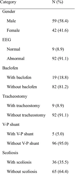
Table 1. Patient Characteristics
A Category N (%) Gender Male 59 (58.4) Female 42 (41.6) EEG Normal 9 (8.9) Abnormal 92 (91.1) Baclofen With baclofen 19 (18.8) Without baclofen 82 (81.2) Tracheostomy With tracheostomy 9 (8.9) Without tracheostomy 92 (91.1) V-P shunt With V-P shunt 5 (5.0) Without V-P shunt 96 (95.0) Scoliosis With scoliosis 36 (35.5) Without scoliosis 65 (64.4)
7 Feeding route Oral 45 (44.6) Nasogastric tube 48 (47.5) Gastrostomy 8 (7.9) Main Diagnosis Epilepsy 67 (66.3) Mitochondrial disease 19 (18.8) Cerebral palsy 8 (7.9) Others 7 (6.9) Brain MRI Normal 25 (24.7) Abnormal 76 (75.2) Total 101 (100)
V-P shunt, ventriculo-peritoneal shunt B
Category Median (IQR)
Disease onset age (month) 3 (0-6)
Examination age (month) 23.4 (10.5-44.1) Duration of disease (month) 15.4 (7.35-30.75)
Number of AED 3 (2-4)
IQR, interquartile range; AED, antiepileptic drug
All patients were under the age of 18. Causes of neurologic impairment were heterogeneous and included perinatal asphyxia, genetic abnormality, cerebral hemorrhage or infarction, hypoxic brain damage, brain tumor, and infections of
8
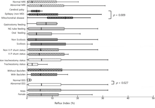
the central nervous system. Substantial numbers of patients had developed neurologic impairment without a definite cause. The main diagnoses of the patients were variable as well, such as Lenox-Gastaut syndrome, infantile spasm, cerebral palsy, mitochondrial disease, Dravet syndrome, and so on. As many patients had complex and combined diagnosis, patients with confirmed mitochondrial disease were categorized to mitochondrial disease group priorly, and patients who have epilepsy but not diagnosed with mitochondrial disease were categorized to epilepsy group. Patients with cerebral palsy were categorized to cerebral palsy group only when diagnosed with neither mitochondrial disease nor epilepsy. When we compared reflux index according to disease category, the mitochondrial disease group (n = 19) had a higher reflex index than did the other epilepsy group (n = 67) or the cerebral palsy group (n = 8) (p = 0.009). The median reflux index values were 14.4, 3.9, and 3.5, respectively. Patient gender, feeding method, scoliosis, tracheostomy, and baclofen use did not lead to statistically significant differences in reflux index. Reflux index was significantly higher in the abnormal EEG group than in the normal EEG group (p = 0.027) (Fig. 1).
9
Fig. 1. Box and whisker plot of reflux index showing the distribution of reflux index according to clinical condition. The box represents the interquartile range, and the line in the box shows the median value. Differences with a p value of < 0.05 are marked.
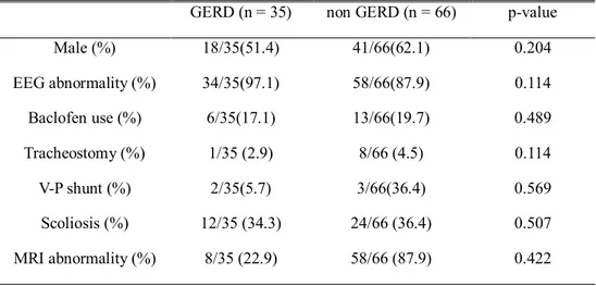
Clinical characteristics between GERD group and non GERD group did not have significant difference (Table2.)
10
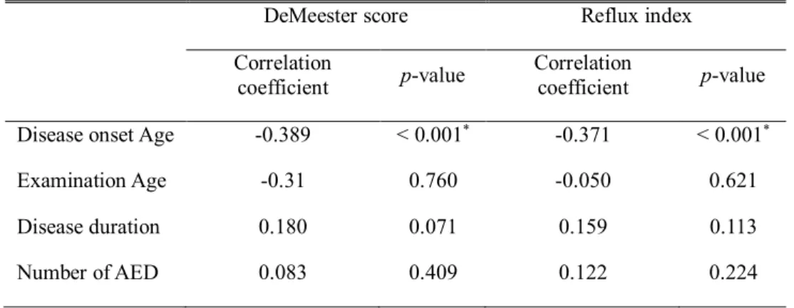
Table 2. Spearman correlation coefficients of gastro-esophageal reflux index and DeMeester score with continuous variables
DeMeester score Reflux index
Correlation
coefficient p-value Correlation coefficient p-value Disease onset Age -0.389 < 0.001* -0.371 < 0.001*
Examination Age -0.31 0.760 -0.050 0.621
Disease duration 0.180 0.071 0.159 0.113
Number of AED 0.083 0.409 0.122 0.224
The age at examination, duration of the disease, and number of antiepileptic drugs did not correlate with reflux index or DeMeester score. However, age of onset of neurological impairment was inversely correlated with DeMeester score (correlation coefficient -0.389; p < 0.001). Reflux index (%) and disease onset age also demonstrated a similar tendency (correlation coefficient -0.371; p < 0.001), which means that early onset of neurologic impairment can aggravate GER (Table 3).
11
Table 3. Comparisons of clinical characteristics between GERD and non-GERD group
GERD (n = 35) non GERD (n = 66) p-value
Male (%) 18/35(51.4) 41/66(62.1) 0.204 EEG abnormality (%) 34/35(97.1) 58/66(87.9) 0.114 Baclofen use (%) 6/35(17.1) 13/66(19.7) 0.489 Tracheostomy (%) 1/35 (2.9) 8/66 (4.5) 0.114 V-P shunt (%) 2/35(5.7) 3/66(36.4) 0.569 Scoliosis (%) 12/35 (34.3) 24/66 (36.4) 0.507 MRI abnormality (%) 8/35 (22.9) 58/66 (87.9) 0.422
When we analyzed reflux index according to disease onset age, patients who had disease onset before 12months showed higher reflux index (8.77 ± 9.33) than patient who had disease onset after 12months of age (4.16 ± 6.66) and the result was statistically significant (p = 0.025).
12
IV. DISCUSSION
GER refers to retrograde passage of gastric contents to the esophagus, pharynx, or oral cavity. GERs can be normal physiological events which occur frequently even in healthy individuals6. However, severe GER can cause troublesome
symptoms or complications and can impair health-related quality of life7. This
condition is called GERD. GERD is much more prevalent in NIP than in the normal population, and can be a significant obstacle to ensuring adequate nutrition. In clinical practice, neurologically impaired children are often malnourished and malnutrition and GER can negatively impact one another8,9.
Therefore, both conditions should be monitored and managed attentively. Furthermore, due to a lack of effective communication, GERD is often diagnosed belatedly. GER is also closely related to respiratory problems. Recurrent pulmonary infections can be caused that give rise to poor prognoses10.
Thus, GERD can decrease a patient’s quality of life both directly and indirectly. If GER can be predicted in NIP, appropriate evaluation and management measures can be taken which will lead to better long-term outcomes.
Although GER is a relatively common problem in both normal infants and NIP, the mechanisms of GER are not fully understood. Transient relaxation of the lower esophageal sphincter (LES) has been reported to be a main mechanism of GER in children11,12. On the other hand, in NIP, absence of LES tone is thought
to be the main mechanism rather than transient relaxation of the LES13. Delayed
gastric emptying and decreased antroduodenal motor function have also been suggested as mechanisms of GER in NIP14,15. As the mechanisms of GER in
NIP are complicated and poorly understood, it is difficult to predict and prevent GERD in such patients. This study was designed to identify the risk factors for the GER in neurologically impaired children.
13
GER. This finding may be because early-onset neurological diseases tend to be related to congenital neurologic anomalies or genetic mutations which bring about major neurologic sequelae, and accordingly, severe systemic complications. In a previous study, 67% of otherwise healthy infants were found to have GER at 4 months of age16. This is due to frequent feeding, short
esophagus, the wide angle between the esophagus and stomach, and the amount of time infants spend in the supine position rather than in an upright position17.
This physiologic GER was alleviated in the course of normal growth, development, and transition to solid food. Only 5% of otherwise healthy children have GER at 12 months of age16. In early-onset NIP, such normal
developmental progress can be halted and GER may persist throughout life. In this study, patients who developed neurological impairment before 12 months of age revealed to have higher reflux index than other patients (8.77 ± 9.33 vs. 4.16 ± 6.66). Therefore, additional attention to GERD and aspiration is necessary especially for patients with infantile-onset neurologic impairment. Although we also hypothesized that a long duration of morbidity might affect the severity of GER, the two were not significantly associated. Disease duration does not seem to be a significant factor for progression of GER in NIP.
The clinical manifestations of neurological diseases are different according to the diagnosis. Therefore, we compared the severity of GER amongst patient with different categories of disease. As the causes and diagnoses of neurologic impairment were heterogeneous, we categorized them as mitochondrial disease, epileptic disorders other than mitochondrial disease, cerebral palsy, and others. Patients with mitochondrial disease showed a higher reflux index than did those with other diseases. Mitochondrial disease is a multi-systemic disorder that is frequently associated gastrointestinal and hepatic manifestations18. Bhardwaj et
al.19 demonstrated that children with mitochondrial disease had frequent
gastrointestinal symptoms, such as abdominal pain, GER, and constipation, which are thought to be related to delayed gastric emptying and small bowel
14
transit time, and which respond poorly to medications. This result suggests that NIP with mitochondrial disease is at additional risk for GER. Not only neurologic impairment but also mitochondrial disease itself may contribute to this result. However, additional randomized controlled studies are required to fully elucidate this matter.
In a previous study, somewhat different characteristics were seen between nasogastric tube fed and orally fed children, and orally fed NIP were at an increased risk for aspiration1. Feeding route and physical characteristics of food
may influence the upper gastrointestinal motility and gastric emptying. However, in the present study, statistical differences were not observed between the gastrostomy, nasogastric tube, and orally fed groups. As patients in the three groups tended to have different clinical conditions, confounding factors may have affected the results.
To compare objective conditions, the patients with ventriculo-peritoneal shunts or tracheostomy or scoliosis were analyzed with the patients who did not have the conditions. As a result, such conditions did not statistically affect reflux activity. In terms of electroencephalography, the normal EEG group had a lower reflux index than did the abnormal EEG group. This means that abnormal EEG can be considered a risk factor for GERD in NIP. However, this does not mean that seizure activity aggravates GERD directly, as the number of antiepileptic drugs, which is an indicator of intractable seizures, is not correlated with reflux activity. Rather, abnormal brain function itself most likely affected reflux activity.
As many patients with neurological problems suffer from tonic posture, baclofen is sometimes used as a muscle relaxant. Because it is as GABA type B receptor agonist, baclofen is known to decrease postprandial acid reflux by reducing transient lower esophageal sphincter relaxation20,21. Kawai et al.
15
acid reflux in neurologically impaired children22. Although we attempted to
investigate the anti-reflux effect of baclofen in this study, no significant differences existed between the baclofen-treated group and the group not taking baclofen. This result may be negatively affected by spasticity itself, the reason for baclofen treatment.
This study has several limitations. First, the study population was heterogeneous. Patients with several different types of neurological impairments were included, and their clinical conditions were variable. Second, the reasons for performing 24 hr esophageal pH monitoring were diverse. Some patients had severe recurrent aspiration pneumonia, whereas others had mild irritability. The decision to perform the test depended individual physicians, which may have introduced a selection bias. Furthermore, the study design was retrospective.
16
V. CONCLUSION
As several complex clinical factors may influence GER, it is difficult to predict the degree of GER just by using objective clinical factors in severely ill, neurologically impaired children. However, as the results of this study show, early onset neurologic impairment, mitochondrial disease, and abnormal EEG can be important risk factors for severe GER. Accordingly, early suspicion and proper evaluations of GER are needed for those patients.
17
REFERENCES
1. Del Buono R, Wenzl TG, Rawat D, Thomson M. Acid and nonacid gastro-oesophageal reflux in neurologically impaired children: investigation with the multiple intraluminal impedance procedure. J Pediatr Gastroenterol Nutr 2006;43:331-5.
2. Del Giudice E, Staiano A, Capano G, Romano A, Florimonte L, Miele E, et al. Gastrointestinal manifestations in children with cerebral palsy. Brain Dev 1999;21:307-11.
3. Trinick R, Johnston N, Dalzell AM, McNamara PS. Reflux aspiration in children with neurodisability--a significant problem, but can we measure it? J Pediatr Surg 2012;47:291-8.
4. Aviv JE, Liu H, Parides M, Kaplan ST, Close LG. Laryngopharyngeal sensory deficits in patients with laryngopharyngeal reflux and dysphagia. The Annals of otology, rhinology & laryngology 2000;109:1000-6.
5. Gossler A, Schalamon J, Huber-Zeyringer A, Hollwarth ME. Gastroesophageal reflux and behavior in neurologically impaired children. J Pediatr Surg 2007;42:1486-90.
6. Sherman PM, Hassall E, Fagundes Neto U, Gold BD, Kato S, Koletzko S, et al. A global, evidence-based consensus on the definition of gastroesophageal reflux disease in the pediatric population. The American journal of gastroenterology 2009;104:1278-95; quiz 96.
7. Madisch A, Kulich KR, Malfertheiner P, Ziegler K, Bayerdörffer E, Miehlke S, et al. Impact of reflux disease on general and diseaserelated quality of life -evidence from a recent comparative methodological study in Germany. Zeitschrift für Gastroenterologie 2003;41:1137-43.
8. Lewis D, Khoshoo V, Pencharz PB, Golladay ES. Impact of nutritional rehabilitation on gastroesophageal reflux in neurologically impaired children. J Pediatr Surg 1994;29:167-9; discussion 9-70.
9. Marchand V, Motil KJ. Nutrition support for neurologically impaired children: a clinical report of the North American Society for Pediatric Gastroenterology, Hepatology, and Nutrition. Journal of pediatric gastroenterology and nutrition 2006;43:123-35.
10. Omari T, Barnett C, Snel A, Davidson G, Haslam R, Bakewell M, et al. Mechanism of gastroesophageal reflux in premature infants with chronic lung disease. Journal of pediatric surgery 1999;34:1795-8.
11. Kawahara H, Nakajima K, Yagi M, Okuyama H, Kubota A, Okada A. Mechanisms responsible for recurrent gastroesophageal reflux in neurologically impaired children who underwent laparoscopic Nissen fundoplication. Surg Endosc 2002;16:767-71.
12. Omari TI, Barnett CP, Benninga MA, Lontis R, Goodchild L, Haslam RR, et al. Mechanisms of gastro-oesophageal reflux in preterm and term infants with reflux disease. Gut 2002;51:475-9.
13. Pensabene L, Miele E, Del Giudice E, Strisciuglio C, Staiano A. Mechanisms of gastroesophageal reflux in children with sequelae of birth asphyxia. Brain Dev 2008;30:563-71.
14. Maxson RT, Harp S, Jackson RJ, Smith SD, Wagner CW. Delayed gastric emptying in neurologically impaired children with gastroesophageal reflux: the
18
role of pyloroplasty. J Pediatr Surg 1994;29:726-9.
15. Miki K, Harada T, Kozaiwa K, Tajiri H, Nagai T, Kawahara H, et al. Antroduodenal motor function and gastro-oesophageal reflux in neurologically impaired children, adolescents and young adults. Eur J Pediatr 1998;157:695-6. 16. Nelson SP, Chen EH, Syniar GM, Christoffel KK. Prevalence of symptoms of
gastroesophageal reflux during infancy. A pediatric practice-based survey. Pediatric Practice Research Group. Archives of pediatrics & adolescent medicine 1997;151:569-72.
17. Bishop W. Pediatric Practice Gastroenterology: Gastroenterology: McGraw Hill Professional; 2010.
18. Rahman S. Gastrointestinal and hepatic manifestations of mitochondrial disorders. Journal of Inherited Metabolic Disease 2013;36:659-73.
19. Bhardwaj J, Wan DQ, Koenig MK, Liu Y, Hashmi SS, Rhoads JM. Impaired gastric emptying and small bowel transit in children with mitochondrial disorders. Journal of pediatric gastroenterology and nutrition 2012;55:194-9. 20. Zhang Q, Lehmann A, Rigda R, Dent J, Holloway RH. Control of transient
lower oesophageal sphincter relaxations and reflux by the GABA(B) agonist baclofen in patients with gastro-oesophageal reflux disease. Gut 2002;50:19-24.
21. van Herwaarden MA, Samsom M, Rydholm H, Smout AJ. The effect of baclofen on gastro-oesophageal reflux, lower oesophageal sphincter function and reflux symptoms in patients with reflux disease. Alimentary pharmacology & therapeutics 2002;16:1655-62.
22. Kawai M, Kawahara H, Hirayama S, Yoshimura N, Ida S. Effect of baclofen on emesis and 24-hour esophageal pH in neurologically impaired children with gastroesophageal reflux disease. J Pediatr Gastroenterol Nutr 2004;38:317-23.
19