저작자표시-비영리-변경금지 2.0 대한민국 이용자는 아래의 조건을 따르는 경우에 한하여 자유롭게 l 이 저작물을 복제, 배포, 전송, 전시, 공연 및 방송할 수 있습니다. 다음과 같은 조건을 따라야 합니다: l 귀하는, 이 저작물의 재이용이나 배포의 경우, 이 저작물에 적용된 이용허락조건 을 명확하게 나타내어야 합니다. l 저작권자로부터 별도의 허가를 받으면 이러한 조건들은 적용되지 않습니다. 저작권법에 따른 이용자의 권리는 위의 내용에 의하여 영향을 받지 않습니다. 이것은 이용허락규약(Legal Code)을 이해하기 쉽게 요약한 것입니다. Disclaimer 저작자표시. 귀하는 원저작자를 표시하여야 합니다. 비영리. 귀하는 이 저작물을 영리 목적으로 이용할 수 없습니다. 변경금지. 귀하는 이 저작물을 개작, 변형 또는 가공할 수 없습니다.
이학 박사학위 논문
Comparison of Multilocus sequence
typing change patterns of
Vancomycin resistant
Enterococcus
faecium
in the past nine years
Ajou University Graduate School
Department of Medical Sciences
Comparison of Multilocus sequence
typing change patterns of
Vancomycin resistant
Enterococcus
faecium
in the past nine years
WEE GYO LEE, Advisor
I submit this thesis as the Doctoral thesis in Medical
Sciences.
The Doctoral thesis of JOON KIM in Medical Sciences
is hereby approved.
Thesis Defense Committee President
___________________
YOUNG AE LIM
Member ___________________
WEE GYO LEE
Member ___________________
SUNG RAN JO
Member ___________________
IL JOONG PARK
Member ___________________
CHANG EUN PARK
Ajou University Graduate School
December, 22th, 2017
-
Abstract-Comparison of Multilocus sequence typing change patterns of Vancomycin
resistant
Enterococcus faecium in the past nine years
Background : To understand the spread of Vancomycin Resistant Enterococcus (VRE) is an important component of hospital infection control measures. Multilocus sequence typing (MLST) is useful in determining the long–term evolutionary process and minimizes differences in experimental results across individuals and laboratories. It is also useful in determining evolutionary origins and backgrounds of bacterial species. This study carries out MLST analysis on vanA Vancomycin resistant Enterococcus faecium isolated from patient specimen in a single university hospital in the past nine years in order to observe changes in genetic evolution over time.
Method : During the years from 2007 to 2015, 45 clinical isolates of
vanA-containing E. faecium were collected from Ajou university hospital in Korea. Species were identified through the VitekII system (BioMerieux, Hazelwood, MO) and antibiotic susceptibility testing was performed by disk diffusion and E-test according to Clinical and Laboratory Standards Institute (CLSI) guidelines. To determine genetic relatedness, Matrix Assisted Laser Desorption Ionization-Time Of Flight mass spectrometry (MALDI-TOF MS) was employed. The multilocus sequence
dalfopristin. 45 clinical isolates were genetically unrelated according to MALDI-TOF MS analysis. MLST showed that the clinical isolates had 7 sequence types (ST), ST17(n=19) being the most common, followed by ST78(n=13), ST192(n=6), ST64(n=4), ST262(n=1), ST414(n=1) and ST981(n=1).
Conclusion : The MLST analysis showed that the sequence types of most isolates belonged to clonal complex 17(CC17). This is consistent with outbreaks in hospitals. The study found that the most common type of separation was changed from ST78 to ST17. We had single observations for ST262, ST414 and ST981 but they appear to be random occurrences. MLST can be useful for speculating the genetic evolution of vanA containing E. faecium isolates.
Table of Contents
ABSTRACT ---ⅰ TABLE OF CONTENTS ---ⅲ LIST OF FIGURES –---ⅴ LIST OF TABLES ---ⅵ Ⅰ.INTRODUCTION ---1Ⅱ. Material and Methods ---5
A. Research Materials 1. Target strains--- 5
B. Research Methods 1. Antimicrobial susceptibility test --- 6
2. Real‐time PCR --- 7
3. MALDI‐TOF M/S --- 9
Ⅳ. DISCUSSION --- 28
Ⅴ. CONCLUSIONS --- 33
Ⅵ. REFERENCE --- 34
LIST OF FIGURES
Fig. 1. Schematic diagram showing the work-flow in a MALDI-TOF MS --- 4
Fig. 2. Algorithms and result sheets of two commercially available MALDI-TOF MS analysis platforms ---10
Fig. 3. Representative image of Multiallign website for compare allelic profiles with 45 strains in this study --- 14
Fig. 4. Representative MALDI-TOF mass spectra of vanA E. faecium strains representing the seven STs established in this study --- 22
Fig. 5. Dendrogram of 45 vanA E. faecium obtained after MALDI-TOF MS analysis - - - 2 3
Fig. 6. eBURST diagram of 45 vavA E. faecium STs of this study --- 26
LIST OF TABLES
Table 1. Criteria of Antisusceptibility test (CLSI 2016 M100-26) --- 6
Table 2. Fluorophore used for Real-time PCR --- 8
Table 3. Oligonucleotide primers used in this study --- 12
Table 4. Antibiotic susceptibility profiles of 45 strains --- 16
Ⅰ. Introduction
Since vancomycin‐resistant enterococci (VRE) were first reported in Europe in 1986, the frequency of isolation has increased worldwide to date, and nowadays it is indigenous to almost all medical inst itut ions. In Korea, the incidence of Enterococcus durans, which is highly resistant to vancomycin, has been reported to have increased up to the present time after being reported by Park et al. [1] in 1992. Although enterococci are relatively weakly pathogenic, infections caused by VRE are often found in long‐term inpatients with impaired immunity, and the t re at ment is not ef fect i v e and t he mort a lit y rat e for V RE bact e rem ia is approximately 37% [2]. In particular, VanA type VRE is highly resistant to vancomycin and is difficult to treat, leading to high mortality from bacteremia [3,4]. The VRE resistant types known to date are VanA, VanB, VanC, VanD, VanE, VanG, VanL, VanM and VanN, and of them, VanA and VanB types account for most of the global epidemic [5‐8]. In Korea, the incidence of VRE is similar to that of the United States, which is spreading to hospital infections. Therefore, it has begun to occur mainly in large hospitals and to occur recently in small hospitals, showing aspects of indigenization [9]. VRE, which is the subject of hospital infection control, is VanA type and VanB type, which are proven resistant, and vanA or vanB gene is transferred among VanC type, so that the same VRE is included in the subject
genetic correlations and gene evolution cannot be detected. Therefore, molecular genetic epidemiological methods other than PFGE were introduced.
Multilocus sequence typing (MLST) analyzes the sequence of 6 to 7 housekeeping genes (450‐500 bp) containing information on the production of proteins involved in essential cell functions and is available on the MLST website (http: // www. mlst.net) for analysis of genetic diversity and type determination (ST). The different sequences of the house keeping genes are distinguished by alleles, which provide an allelic profile and serve as identification markers for strain typing. In the case of E. faecium, seven genes including adk (adenylate kinase), atpA (ATP synthase, alpha subunit), ddl (D‐alanine:D‐alanine ligase), gyd (glyceraldehyde‐3‐phosphate dehydroge nase), gdh (glucose‐6‐phosphate dehydrogenase), purK (phosphoribosylaminoimidazol e carboxylase ATPase subunit), pstS (phosphate ATP‐binding cassette transporter), etc. are used. Bacterial identification by MLST has sufficient resolution to distinguish even very closely related strains. The MLST method reflects the long‐term dynamics of genes and is a method for estimating the epigenetic origins and evolutionary backgrounds of the same strains without error between experimenter and laboratory [11].
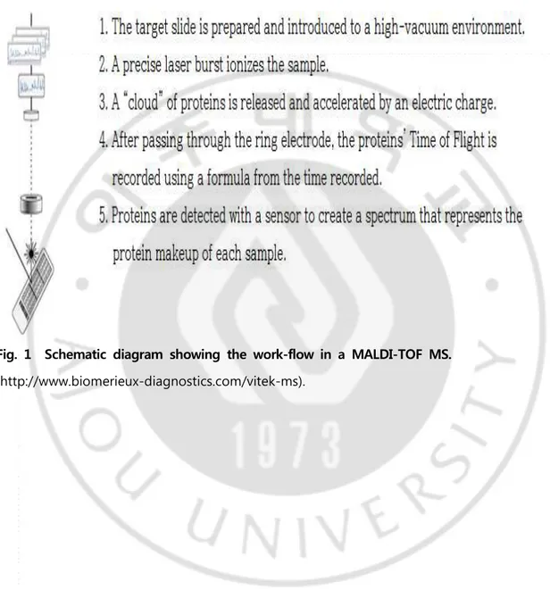
Matrix assisted laser desorption ionization‐time of flight mass spectrometry (MALDI-TOF MS) was developed for the analysis of polymeric materials in the 1980s. This method is known to be suitable for biopolymers and synthetic polymers which are massive and unstable in heat, by vaporization/ionization without decomposition of the sample [12,13,14]. The principle of analysis is to detect the ion of the analysis target itself and to predict the mass of the analysis target. It uses the post‐source‐decay method to spontaneously proceed with peptide degradation and ionization in the flight tube, and peptides or proteins can be
identified by providing direct information about the sequence of an unknown peptide or protein (Fig. 1).
MALDI‐TOF MS can be divided into a laser desorption ionization stage, which irradiates the sample with a strong pulsed laser to disintegrate the sample‐matrix to release ions, and a Time of Flight (TOF) stage to measure ion flight time. The TOF method can be classified into linear TOF using the principle that light ions arrive at the detector before heavy ions and reflectron TOF using the principle that high‐ speed ions fly longer distances in the reflectron [15]. By using MALDI‐TOF MS analysis, the strains can be identified by comparing the protein information of the bacteria to be known with the information of each strain already constructed. It has been proven that the efficiency of the classification and identification of the microorganisms in the hospital is high, and it has been used to identify S. aureus, E. coli, Klebsiella, and Salmonella other than Enterococci, and to verify the classification model [16]. Thus, on the target of vanA vancomycin‐resistant E. faecium that was isolated for 9 years in a university in Korea, this study attempted to compare the results of analyzing MALDI‐TOF M/S and MLST and investigate the characteristics of house-keeping gene variation. Therefore, this study tried to find out the prevalence of strains belonging to the CC17 in hospital and to use it as a useful data for management to reduce it.
Fig. 1 Schematic diagram showing the work-flow in a MALDI-TOF MS. (http://www.biomerieux-diagnostics.com/vitek-ms).
Ⅱ. MATERIALS AND METHODS
A. Research Materials
1. Target strains
From a vanA vancomycin‐resistant E. faecium isolated from a urine culture study commissioned to the Department of Diagnostic Medicine, Ajou University hospital for 9 years between 2007 and 2015, 5 strains were collected every year from 2007 to 2015, and a total of 45 strains (strain code name vanA 01 ~ vanA 45) were tested. To determine the long‐term dynamics of the gene of interest, the target strains were selected based on similar conditions, if possible. The isolated strains were collected from urine specimens that were relatively easy to obtain, and the 45 strains of vanA vancomycin‐resistant E. faecium isolated at 2‐3 month intervals a year from 2007 to 2015 were collected.
Each strain was tested for vancomycin resistance using brain heart infusion (BHI) agar containing 6 ㎍/mL vancomycin. All strains identified as VRE were identified
using the Vitek II system (BioMerieux, Hazelwood, MO) and biochemical techniques. E. faecium BM4147 was used as a positive control strain and vancomycin susceptible E. faecalis (ATCC 29212) and vancomycin susceptible E. faecium 2 strains were used as negative control strains.
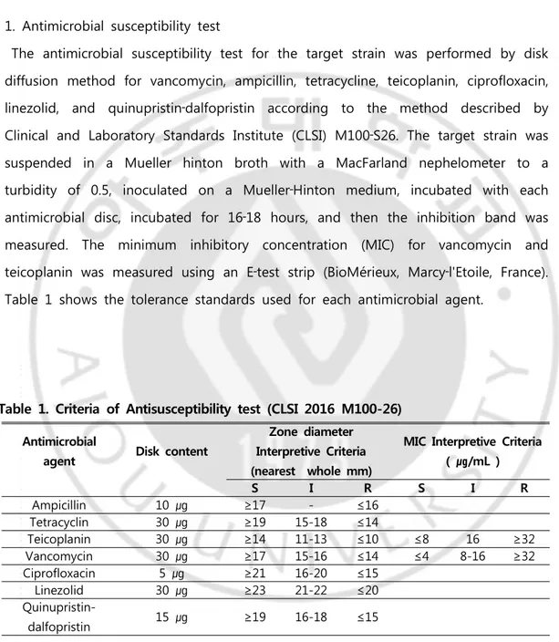
Antimicrobial
agent Disk content
Zone diameter Interpretive Criteria (nearest whole mm)
MIC Interpretive Criteria ( ㎍/mL ) S I R S I R Ampicillin 10 ㎍ ≥17 - ≤16 Tetracyclin 30 ㎍ ≥19 15-18 ≤14 Teicoplanin 30 ㎍ ≥14 11-13 ≤10 ≤8 16 ≥32 Vancomycin 30 ㎍ ≥17 15-16 ≤14 ≤4 8-16 ≥32 Ciprofloxacin 5 ㎍ ≥21 16-20 ≤15 Linezolid 30 ㎍ ≥23 21-22 ≤20 Quinupristin-dalfopristin 15 ㎍ ≥19 16-18 ≤15 B. Research Methods
1. Antimicrobial susceptibility test
The antimicrobial susceptibility test for the target strain was performed by disk diffusion method for vancomycin, ampicillin, tetracycline, teicoplanin, ciprofloxacin, linezolid, and quinupristin‐dalfopristin according to the method described by Clinical and Laboratory Standards Institute (CLSI) M100‐S26. The target strain was suspended in a Mueller hinton broth with a MacFarland nephelometer to a turbidity of 0.5, inoculated on a Mueller‐Hinton medium, incubated with each antimicrobial disc, incubated for 16‐18 hours, and then the inhibition band was measured. The minimum inhibitory concentration (MIC) for vancomycin and teicoplanin was measured using an E‐test strip (BioMérieux, Marcy‐l'Etoile, France). Table 1 shows the tolerance standards used for each antimicrobial agent.
2. Real‐time PCR
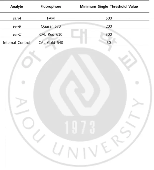
Real‐time PCR was performed according to the manufacturer's method using the Anyplex™ VanR Real‐time Detection kit (Seegene, Sankt Ingbert, Germany) to confirm the vancomycin‐resistant genotype of the target strain. The method was as follows: Brain heart infusion broth containing Vancomycin 6 ㎍/mL was inoculated with target strains and cultured at 35°C for more than 1 day. The DNA extraction solution in the kit was vortexed and added 200 µL into a 1.5 mL microcentrifuge tube. 50 μL of cultured broth was added to a 1.5 mL microcentrifuge tube. Tightly closed microcentrifuge tube using cap lock were vigorously vortexed and boiled for 10 minutes in heat block (100°C). After incubation at room temperature for 5 minutes, the tube centrifuged at 13,000 rpm for 5 minutes. 100 μL of supernatant transferred to a new tube, and used 3 μL for PCR. For PCR, added 17 μL of PCR Mastermix [Seegene, Sankt Ingbert, Germany: 4 µL 5X Van OM, 3µL 8‐ MOP solution, 10 µL 2X detection mix] and 3 μL of sample DNA in a PCR tube and mixed softly and briefly centrifuged. PCR was performed at 95°C for 2 min, 1 cycle at 95°C for 30 sec, and 45 cycles at 60°C for 30 sec using the CFX96™ Real‐ time PCR System (Bio‐Rad Laboratories, Inc, Hercules, CA, USA.). The target analyte and Fluorophore used are shown in Table 2.
Analyte Fluorophore Minimum Single Threshold Value
vanA FAM 500
vanB Quasar 670 200
vanC CAL Red 610 300
Internal Control CAL Gold 540 50
3. MALDI‐TOF M/S
MALDI‐TOF M/S was used to analyze genetic relatedness of target strains. A single colony was selected on a blood agar culture medium (KOMED, Seoul, Korea) cultured at 37°C for 24 hours and thinly coated on a target slide. Then, 1 μL of α‐cyano‐4‐hydroxycinnamic acid (CHCA) matrix (BioMérieux, La Balme‐les‐Grottes, France) and prepared a sample slide that was dried at room
temperature. The prepared sample slides were loaded into a VITEK MS (BioMérieux, Marcy l'Etoile, France) system and spectra were obtained at a range of 2,000‐20,000 Da mass spectrometry using the Research Use Only Mode (RUO) software Lanchpad V2.84.
Each strain was repeated for analysis for three times. The spectra obtained from the instrument were identified using the Spectral ARchive And Microbial Identification System (SARAMIS) V 4.13 database and the VITEK MS RUO system was calibrated using the E.coli ATCC 8739 standard strain before and after the test. To analyze the correlation of 45 Enterococcus faecium strains analyzed by MLST, all spectra obtained from the equipment were exported to the ASCII format peak list through the processing process, and the peak list was to be taken to the SARAMIS database for further analysis (Fig. 2). The software that compared the mass spectra generates a score value based on the similarities between the test strain and stored data sets. This score value provides information about the
Fig. 2 Algorithms and result sheets of two commercially available MALDI-TOF analysis platforms[17].
4. Multilocus sequence typing
(A) Genomic DNA extract
45 strains were streaked on blood agar plates and incubated at 37 °C for 18-24 hours. 5-15 colonies were collected into 200 µL sterilized purified water, centrifuged at 12,000 rpm for 5 min. The precipitate was used to extract genome DNA with Quick Gene DNA whole blood kit (KURABO Industries Ltd, japan) according to operating instruction.
(B) Gene amplification
PCR was performed according to the method described in website (http://efaecium.mlst.net.) The total amount of the PCR reaction was adjusted to
50 μL, and the reaction was carried out using 1.25 U of TaKaRa Ex TaqTM, 2 × buffer (25 mM TAPS, 50 mM KCl, 2 mM MgCl2, 1 mM 2‐mercaptoethanol), 0.4
mM dNTPs mixture, 10 pM primer 1 μL and 10 ng of purely isolated chromosomal DNA.
All PCR amplifications were carried out in a GeneAmp PCR System 9600 (Perkin‐ Elmer Corp., Norwalk, CT.) under the following conditions: Initial denaturation at 95 °C for 15 min, 35 cycles of 30s at 94 °C, 30s at 52 °C, 30s at 72 °C, followed
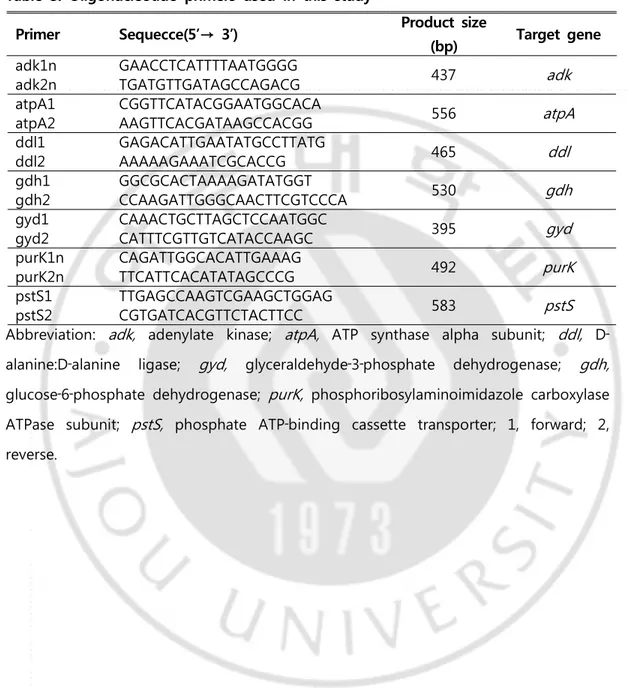
Primer Sequecce(5’→ 3’) Product size (bp) Target gene adk1n GAACCTCATTTTAATGGGG 437 adk adk2n TGATGTTGATAGCCAGACG atpA1 CGGTTCATACGGAATGGCACA 556 atpA atpA2 AAGTTCACGATAAGCCACGG ddl1 GAGACATTGAATATGCCTTATG 465 ddl ddl2 AAAAAGAAATCGCACCG gdh1 GGCGCACTAAAAGATATGGT 530 gdh gdh2 CCAAGATTGGGCAACTTCGTCCCA gyd1 CAAACTGCTTAGCTCCAATGGC 395 gyd gyd2 CATTTCGTTGTCATACCAAGC purK1n CAGATTGGCACATTGAAAG 492 purK purK2n TTCATTCACATATAGCCCG pstS1 TTGAGCCAAGTCGAAGCTGGAG 583 pstS pstS2 CGTGATCACGTTCTACTTCC
Table 3. Oligonucleotide primers used in this study
Abbreviation: adk, adenylate kinase; atpA, ATP synthase alpha subunit; ddl, D‐ alanine:D‐alanine ligase; gyd, glyceraldehyde‐3‐phosphate dehydrogenase; gdh, glucose‐6‐phosphate dehydrogenase; purK, phosphoribosylaminoimidazole carboxylase ATPase subunit; pstS, phosphate ATP‐binding cassette transporter; 1, forward; 2, reverse.
(C) Housekeeping gene sequencing
Sequencing reactions were performed in the DNA Engine Tetrad 2 Peltier Thermal Cycler (BIO-RAD) using the ABI BigDye(R) Terminator v3.1 Cycle Sequencing Kit (Applied Biosystems), following the protocols supplied by the manufacturer. Single-pass sequencing was performed on each template using primer. The fluorescent-labeled fragments were purified by the method that Applied Biosystems recommends as it removes the unincorporated terminators and dNTPs. The samples were injected to electrophoresis in an ABI 3730xl DNA Analyzer (Applied Biosystems).
(D) Database analysis for allele profile and ST identification
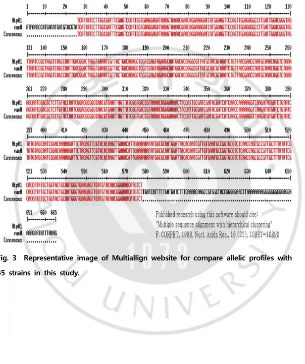
To confirm the sequence type (ST), PCR products of 7 house‐keeping genes were submitted to Macrosen (Seoul, Korea) for sequencing. Each of the analyzed sequences was submitted to the Multiallign website (http://multalin.toulouse.in.ra.
fr/multalin) and PubMLST (https://pubmlst.org/databases/) to confirm the respective allele number and the final ST was confirmed for E. faecium (Fig. 3).
Each strain was analyzed using the eBURST program (https://eburst.mlst.net/v3/ml st_datasets/) on the PubMLST website with the nucleotide sequence of each gene and corresponding ST.
Fig. 3 Representative image of Multiallign website for compare allelic profiles with 45 strains in this study.
Ⅲ. RESULTS
1. Antimicrobial susceptibility test and resistant genotype
The antimicrobial susceptibility tests performed on 45 strains according to the CLSI standard showed that the vancomycin MIC values were all highly resistant to ≥256 ㎍/mL. The MIC values of teicoplanin were moderately resistant to vanA 03 and 35 strains at 16 ㎍/mL, vanA 04, 10, 17 and 39 strains at 8 ㎍/mL and vanA 18 strain at 4 ㎍/mL, respectively. vanA 06, 11, 12, 31, and 41 strains showed resistance at 32 ㎍/mL, and the remaining 33 strains were resistant at 64 ㎍/mL. Ampicillin 10 ㎍/mL and ciprofloxacin 5 ㎍/mL showed resistance to all strains, all strains were susceptible to linezolid 30 ㎍/mL and quinupristin‐dalfopristin 15 ㎍ /mL, and tetracyclin 30 ㎍/mL was susceptible to 34 strains, resistant to 10 strains, and moderately resistant to 1 strain (Table 4).
To confirm the resistance genotypes, real‐time PCR with three fluorophores for vanA, vanB and vanC genes was performed it was observed that all 45 strains had vanA resistant genes, and that vanB and vanC genes were negative.
strain MIC (㎍/mL) Resistance to antibiotics Year VA TP AM TE CIP LZD SYN vanA01 >256 64 R S R S S 2007 vanA02 >256 64 R S R S S 2007 vanA03 >256 16 R S R S S 2007 vanA04 >256 8 R R R S S 2007 vanA05 >256 64 R S R S S 2007 vanA06 >256 32 R S R S S 2008 vanA07 >256 64 R S R S S 2008 vanA08 >256 64 R S R S S 2008 vanA09 >256 64 R S R S S 2008 vanA10 >256 8 R R R S S 2008 vanA11 >256 32 R S R S S 2009 vanA12 >256 32 R I R S S 2009 vanA13 >256 64 R R R S S 2009 vanA14 >256 64 R S R S S 2009 vanA15 >256 64 R S R S S 2009 vanA16 >256 64 R S R S S 2010 vanA17 >256 8 R S R S S 2010 vanA18 >256 4 R S R S S 2010 vanA19 >256 64 R R R S S 2010 vanA20 >256 64 R S R S S 2010 vanA21 >256 64 R S R S S 2011 vanA22 >256 64 R S R S S 2011 vanA23 >256 64 R S R S S 2011 vanA24 >256 64 R S R S S 2011 vanA25 >256 64 R S R S S 2011 vanA26 >256 64 R R R S S 2012 vanA27 >256 64 R R R S S 2012 vanA28 >256 64 R R R S S 2012 vanA29 >256 64 R S R S S 2012 vanA30 >256 64 R S R S S 2012 vanA31 >256 32 R R R S S 2013 vanA32 >256 64 R S R S S 2013 vanA33 >256 64 R S R S S 2013 vanA34 >256 64 R S R S S 2013 vanA35 >256 16 R S R S S 2013 vanA36 >256 64 R S R S S 2014 vanA37 >256 64 R S R S S 2014 vanA38 >256 64 R S R S S 2014 vanA39 >256 8 R R R S S 2014 vanA40 >256 64 R S R S S 2014 vanA41 >256 32 R S R S S 2015 vanA42 >256 64 R S R S S 2015
vanA43 >256 64 R R R S S 2015
vanA44 >256 64 R S R S S 2015
vanA45 >256 64 R S R S S 2015
Abbreviation: MIC, minimum inhibitiory concentration; VA, vancomycin; TP, teicoplanin; AM, ampicillin; TE, tetracyclin; CIP, ciprofloxacin; LZD, linezolid; SYN, quinupristin-dafopristin; ST, sequence type.
2. MALDI‐TOF M/S
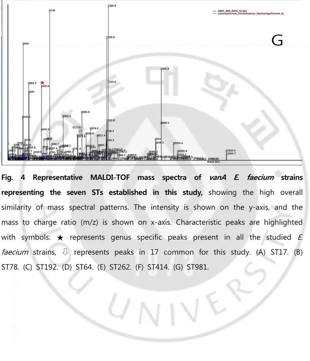
The E. faecium 45 strains isolated from the urine specimens were identified as E. faecium in the VITEK MS RUO system, and were consistent with biochemical differentiation tests other than VITEK 2 Gram‐positive (GP) Identification card (BioMerieux, Hazelwood, MO). All of the obtained spectra showed good resolution, with a variety of peaks and specific spectral profiles for each strains. Total mass lists for each of the 45 strains were generated with the SARAMIS system by calculating the arithmetic means for the m/z values of the three replicate spectra acquired by MALDI‐TOF M/S. The peak measured for each strain showed a peak mass spectra of 227 on average from a minimum of 179 peaks (vanA 02) to a maximum of 257 peaks (vanA 31). A total of 18 common peaks were identified, of which m/z 4429 Da±1 peaks found in both 45 strains and control strains, was reported by Quintela et al.[19] (m/z 4426 Da±1). It was a genus specific biomarker that confirmed its presence in all Enterococcus spp.
The other 17 major peak masses were m/z 3884 Da±1, 4776 Da±1, 5354 Da±2, 5972 Da±2, 5989 Da±3, 6038 Da±2, 6052 Da±2, 6169 Da±1, 6182 Da±2, 6341 Da±2, 6889 Da±2, 7288 Da±1, 8120 Da±3, 8162 Da±2, 9062 Da±2, 9553 Da±1, 10074 Da±2, etc. (Fig. 4). The results of the genotyping analysis based on
the spectra of 45 strains obtained using MALDI‐TOF MS showed that the strains showed genetic diversity and were not associated between strains (Fig. 5).
Fig. 4 Representative MALDI-TOF mass spectra of vanA E. faecium strains representing the seven STs established in this study, showing the high overall similarity of mass spectral patterns. The intensity is shown on the y-axis, and the mass to charge ratio (m/z) is shown on x-axis. Characteristic peaks are highlighted with symbols: ★ represents genus specific peaks present in all the studied E. faecium strains, ⇩ represents peaks in 17 common for this study. (A) ST17. (B) ST78. (C) ST192. (D) ST64. (E) ST262. (F) ST414. (G) ST981.
3. Multilocus sequence typing analysis
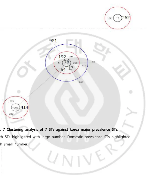
The MLST of vanA E. faecium isolated showed seven STs (Table 5). The atpA and pstS genes have three allelic numbers of 1, 7, 15 and 1, 7, and 20, respectively. The two allelic numbers of ddl and gyd are 1 and 5 and adk gene have 1 and 3. The remaining gdh and purK genes are 1 allelic number. To confirm the diversity of the clones, the seven STs were classified into n-3, or more mathes by the eBURST program (Fig. 6), all belonging to Clonal Complex 17 (CC17). ST17 (19 strains), ST78 (13 strains) and ST192 (6 strains) were common types in seven STs. The most common ST17 was distributed evenly over 9 years, and ST78 and ST192 were also uniformly distributed over a period of 9 years. In addition, ST64 (4 strains), ST262 (1 strain), ST414 (1 strain) and ST981 (1 strain) were isolated, and ST64 (4 strains) was recently isolated. This study compared seven STs with the major ST types isolated from domestic prevalence strains by the eBURST programs (Fig. 7).
ST(allelic profile) (atpA-ddl-gdh-gyd-pstS -purK-adk) No. of isolates 2007 2008 2009 2010 2011 2012 2013 2014 2015 Total (n=45) 17 (1-1-1-1-1-1-1) 3 1 3 3 1 2 2 3 1 19 78 (15-1-1-1-1-1-1) 1 4 2 1 2 0 2 0 1 13 192 (15-1-1-1-7-1-1) 1 0 0 1 1 2 0 0 1 6 64 (7-1-1-1-1-1-1) 0 0 0 0 0 0 1 2 1 4 262 (7-1-1-5-7-1-1) 0 0 0 0 0 1 0 0 0 1 414 (15-5-1-1-20-1-1) 0 0 0 0 1 0 0 0 0 1 981 (15-5-1-1-7-1-3) 0 0 0 0 0 0 0 0 1 1
Table 5. Distribution of sequence types of 45 strains
Abbreviation: ST, sequence type.
Fig. 6 eBURST diagram of 45 vanA E. faecium STs of this study. (n-3, more matches condition).
Fig. 7 Clustering analysis of 7 STs against korea major prevalence STs.
Each STs highlighted with large number. Domestic prevalence STs highlighted with small number.
Ⅳ. DISCUSSION
Of vanA E. faecium target strains, the MIC for vancomycin was ≥256 ㎍/mL in all 45 strains, which indicates high resistance. On the other hand, the MIC values of teicoplanin were lower than those of vanA 04,10,17,39 strains at 8 ㎍/mL, and vanA 18 strain at 4 ㎍/mL, respectively, and the susceptible VanB phenotype‐vanA genotype. In general, vancomycin genotype and phenotype are consistent, but VanB phenotype‐vanA genotype VRE, which is resistant to vancomycin and susceptible to teicoplanin, is frequently reported. The VanB phenotype‐vanA genotype VRE occurring in Korea is the main cause of the vanX, vanY and vanZ gene loss associated with the insertion of IS1216V [20‐22], unlike foreign countries where most of the vanS domain is mutated in Tn1546. On the other hand, vanA 03 and 35 strains were moderately resistant to 16 ㎍/mL, and all other strains were resistant to teicoplanin, VanA phenotype‐vanA genotype. All strains showed resistance to ampicillin 10 ㎍/mL and ciprofloxacin 5 ㎍/mL. All strains were susceptible to linezolid 30 ㎍/mL and quinupristin‐dalfopristin 15 ㎍/mL. Although enterococci resistant to linezolides have been found and reported in the early 2000s [23], they have not been found in this study.
MALDI‐TOF MS is capable of analyzing samples with not dissolve or little volatility, and unlike conventional mass spectrometry, it is not necessary to use extreme conditions requiring modification of materials such as pyrolysis, and since the column is not used, it has the advantage that it is relatively easy to manipulate [14]. In addition, microbiological identification using MALDI‐TOF MS allows for low proficiency, low analysis cost, and rapid identification compared to other commercial identification systems (VITEK 2 system, 16s rRNA gene analysis) [24].
The analysis of genotypes of 45 strains using MALDI‐TOF MS showed an average various 227 peaks mass from at least 179 peaks (vanA 02) to a maximum of 257 peaks (van A31). A total of 18 common peaks were identified, and a peak of m/z 4429 Da±1, found in both 45 strains and control strains, was a genus specific biomarker that could be detected in all Enterococcus spp. The other 17 major peaks were m/z 3884 Da±1, m/z 4776 Da±1, m/z 5354 Da±2, m/z 5972 Da±2, m/z 5989 Da±3, m/z 6038 Da±2, m/z 6052 Da±2, m/z 6169 Da ±1, m/z 6182 Da±2, m/z 6341 Da±2, m/z 6889 Da±2, m/z 7288 Da±1, m/z 8120 Da±3, m/z 8162 Da±2, m/z 9062 Da±2, m/z 9553 Da±1, m/z 10074 Da±2, etc. Two peaks of m/z 6052 Da±2 and m/z 6889 Da±2 were species specific biomarkers identified only in E. faecium [25]. However, 45 strains showed genetic diversity and showed no relationship between strains[26-28]. This suggests that, as it is typical in infection, the vancomycin‐resistant genes in the enterococcal host were transferred horizontally.
MLST analysis is a method to determine the nucleotide sequence of seven house‐ keeping genes from bacterial chromosomal DNA, input it into the MLST database, and then determine the Sequence Type (ST) by combining allelic profiles. The variation of the gene sequence of the house‐keeping gene was reflected, thus indicating a long‐term evolution [29]. MLST can compensate for the disadvantages of PFGE, which reflects short‐term gene mutations by reflecting long‐term gene
widely used to determine the long‐term variation of VRE gene expression. A number of MLST analysis studies of VRE have been undertaken during about 10 years after Homan's report [30‐34]. Clonal complex 17 (CC17) is an E. faecium resistant to ampicillin, which causes, epidemics in six continents around the world.
E. faecium, belonging to CC17, is highly resistant to ampicillin and fluoroquinolone and has a novel putative pathogenicity island, so it is presumed to have acquired vancomycin resistance after being indwelled in hospitals for a long time [36,37]. The discovery of these global epidemic strains was made possible by MLST analysis. The primary founder of CC17 was ST22, but the distribution was epidemic around ST17, a secondary founder rather than ST22. Representative types belonging to CC17 are ST78, ST17, ST64, etc., and types with single locus variation (SLV) and types with double locus variation (DLV) occupy most of the global epidemic. In particular, enterococci belonging to CC17 are known to have a high vancomycin‐ resistant transposon acquisition rate [30]. For this reason, worldwide, CC17 vancomycin‐resistant enterococci (VRE) account for the vast majority of hospital infection epidemics [31‐33]. In the MLST results of this study, 19 strains (42%) were the most common as ST17 type, 13 strains(29%) were ST78 type, 6 strains(13%) were ST192 type, 4 strains(8%) were ST64 type, and ST262 type(2%), ST414 type(2%) and ST981 type(2%) were one strain each (Table 5). The most common ST17 type was distributed evenly over 9 years, and the ST192 type and ST78 type were found to be uniformly distributed over a period of 9 years. Other ST64 type (4 strains), ST262 type (1 strain), ST414 (1 strain) and ST981 (1 strain) were confirmed, and ST64 (4 strains) has recently been able to identify a large number of isolated cases, and it is necessary to continuously observe whether it is due to a temporary epidemic. All seven sequence types in this study belonged to CC17. Of
these, ST17, ST64, and ST78 are the main types, ST192 is the SLV of ST78, and the point mutation of the pstS gene is caused [35], and ST262 and ST414 are DLV of ST192. ST981 is DLV of ST192 and ST414. As the strains of this study, even after a period of 9 years, show that most of the CC17 lineages were found, it is suggested that CC17 survived predominantly in the hospital environment.
The MLST type for the VRE domestically isolated is ST17, ST18, ST32, ST78, ST64, ST117, ST132, ST192, ST202, ST203, ST204, ST205, ST206, ST207, ST233, ST262, ST414, etc., and all of the types found in this study were of the previously reported ST type [34,35]. However, in a study the authors conducted on VRE isolated from the same hospital between 2002 and 2004, ST78 (43%), ST203 (26%), ST205 (5%), ST17 (3%) and ST18 and ST32 type were one strain, respectively. In this study, which isolated VRE from 2007‐2015, ST17 (43%), ST78 (29%), ST192 (14%) and ST64 (9%) and other ST262, ST414 and ST981 were 1 strain, respectively, and it was found that the change occurred with time [38]. The most commonly isolated type was changed from ST78 to ST17, and ST203, which accounted for 26%, disappeared [39], indicating that ST192 occupied the site. In addition, ST type showing sporadic occurrence also showed changes. From the above results, MLST analysis showed that the predominant type of ampicillin‐resistant VRE in the hospital changed with the duration of the MLST type. In addition, the ST17 type isolated in this study is a type that is often isolated in most countries, including
in the hospital. In the future, it is necessary to use MLST to continuously analyze the variation patterns of strains in hospitals and to compare epidemic strains. Since the VRE was first discovered in 1986, it is now difficult to eradicate it in hospitals. In the future, by analyzing using MLST and searching early, it will be possible to identify the strains belonging to CC17 in advance and that efforts should be made to reduce this.
Ⅴ. CONCLUSIONS
Recently, the MALDI‐TOF M/S method has been widely used for genetic analysis of experimental strains by replacing PFGE. The MLST method reflects the long‐term dynamics of genes and is a method for estimating the epigenetic origins and evolutionary backgrounds of the same strains without error between experimenter and laboratory. For vanA vancomycin‐resistant E. faecium isolated in a domestic university hospital for 9 years, this study was conducted to carry out MLST analysis to investigate gene mutation by period. The antimicrobial susceptibility test of E. faecium 45 strains with the vanA gene collected at Ajou University Hospital from 2007 to 2015 showed high resistance to ampicillin, teicoplanin, ciprofloxacin and vancomycin antibiotics, whereas linezolid and quinupristin‐dalfopristin were all susceptible.
In the results of genetic typing of 45 strains using MALDI‐TOF MS, 18 common peak masses including genus and species specific peak mass were confirmed, but genetic diversity indicated that there was no association between strains as similar peak patterns were observed. The results of MLST analysis showed 7 sequence types (ST). ST17 type was the most common with 19 strains, ST78 type 13 strains, ST192 type 6 strains, ST64 type 4 strains, and ST262 type, ST414 type and ST981 type were one strain respectively. In the MLST analysis, all strains belonged to
Ⅵ. REFERENCES
1. Park JW, Kim YR, Shin WS, Kang MW, Han KJ, Shim SI. Susceptibility test of vancomycin-resistant enterococci. Korean J Infect Dis 1992;24:133-37
2. Lautenbach E, Bilker WB, Brennan PJ. Enterococcal bacteremia: Risk factors for vancomycin resistant and predictors of mortality. Infect Control Hosp Epidemiol 1999;20:318-23
3. Perichon B, Reynolds P, Courvalin P. VanD-type glycopeptide-resistant Enterococcus faecium BM4339. Antimicrob Agents Chemother 1997;41:2016-18
4. Edmond MB, Ober JF, Dawson JD, Weinbaum DL, Wenzel RP. Vancomycin resistant enterococcal bacteremia natural history and attributable mortality. Clin Infect Dis 1996;23:1234-39
5. Boyd DA, Willey BM, Fawcett D, Gillani N, Mulvey MR. Molecular characterization of Enterococcus faecalis N06-0364 with low-level vancomycin
resistance harboring a novel D-Ala-D-Ser gene cluster, vanL. Antimicrob Agents Chemother 2008;52:2667-72
6. McKessar SJ, Berry AM, Bell JM, Turnidge JD, Paton JC. Genetic characterization
of vanG, a novel vancomycin resistance locus of Enterococcus faecalis. Antimicrob Agents Chemother 2000;44:3224-28
7. Xu X, Lin D, Yan G, Ye X, Wu S, Guo Y, et al. vanM, a new glycopeptide resistance gene cluster found in Enterococcus faecium. Antimicrob Agents Chemother 2010;54:4643-47
8. Lebreton F, Depardieu F, Bourdon N, Fines-Guyon M, Berger P, Camiade S, et al. D-Ala-d-Ser VanN-type transferable vancomycin resistance in Enterococcus faecium. Antimicrob Agents Chemother 2011;55:4606-12
9. Lee WG, Jernigan JA, Rasheed JK, Anderson GJ, Tenover FC. Possible horizontal transfer of the vanB2 gene among genetically diverse strains of vancomycin- resistant Enterococcus faecium in a Korean hospital. J Clin Microbiol 2001;39: 1165-68
10. Lee WG. Resistance mechanism and epidemiology of vancomycin-resistant enterococci. Korean J Microbiol 2008;11:71-77
11. Homan WL, Tribe D, Poznanski S, Li M, Hogg G, Spalburg E, et al. Multilocus sequence typing scheme for Enterococcus faecium. J Clin Microbiol 2002;40: 1963-71
14. Krishnamurthy T. Ross PL. Rapid identification of bacteria by direct matrix- assisted laser desorption/ionization mass spectrometric analysis of whole cells. Rapid Commun. Mass Spectrom. 1996;10:1992-96
15. Kim HW, Ham JS, Seal KH, Han SA, Park BY, Oh MH. MALDI-TOF MS system for the identification of microorganisms in milk and dairy products. Korean J. Dairy Sci. Technol. 2012;30:131-37
16. Hsieh SY, Tseng CL, Lee YS, Kuo AJ, Sun CF, Lin YH, and et al. High efficient classification and identification of human pathogenic bacteria by MALDI-TOF MS. Mol Cell Proteomics. 2008;27:448-56
17. Kim TS, Lee KH, Hong YJ, Hwang SM, Park JS, Park KU, et al. MALDI-TOF MS: Its application in the clinical laboratory and a paradigm shift in clinical microbiology. Lab Med Online 2015;5:176-87
18. Urwin R, Maiden MC. Multilocus sequence typing: a tool for global epidemiolo gy. Trends Microbiol. 2003;11:479-87
19. Quintela-Baluja M, Bohme K, Fernández-No IC, Morandi S, Alnakip ME, Caamaño-Antelo S, et al. Characterization of different food-isolated Enterococcus strains by MALDI-TOF mass fingerprinting. Electrophoresis 2013;34:2240-50
20. Lauderdale TL, McDonald LC, Shiau YR, Chen PC, Wang HY, Lai JF, et al. Vancomycin resistant enterococci from humans and retail chickens in Taiwan with unique VanB phenotype-vanA genotype incongruence. Antimicrob Agents Chemother 2002;46:525-27
21. Eom JS, Hwang IS, Hwang BY, Lee JG, Lee YJ, Cheong HJ, et al. Emergence of vanA genotype vancomycin-resistant enterococci with low or moderate levels of teicoplanin resistance in Korea. J Clin Microbiol 2004;42:1785-86
22. Lee WG, Huh JY, Cho SR, Lim YA. Reduction in glycopeptide resistance in vancomycin-resistant enterococci as a result of vanA-cluster rearrangements. Antimicrob Agents Chemother 2004;48:1379-81
23. Kainer MA, Devasia RA, Jones TF, Simmons BP, Melton K, Chow S, et al. Response to emerging infection leading to outbreak of linezolid-resistant enterococci. Emerg Infect Dis 2007;13:1024-30
24. Szabados F, Woloszyn J, Richter C, Kaase M, Gatermannm S. Identification of molecularly defined Staphylococcus aureus strains using matrix-assisted laser desorption/ionization time of flight mass spectrometry and the Biotyper 2.0
26. Schlebusch S, Price GR, Gallagher RL, Horton-Szar V, Elbourne LD, Griffin P, et al. MALDI-TOF MS meets WGS in a VRE outbreak investigation. Eur J Clin
Microbiol Infect Dis 2017;36:495-99
27. Nowakiewicz A, Zióoł.kowska G, Zię.ba P, Gnat S, Troś.ciań.czyk A, Adaszek Ł. Characterization of multidrug resistant E. faecalis strains from pigs of local origin by ADSRRS-fingerprinting and MALDI-TOF MS; Evaluation of the compatibility of methods employed for multidrug resistance analysis. PLoS One 2017;12:e0171160.
28. McCarthy KL, Kidd TJ, Paterson DL. Molecular epidemiology of Pseudomonas aeruginosa bloodstream infection isolates in a non-outbreake setting. J Med Microbiol 2107;66:154-59
29. Enright MC, Spratt BG. Multilocus sequence typing. Trends Microbiol 1999;7: 482-87
30. Willems RJL. Genetic evolution of E. faecium and E. faecalis : similarities and differences. Oral communication S1:6. In: 4th ASM Conference on Enterococci, Cartagena, Colombia. American Society for Microbiology, 2014
31. Galloway-Pen˜a JR, Nallapareddy SR, Arias CA, Eliopoulos GM, Murray BE. Analysis of clonality and antibiotic resistance among early clinical isolates of Enterococcus faecium in the United States. J Infect Dis 2009;200:1566–73
32. Gordts B, Van Landuyt H, Ieven M, Vandamme P, Goossens H. Vancomycin-resistant enterococci colonizing the intestinal tracts of
hospitalized patients. J Clin Microbiol 1995;33:2842–46
33. Mato R, de Lencastre H, Roberts RB, Tomasz A. Multiplicity of genetic backgrounds among vancomycin-resistant Enterococcus faecium isolates recovered from an outbreak in a New York City hospital. Microb Drug Resist 1996;2:309–17
34. Oh JY, Her SH, Seol SY, Lee YC, Lee JC, Kim JM, et al. Antimicrobial resistance and multilocus sequence typing of vancomycin resistant Enterococcus faecium
isolated from clinical specimens. Journal of Bacteriology and Virology 2008;38:19-27
35. Ko KS, Baek JY, Lee JY, Oh WS, Peck KR, Lee N, et al. Molecular characterization of vancomycin-resistant Enterococcus faecium isolates from
Korea. J Clin Microbiol 2005;43:2303-06
36. Willems RJ, Top J, van Santen M, Robinson DA, Coque TM, Baquero F, et al. Global spread of vancomycin-resistant Enterococcus faecium from distinct nosocomial genetic complex. Emerg Infect Dis 2005;11:821-28
isolated from hospitalized patients in Korea. Letters in Applied Microbiology 2006;43:274-79
39. Klare I, Konstabel C, Mueller-Bertling S, Werner G, Strommenger B, Kettlitz C, et al. Spread of ampicillin/vancomycin-resistant Enterococcus faecium of the epidemic-virulent clonal complex-17 carrying the genes esp and hyl in German hospitals. Eur J Clin Microbiol Infect Dis 2005;24:815-25
40. Leavis HL, Willems RJ, Top J, Bonten MJ. High-level ciprofloxacin resistance from point mutations in gyrA and parC confined to global hospital-adapted clonal lineage CC17 of Enterococcus faecium. J Clin Microbiol 2006;44:1059-64
41. Yang JX, Li T, Ning YZ, Shao DH, Liu J, Wang SQ, et al. Molecular characterization of resistance, virulence and clonality in Vancomycin-resistant
Enterococcus faecium and Enterococcus faecalis: a hospital-based study in Beijing, China. Infect Genet Evol 2015;33:253-60
국문요약 배경 : 반코마이신 내성 장알균의 유행을 이해하는 것은 의료 관련 감염관리에 있어서 중요한 요인이다. MLST법은 유전자의 장기적 역학 변동을 반영하고 실험자와 실험실간 의 오차 없이 동일 균종들의 역학적 기원 및 진화적 배경들을 추정할 수 있는 방법이 다. MALDI-TOF MS 분석법은 최근 들어 PFGE법을 대체하여 실험균주의 유전적 형별분 석에 많이 사용되어지고 있다. 이에 본 연구는 국내 일개 대학병원에서 9년간 분리된 vanA 반코마이신 내성 E. faecium을 대상으로 MLST 분석을 시행하여 기간에 따른 유 전자 변이를 알아보고자 하였다. 대상 및 방법 : 2007년부터 2015년까지 아주대학교병원에서 수집된 vanA 유전자를 지
닌 E. faecium 45주를 대상으로 하였다. 수집된 균주는 VitekⅡ system (BioMerieux, Hazelwood, MO)및 생화학적 감별 시험으로 동정하였고, CLSI 기준에 따라 디스크확산 법과 E-test 방법으로 항균제 감수성 검사를 실시하였다. MALDI-TOF 질량 분석법을 이 용하여 각 균주의 유전적 연관성을 분석하였다. 그리고 MLST 분석을 이용하여 vanA 반코마이신 내성 E. faecium 클론의 다양성에 대하여 분석을 실시하였다.
결 과: 모든 균주는 ampicillin, teicoplanin, ciprofloxacin 과 vancomycin 항균제에 대해 고도의 내성을 갖고 있었으며 반면에 linezolid 와 quinupristin-dalfopristin에는 모두가 감수성 이였다. MALDI-TOF MS를 이용한 45균주의 유전적 형별분석 결과에서는 유전
병원 유행에서 흔히 관찰되는 결과이다. 본 연구에서는 가장 흔하게 분리되는 유형이 ST78에서 ST17로 바뀌었음을 알 수 있었다. 각각 1주씩 분리된 ST262와 ST414형 그리 고 ST981형은 산발적 발생임을 알 수 있었다. 이에 MLST 분석은 vanA VRE 유전자의 진화 양상을 추정 할 수 있는 효과적인 방법이라 사료된다.
![Fig. 2 Algorithms and result sheets of two commercially available MALDI-TOF analysis platforms[17]](https://thumb-ap.123doks.com/thumbv2/123dokinfo/4686828.3417/20.774.68.680.160.895/algorithms-result-sheets-commercially-available-maldi-analysis-platforms.webp)