INTRODUCTION
Gallbladder cancer (GBC) is a relatively rare disease that has been traditionally considered an incurable malignancy, ow-ing to the advanced stage at diagnosis.1,2 Currently, radical
sur-gery is the only curative option for GBC, but the extent of
opera-tion for T1/T2 GBC remains controversial. Generally, simple cholecystectomy alone (including laparoscopic cholecystec-tomy) is considered adequate for the pathologic stage of T1a GBC, as reflected by the current National Comprehensive Cancer Network guidelines (version 2017). However, for GBC of stage T1b or greater, radical cholecystectomy is recommended, which includes en bloc hepatic resection (segments IVB and V) and lymphadenectomy to clear all lymph nodes in the porta hepatis with/without bile duct resection. Nevertheless, the surgical extent for T1b and T2 GBC has been highly debated, and evidence of increased survival after radical cholecystecto-my in patients with T1b GBC is lacking.3 Moreover, guidelines
for surgical extent also vary greatly among institutions, and the global prevalence of GBC is imbalanced with high incidence rates reported in South America and Asia.4 However, the
high-er recurrence rate reported with simple cholecystectomy in cas-es with stage T1b suggcas-ests that more radical operations should
Oncologic Outcomes of Extended Lymphadenectomy
without Liver Resection for T1/T2 Gallbladder Cancer
Jae Uk Chong
1,2and Woo Jung Lee
1,31Division of Hepatobiliary and Pancreatic Surgery, Department of Surgery, Yonsei University College of Medicine, Seoul; 2Department of Surgery, National Health Insurance Service Ilsan Hospital, Goyang;
3Pancreatobiliary Cancer Center, Yonsei Cancer Center, Severance Hospital, Seoul, Korea.
Purpose: This study provides a standardized operative strategical algorithm that can be applied to patients with T1/T2 gallblad-der cancer (GBC). Our aim was to determine the oncologic outcome of radical cholecystectomy with para-aortic lymph node dis-section without liver redis-section in T1/T2 GBC.
Materials and Methods: From January 2005 to December 2017, 164 patients with GBC underwent operations by a single surgeon at Severance Hospital. A retrospective review was performed for 113 of these patients, who were pathologically determined to be in stages T1 and T2 according to American Joint Committee on Cancer 7th guidelines.
Results: Of the 113 patients, 109 underwent curative resection for T1/T2 GBC; four patients who underwent palliative operations without radical cholecystectomies were excluded from further analyses. For all T1b and T2 lesions, radical cholecystectomy with para-aortic lymph node dissection was performed without liver resection. There were four GBC-related mortalities, and 5-year disease-specific survival was 97.0%. The median follow-up was 50 months (range: 5–145 months). In all T stages, the median was not reached for survival analysis. Five-year disease-specific survival for T1a, T1b, and T2 were 100%, 94.1%, and 97.1%, respectively. Five-year disease-free survival for T1a, T1b, and T2 were 100%, 87.0%, and 91.8%, respectively.
Conclusion: Our results suggest that the current operative protocol can be applied to minimal invasive operations for GBC with similar oncologic outcomes as open approach. For T1/T2 GBC, radical cholecystectomy, including para-aortic lymph node dis-section, can be performed safely with favorable oncologic outcomes.
Key Words: Gallbladder neoplasms, cholecystectomy, lymph node dissection, survival analysis
pISSN: 0513-5796 · eISSN: 1976-2437
Received: February 11, 2019 Revised: October 15, 2019 Accepted: October 22, 2019
Corresponding author: Woo Jung Lee, MD, PhD, Division of Hepatobiliary and Pancreatic Surgery, Department of Surgery, Yonsei University College of Medicine, 50-1 Yonsei-ro, Seodaemun gu, Seoul 03722, Korea.
Tel: 82-2-2228-2120, Fax: 82-2-313-8289, E-mail: wjlee@yuhs.ac •The authors have no potential conflicts of interest to disclose. © Copyright: Yonsei University College of Medicine 2019
This is an Open Access article distributed under the terms of the Creative Com-mons Attribution Non-Commercial License (https://creativecomCom-mons.org/licenses/ by-nc/4.0) which permits unrestricted non-commercial use, distribution, and repro-duction in any medium, provided the original work is properly cited.
Yonsei Med J 2019 Dec;60(12):1138-1145 https://doi.org/10.3349/ymj.2019.60.12.1138
be considered.5
With regard to the extent of radical cholecystectomy, the pol-icy for T1/T2 GBC at Severance Hospital traditionally includ-ed liver resection. However, our earlier experiences showinclud-ed that recurrences in the liver rarely occur, and we opted not to resect the liver during radical cholecystectomy.6 We
encoun-tered recurrences in distant lymph nodes, especially para-aortic lymph nodes after radical cholecystectomy; therefore, begin-ning in 2005, we routinely extended lymphadenectomies to include the para-aortic lymph nodes in patients with T1/T2 GBC. Moreover, our experiences with complications in the common bile duct after radical cholecystectomy have led us to include the insertion of a T-tube as the final step of operation. The present study analyzes the oncologic outcomes of patients at Severance Hospital undergoing these treatments with ex-tended lymphadenectomy and without liver resection.
MATERIALS AND METHODS
Patients and study designFrom January 2005 to December 2017, 164 patients with GBC underwent operation by a single surgeon (WJL) at Severance Hospital. We retrospectively reviewed the cases of 113 patients who underwent extended cholecystectomy without hepatecto-my and were pathologically determined to be at stages T1 and T2. Pathological results, including T and N stages, were defined according to the American Joint Committee on Cancer (AJCC) cancer staging manual, 7th edition. Patients who underwent ad-ditional hepatectomy under the suspicion of T3 tumor were ex-cluded as they were not within our treatment algorithm. Four patients who underwent palliative operations without radical cholecystectomy were also excluded. This study was reviewed and approved by the Institutional Review Board of Yonsei Uni-versity College of Medicine, Seoul, Republic of Korea (IRB: 4-2017-0262).
Preoperative evaluation
Patients with suspected GBC from abdominal ultrasonography and/or computed tomography underwent positron emission tomography to assess distant metastases. Endoscopic ultraso-nography was used to assess tumor invasion depth. In cases with incidentally diagnosed GBC, positron emission tomogra-phy was performed after laparoscopic cholecystectomy to eval-uate possible metastatic lesions. Carbohydrate antigen 19-9 was evaluated as the tumor marker.
Operative strategy and adjuvant treatment
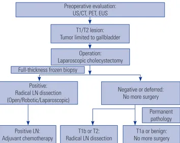
A schematic detailing the treatment algorithm is shown in Fig. 1. Three different approaches to radical cholecystectomy were employed: open or minimally invasive (laparoscopic and ro-botic). If preoperative evaluation suggested a T2 lesion with suspicious lymph node metastasis, the operation was
per-formed using an open approach. For T1 and T2 lesions with-out suspicious lymph node metastasis, laparoscopic cholecys-tectomy was performed initially. During cholecyscholecys-tectomy, the cystic plate was completely removed with the gallbladder to avoid exposing the subserosal layer. If the results of a full-thick-ness frozen biopsy were positive for malignancy, radical lymph node dissection was performed. The extents of lymphadenec-tomy were hepatoduodenal (#12), common hepatic artery (#8), retropancreatic (#13), and para-aortic (#16). T-tubes were rou-tinely inserted into the common bile duct. As noted above, liver resection was not performed for all T1 and T2 lesions. If the fro-zen biopsy results were negative or indeterminate, no further operation was performed until the pathology was confirmed. If the results confirmed a T1b or T2 lesion, radical lymph node dissection was subsequently performed. Cases with T1b and T2 lesions referred from other hospitals after laparoscopic chole-cystectomy underwent reoperation via an open approach.
The role of adjuvant chemotherapy remains unclear, with some reports of benefit in node-positive GBC or R1 disease.7,8
Therefore, adjuvant chemotherapy was only considered in cas-es of lymph node metastasis or gallbladder perforation during operation. Postoperative complications were graded according to Clavien–Dindo classifications.9
Statistical analysis
All statistical analyses were performed using Statistical Pack-age for Social Sciences version 20 (IBM Corp., Armonk, NY, USA). For each quantitative variable, Shapiro–Wilk test was used as a test of normality. Disease-specific survival and dis-ease-free survival were analyzed using Kaplan–Meier method and compared by log-rank tests. Cox proportional hazards model was used for multivariate survival analysis. Statistical significance was set as p value <0.05.
Permanent pathology Full-thickness frozen biopsy
Preoperative evaluation: US/CT, PET, EUS
T1/T2 lesion: Tumor limited to gallbladder
Operation: Laparoscopic cholecystectomy
Positive LN:
Adjuvant chemotherapy Radical LN dissectionT1b or T2: No more surgeryT1a or benign: Positive:
Radical LN dissection (Open/Robotic/Laparoscopic)
Negative or deferred: No more surgery
Fig. 1. Treatment algorithm for gallbladder carcinoma. US, ultrasonogra-phy; CT, computed tomograultrasonogra-phy; PET: positron emission tomograultrasonogra-phy; EUS, endoscopic ultrasonography; LN, lymph node.
RESULTS
Clinicopathologic characteristics
For the 109 patients included in our analysis, the mean age was 65±9 years, and 47 patients (43%) were male. Forty-five patients (41%) were diagnosed with incidental GBC. Among them, 11 patients (24%) were diagnosed with T1a GBC and did not undergo further operations. The mean level of carbo-hydrate antigen 19-9 was 13.0±16.8 U/mL. The median follow-up was 50 months (range, 5–145 months). The mean number of retrieved lymph nodes was 14±9 lymph nodes. Node-posi-tive GBC was found in 14 patients (13%). Among them, 12 pa-tients underwent adjuvant chemotherapy and two papa-tients re-fused adjuvant therapy.
Comparison of clinicopathologic outcomes between T stages
Clinicopathologic characteristics according to T stage are
shown in Table 1. Clinical characteristics did not differ among T stages. There were no differences in terms of the locations of tumors, i.e., hepatic vs. peritoneal sides (p=0.805); however, tumors involving hepatic side were more frequent (n=63, 58%). With regard to histologic differentiation, higher T stages were significantly associated with poorly differentiated GBC (p= 0.004). Two patients in T2 group with poorly differentiated GBC showed combined neuroendocrine features in the patho-logic report. Significantly higher lymph node metastasis was noted in T2 group than those in T1b and T1a groups (21% vs. 4% and 0%, respectively, p<0.001). The numbers of retrieved lymph nodes were higher for T1b and T2 groups than for T1a group (p<0.001) (Table 1). In T1a group, six patients who were diagnosed with incidental GBC did not undergo lymph node dissection, whereas 15 patients did as a higher T stage was sus-pected.
There were no significant differences between stages in terms of complications (p=0.467). All grade I complications were ei-Table 1. Clinicopathologic Characteristics according to T Stage
Variables T1a (n=21) T1b (n=26) T2 (n=62) p value
Age (yr) 62 (40–81) 67 (35–82) 65 (47–81) 0.331
Male 9 (43) 10 (38) 28 (45) 0.836
CA 19-9 (U/mL) 10.1 (1.2–83.6) 6.7 (0.6–51.7) 8.4 (0.1–96.8) 0.380 Incidental gallbladder cancer 11 (52) 12 (46) 22 (35) 0.359 Tumor size (cm) 2.0 (0.1–4.5) 2.0 (0.1–4.2) 2.2 (0.3–8.5) 0.160 Tumor location 0.805 Hepatic side 11 (52) 16 (62) 36 (58) Peritoneal side 10 (48) 10 (38) 26 (42) Histologic differentiation 0.004 Well 19 (90) 21 (81) 31 (50) Moderate 2 (10) 4 (15) 23 (37) Poor 0� 1 (4) 8 (13) N stage <0.001 Nx 6 (29) 0� 0� N0 15 (71) 25 (96) 49 (79) N1 0� 1 (4) 11 (18) N2 0� 0� 2 (3) 0.273 Lymphovascular invasion 0� 2 (8) 9 (15) 0.881 Perineural invasion 1 (5) 1 (4) 3 (5) <0.001
Median number of retrieved LNs 2 (0–19) 16 (2–28) 18 (3–35)
Operation method <0.001 Laparoscopic 16 (76) 5 (19) 10 (16) Robotic 3 (14) 4 (15) 5 (8) Open 2 (10) 17 (65) 47 (76) Complications (Clavien-Dindo) 0.467 I 0� 1 (4) 7 (11) II 1 (5) 0� 4 (6) III 1 (5) 2 (8) 3 (5) Adjuvant therapy 0� 1 (4) 11 (18) 0.028 LN, lymph node.
ther fever or wound seroma. Grade II complications were chy-lous leakage (two cases), which were medically managed, and abscess formation (three cases), which were managed with antibiotics. Grade III complications were postoperative bile leakage requiring endoscopic retrograde biliary drainage (five cases), and intra-abdominal abscess formation requiring per-cutaneous catheter drainage (one case).
Operative technique and postoperative complications Open radical cholecystectomies were performed in 66 patients (61%), whereas the approaches were laparoscopic for 31 pa-tients (28%) and robotic for 12 papa-tients (11%). The median (range) operation times for open, laparoscope, and robotic rad-ical cholecystectomies were 183 (95–340), 73 (30–173), and 231 (172–278) min, respectively (p<0.001). There were three cases of open conversion during minimally invasive approaches: one from phrenic artery injury (laparoscopic approach) and two from renal vein injury and aortic wall bleeding (robotic ap-proach).
There were no significant differences in complications be-tween operative techniques (p=0.066). For minimally invasive approaches, there were no complications in laparoscopic group, but three complications (24%) were observed in robotic group: grade I for fever, which was treated conservatively; grade II for intra-abdominal abscess formation requiring antibiotics; and grade IIIA requiring endoscopic retrograde biliary drainage due to postoperative bile leakage. There were 13 complications (21%) in patients undergoing an open approach: six cases were grade I for fever and wound seroma, four in grade II (including two cases of postoperative chylous leakage and two cases of intra-abdominal abscess formation requiring pharmacologic treatments), and three in grade IIIA (including two cases of
endoscopic retrograde biliary drainage due to postoperative bile leakage and one case of percutaneous catheter drainage for intra-abdominal abscess formation).
Oncologic outcomes
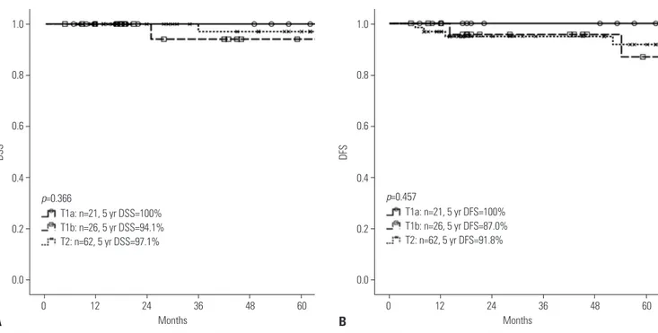
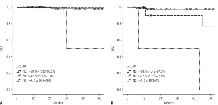
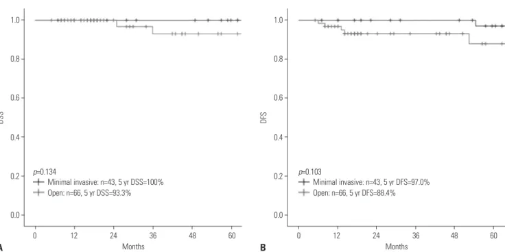
There were four GBC-related mortalities during the study pe-riod, and the 5-year disease-specific survival rate was 97%. There were no significant differences according to T stage in disease-specific survival (p=0.366; Fig. 2A) or disease-free sur-vival (p=0.457; Fig. 2B). In all T stages, the median sursur-vival was not reached for analysis. In N2 disease, significantly poor on-cologic outcomes (5-year disease-specific survival: 50%, 5-year disease-free survival: 0%; Fig. 3) were observed. Oncologic out-comes were not associated with the tumor location (Fig. 4) nor the type of surgical approach (Fig. 5).
Recurrence pattern
The details on recurrence for each GBC stage are shown in Ta-ble 2. Overall, there were six cases of recurrence, with a 5-year disease-free survival rate of 92.4%. In cases with T1a GBC, no lymph node metastasis was found in 15 patients who under-went lymph node dissection. Among the 26 patients with T1b lesions, there was only one patient (4%) with lymph node me-tastasis, and two patients (8%) had recurrence. For T2 GBC, 13 patients (22%) had lymph node metastasis, two of whom were staged N2: one patient had only single lymph node metastasis in retropancreatic lymph nodes from total of 26 lymph nodes investigated, and the other patient had one metastatic para-aortic lymph node and four metastatic hepatoduodenal lymph nodes from a total of 20 lymph nodes investigated. These pa-tients with N2 staging showed recurrences. Two papa-tients show-ing poorly differentiated adenocarcinoma with neuroendo-1.0 0.8 0.6 0.4 0.2 0.0 1.0 0.8 0.6 0.4 0.2 0.0 0 12 24 36 48 60 0 12 24 36 48 60 T1a: n=21, 5 yr DSS=100% T1b: n=26, 5 yr DSS=94.1% T2: n=62, 5 yr DSS=97.1% T1a: n=21, 5 yr DFS=100% T1b: n=26, 5 yr DFS=87.0% T2: n=62, 5 yr DFS=91.8% Months Months DSS DFS A B p=0.366 p=0.457
crine features had recurrence within 1 year after the operation.
DISCUSSION
Historically, GBC has been associated with pessimistic onco-logic outcomes. In 1924, Blalock10 wrote that “in malignancy
of the gallbladder, when a diagnosis can be made without ex-ploration, no operation should be performed, in as much as it only shortens the patient’s life.” With improvements in radiol-ogy and the invention of laparoscopic operations, early diag-noses of GBC have become more frequent. However, the cu-rative options are limited to radical operations, as GBC has a propensity for early dissemination and is proximal to the liver and major vasculature. Recently, favorable outcomes after less radical cholecystectomies have been reported in patients with T1/T2 GBC, with some reports on the safety and non-inferior oncologic outcomes of laparoscopic approach.11-14 However,
there is no consensus on the extent of surgery during radical cholecystectomy except for T1a GBC, in which laparoscopic simple cholecystectomy is adequate. Further lymph node dis-section may still be indicated, as frozen biopsies during opera-tions cannot always discriminate between T1 and T2 GBC.
Clarification of the oncologic outcomes for GBC is difficult due to the heterogeneity of operative techniques. For example, the extent of surgery differs among surgeons and institutions. Therefore, to compare oncologic outcomes based on the extent of surgery, we analyzed cases involving a single surgeon (WJL) at Severance Hospital, where laparoscopic operations for GBC have been performed since 1993. At that time, if a patient was diagnosed with incidental T1a GBC, routine follow-up was scheduled. For those with T1b GBC or higher, open radical
cho-lecystectomies were performed, unless the patients refused for personal reasons. Our previous reports on patients who did not undergo further radical cholecystectomy due to personal rea-sons or the patients’ general condition showed that the 5-year survival rates of GBC patients with T1a, T1b, and T2 lesions were 100%, 75%, and 56.2%, respectively.15 Two cases of
recur-rences in para-aortic lymph nodes were noted after radical cholecystectomy, including only regional lymphadenectomy, in patients with T2 GBC at 1 month and 5 months. There was no recurrence in the liver bed, which extends from the cystic plate, in any of the patients. Starting in 2005, with the introduc-tion of robotic system at Severance Hospital, minimally invasive radical cholecystectomies, including the robotic approach, were also performed. Our results have shown no differences in onco-logic outcomes among different surgical approaches. Therefore, for T1/T2 GBC, having a consistent and reasonable operative principle is more important than the mode of surgical approach.
Our anecdotal experiences have indicated that the dissection of regional lymph nodes along with para-aortic lymph nodes may reduce recurrence. Para-aortic lymph nodes are the final destination in the abdominal lymphatic route from the gallblad-der via the cholecysto-retropancreatic pathway and the retro-portal node.16 However, evidence on the role of extensive
lymph-adenectomy is lacking. Tsukada, et al.17 reported that in T2 GBC,
metastases in para-aortic lymph nodes were discovered in 12% of cases, whereas Ogura, et al.18 reported lymph node metastases
in 44.3%, with 2.5% of T1a and 15.6% of T1b cases also showing metastases. Nevertheless, para-aortic lymphadenectomy did not seem to provide any survival benefit.19 In contrast, our findings
suggest a benefit of extensive lymphadenectomy on the onco-logic outcomes of GBC patients without pre-existing lymph node metastases. 1.0 0.8 0.6 0.4 0.2 0.0 1.0 0.8 0.6 0.4 0.2 0.0 0 12 24 36 48 60 0 12 24 36 48 60 N0: n=89, 5 yr DSS=98.2% N1: n=12, 5 yr DSS=100% N2: n=2, 5 yr DSS=50% N0: n=89, 5 yr DFS=97.6% N1: n=12, 5 yr DFS=77.1% N2: n=2, 5 yr DFS=0% Months Months DSS DFS A B p<0.001 p<0.001
One recent meta-analysis of T1 GBC found comparable sur-vival outcomes between simple and extended cholecystecto-mies,20 whereas another meta-analysis reported conflicting
results and suggested an improved prognosis associated with liver resection and lymph node resection for stage T1b GBC or higher.21 However, the authors of both meta-analyses noted that
a lack of essential data for comparison and a lack of standard-ization in operative extent and definition can limit the inter-pretation of the results. Additionally, since liver parenchyme is not involved in T1/T2 GBC, the complete excision of the cystic plate along with the gallbladder may be sufficient. Indeed, our
experiences after the implementation of these principles in 2005 have indicated better oncologic outcomes for GBC, espe-cially for T2 tumors. The 5-year disease-specific survival rate was 97.1% at Severance Hospital, compared to rates ranging from 69.4% to 90.2% in previous studies.6,14,22 Although this
study found one recurrence in the liver, it was bi-lobular and distant from the cystic plate. Horiguchi, et al.23 also reported no
difference in disease-free survival rates between patients re-ceiving cystic bed resection only and those also undergoing liver resection. Therefore, the complete excision of the cystic plate should be adequate for T1/T2 GBC.
1.0 0.8 0.6 0.4 0.2 0.0 1.0 0.8 0.6 0.4 0.2 0.0 0 12 24 36 48 60 0 12 24 36 48 60 Peritoneal side: n=46, 5 yr DSS=96.3% Hepatic side: n=63, 5 yr DSS=97.5% Peritoneal side: n=46, 5 yr DFS=97.8% Hepatic side: n=63, 5 yr DFS=88.5% Months Months DSS DFS A B p=0.417 p=0.195 1.0 0.8 0.6 0.4 0.2 0.0 1.0 0.8 0.6 0.4 0.2 0.0 0 12 24 36 48 60 0 12 24 36 48 60 Peritoneal side: n=26, 5 yr DSS=91.7% Hepatic side: n=36, 5 yr DSS=100% Peritoneal side: n=26, 5 yr DFS=96.2% Hepatic side: n=36, 5 yr DFS=89.1% Months Months DSS DFS C D p=0.672 p=0.523
Nortably, the percentage of incidental GBC cases following cholecystectomy in the present report was higher than those reported for laparoscopic cholecystectomies, which ranged from 0.2% to 1.1%.24 This may be due to the fact that Severance
Hospital is a tertiary referral hospital. Additionally, our results indicate that the location of the tumor, whether on the hepatic or peritoneal side, does not influence the oncologic outcome, even though the current 8th AJCC staging system further di-vides T2 stage accordingly.
In terms of recurrence, six incidences were observed in our study group. Two cases of recurrence showed that GBC was poorly differentiated with neuroendocrine features, which is known to behave more aggressively.25 Among the remaining
four cases, one patient was initially diagnosed with T1b lesion without lymph node metastasis, and had recurrence at the para-aortic lymph node 14 months later. This may have resulted from an incomplete para-aortic lymph node dissection; therefore,
radical cholecystectomy in T1b should be sought more actively. Regarding postoperative complications, some patients showed postoperative bile leakage. These complications may have re-sulted from complete and radical dissections of hepatoduo-denal lymph nodes, which completely expose the common bile duct and disrupt its blood supply. We now routinely insert T-tubes to prevent such postoperative biliary complications.
The limitations of the present study include the limited num-ber of patients analyzed, as well as the inherent selection bias resulting from the retrospective design and selection criteria of the study. Five patients who underwent additional hepa-tectomy under the suspicion of T3 tumor were excluded from this study. The operative finding showed that the possibility of acute inflammation and liver invasion could not be completely excluded. Therefore, the current treatment algorithm should be applied with discretion in patients with acute inflammation. Furthermore, these results were based on the experience of a Table 2. Lymph Node Status and Recurrence Patterns according to T Stage
T stage (n=112) Nodal status Number of recurrence Site of recurrence Time of recurrence Survival months Status T1a (n=21) Nx* (n=6, 29%) None
-N0 (n=15, 71%) None
T1b (n=26) N0 (n=25, 96%) 1 (4%) Para-aortic LN 14 25 Dead
N1 (n=1, 4%) 1 (100%) Spine 53 60 Dead
T2 (n=62) N0 (n=49, 79%) 1 (2%) Liver‡ 6 20 Alive
N1 (n=11, 18%) 1 (9%) Regional LN, Supraclavidular LN 13 15 Alive N2† (n=2, 3%) 2 (100%) Common bile duct 52 65 Dead
Paraortic LN‡ 8 36 Dead
LN, lymph node.
*Nx represents without lymph node dissection for examination; †N2 represents positive para-aortic lymph nodes; ‡Histologic grade showed poorly differentiated
adenocarcinoma with neuroendocrine features. 1.0 0.8 0.6 0.4 0.2 0.0 1.0 0.8 0.6 0.4 0.2 0.0 0 12 24 36 48 60 0 12 24 36 48 60 Minimal invasive: n=43, 5 yr DSS=100% Open: n=66, 5 yr DSS=93.3% Minimal invasive: n=43, 5 yr DFS=97.0% Open: n=66, 5 yr DFS=88.4% Months Months DSS DFS A B p=0.134 p=0.103
single surgeon (WJL). Nevertheless, the preventive role of ex-tensive lymphadenectomy, including para-aortic lymph node dissection, should be given further attention, and more sur-geons should consider our operative principle to enable a large-scale cohort study.
A complete R0 resection in GBC is the standard of care in patients with localized disease. However, there is no consen-sus on the extent of radical cholecystectomy for T1/T2 GBC, partly due to the low incidence of GBC that impedes random-ized controlled trials to establish optimal treatment modalities. Here, we report on our experiences with an operative principle at Severance Hospital, which indicates that extended lymph-adenectomy with para-aortic lymph node dissection without liver resection for T1/T2 GBC produces favorable oncologic outcomes.
AUTHOR CONTRIBUTIONS
Conceptualization: Woo Jung Lee. Data curation: All authors. Formal analysis: Jae Uk Chong. Funding acquisition: Woo Jung Lee. Investi-gation: All authors. Methodology: All authors. Project administration:
Woo Jung Lee. Resources: All authors. Software: All authors. Supervi-sion: Woo Jung Lee. Validation: All authors. Visualization: All authors.
Writing—original draft: All authors. Writing—review & editing: All authors.
ORCID iDs
Jae Uk Chong https://orcid.org/0000-0001-9713-1653 Woo Jung Lee https://orcid.org/0000-0001-9273-261X
REFERENCES
1. Hueman MT, Vollmer CM Jr, Pawlik TM. Evolving treatment strat-egies for gallbladder cancer. Ann Surg Oncol 2009;16:2101-15. 2. Lazcano-Ponce EC, Miquel JF, Muñoz N, Herrero R, Ferrecio C,
Wistuba II, et al. Epidemiology and molecular pathology of gall-bladder cancer. CA Cancer J Clin 2001;51:349-64.
3. Lee SE, Kim KS, Kim WB, Kim IG, Nah YW, Ryu DH, et al. Practi-cal guidelines for the surgiPracti-cal treatment of gallbladder cancer. J Korean Med Sci 2014;29:1333-40.
4. Randi G, Franceschi S, La Vecchia C. Gallbladder cancer world-wide: geographical distribution and risk factors. Int J Cancer 2006;118:1591-602.
5. Lee SE, Jang JY, Lim CS, Kang MJ, Kim SW. Systematic review on the surgical treatment for T1 gallbladder cancer. World J Gastro-enterol 2011;17:174-80.
6. Kim DH, Kim SH, Choi GH, Kang CM, Kim KS, Choi JS, et al. Role of cholecystectomy and lymph node dissection in patients with T2 gallbladder cancer. World J Surg 2013;37:2635-40.
7. Hoehn RS, Wima K, Ertel AE, Meier A, Ahmad SA, Shah SA, et al. Adjuvant therapy for gallbladder cancer: an analysis of the Na-tional Cancer Data Base. J Gastrointest Surg 2015;19:1794-801. 8. Ma N, Cheng H, Qin B, Zhong R, Wang B. Adjuvant therapy in the
treatment of gallbladder cancer: a meta-analysis. BMC Cancer 2015;15:615.
9. Dindo D, Demartines N, Clavien PA. Classification of surgical complications: a new proposal with evaluation in a cohort of 6336 patients and results of a survey. Ann Surg 2004;240:205-13. 10. Blalock A. A statistical study of eight hundred and eighty-eight
cases of biliary tract disease. Johns Hopkins Hosp Bull 1924;35: 391-409.
11. Itano O, Oshima G, Minagawa T, Shinoda M, Kitago M, Abe Y, et al. Novel strategy for laparoscopic treatment of pT2 gallbladder carcinoma. Surg Endosc 2015;29:3600-7.
12. Lee H, Choi DW, Park JY, Youn S, Kwon W, Heo JS, et al. Surgical strategy for T2 gallbladder cancer according to tumor location. Ann Surg Oncol 2015;22:2779-86.
13. Zimmitti G, Manzoni A, Guerini F, Ramera M, Bertocchi P, Aroldi F, et al. Current role of minimally invasive radical cholecystecto-my for gallbladder cancer. Gastroenterol Res Pract 2016. Article ID: 7684915
14. Yoon YS, Han HS, Cho JY, Choi Y, Lee W, Jang JY, et al. Is laparos-copy contraindicated for gallbladder cancer? A 10-year prospec-tive cohort study. J Am Coll Surg 2015;221:847-53.
15. Kang CM, Lee WJ, Choi GH, Kim JY, Kim KS, Choi JS, et al. Does “clinical” R0 have validity in the choice of simple cholecystectomy for gallbladder carcinoma? J Gastrointest Surg 2007;11:1309-16. 16. Ito M, Mishima Y. Lymphatic drainage of the gallbladder. J
Hepa-tobiliary Pancreat Surg 1994;1:302-8.
17. Tsukada K, Hatakeyama K, Kurosaki I, Uchida K, Shirai Y, Muto T, et al. Outcome of radical surgery for carcinoma of the gallbladder according to the TNM stage. Surgery 1996;120:816-21.
18. Ogura Y, Mizumoto R, Isaji S, Kusuda T, Matsuda S, Tabata M. Radical operations for carcinoma of the gallbladder: present sta-tus in Japan. World J Surg 1991;15:337-43.
19. Kondo S, Nimura Y, Hayakawa N, Kamiya J, Nagino M, Uesaka K. Regional and para-aortic lymphadenectomy in radical surgery for advanced gallbladder carcinoma. Br J Surg 2000;87:418-22. 20. Lee H, Kwon W, Han Y, Kim JR, Kim SW, Jang JY. Optimal extent
of surgery for early gallbladder cancer with regard to long-term survival: a meta-analysis. J Hepatobiliary Pancreat Sci 2018;25: 131-41.
21. Sternby Eilard M, Lundgren L, Cahlin C, Strandell A, Svanberg T, Sandström P. Surgical treatment for gallbladder cancer–a system-atic literature review. Scand J Gastroenterol 2017;52:505-14. 22. Jung W, Jang JY, Kang MJ, Chang YR, Shin YC, Chang J, et al.
Ef-fects of surgical methods and tumor location on survival and re-currence patterns after curative resection in patients with T2 gall-bladder cancer. Gut Liver 2016;10:140-6.
23. Horiguchi A, Miyakawa S, Ishihara S, Miyazaki M, Ohtsuka M, Shimizu H, et al. Gallbladder bed resection or hepatectomy of segments 4a and 5 for pT2 gallbladder carcinoma: analysis of Jap-anese registration cases by the study group for biliary surgery of the Japanese Society of Hepato-Biliary-Pancreatic Surgery. J Hep-atobiliary Pancreat Sci 2013;20:518-24.
24. Pitt SC, Jin LX, Hall BL, Strasberg SM, Pitt HA. Incidental gallblad-der cancer at cholecystectomy: when should the surgeon be sus-picious? Ann Surg 2014;260:128-33.
25. Eltawil KM, Gustafsson BI, Kidd M, Modlin IM. Neuroendocrine tumors of the gallbladder: an evaluation and reassessment of management strategy. J Clin Gastroenterol 2010;44:687-95.