Squamous cell carcinoma (SCC) is the most common prima-ry malignancy of the orophaprima-rynx, followed by lymphoma.1 Al-though pretreatment imaging evaluation of these two malignant tumors is critical for tumor staging and treatment planning, it is not easy to differentiate these tumors on the basis of convention-al magnetic resonance (MR) imaging convention-alone, because both SCC and lymphoma of the oropharynx usually have similar imaging
characteristics.2
Previous studies have reported that advanced MR imaging techniques, such as diffusion-weighted imaging2-5 and dynam-ic contrast-enhanced MR imaging,6-8 may be helpful in the dif-ferentiation of head and neck cancer. However, these MR imag-ing sequences are time consumimag-ing and may not be performed routinely in daily practice. However, conventional MR imaging with T2-weighted images (T2WIs) and contrast-enhanced T1-weighted images (CE-T1WIs) are almost always available.
A recently introduced radiomics model, which is based on the extraction of high-dimensional quantitative imaging fea-tures, can potentially provide underlying pathophysiological information that is difficult to ascertain during visual inspec-tion.9,10 Accordingly, several previous studies have applied ra-diomics on head and neck cancers:11-13 One of these studies found that seven texture features predicted tumor suppressor protein p53 status of oropharyngeal SCC with an accuracy of 81.3%.11 Another study showed that radiomic features
extract-Squamous Cell Carcinoma and Lymphoma
of the Oropharynx: Differentiation Using
a Radiomics Approach
Sohi Bae
1, Yoon Seong Choi
2, Beomseok Sohn
2, Sung Soo Ahn
2,
Seung-Koo Lee
2, Jaemoon Yang
2, and Jinna Kim
21Department of Radiology, National Health Insurance Service Ilsan Hospital, Goyang;
2Department of Radiology and Research Institute of Radiological Science, Yonsei University College of Medicine, Seoul, Korea.
The purpose of this study was to evaluate the diagnostic performance of magnetic resonance (MR) radiomics-based machine learning algorithms in differentiating squamous cell carcinoma (SCC) from lymphoma in the oropharynx. MR images from 87 patients with oropharyngeal SCC (n=68) and lymphoma (n=19) were reviewed retrospectively. Tumors were semi-automatically segmented on contrast-enhanced T1-weighted images registered to T2-weighted images, and radiomic features (n=202) were ex-tracted from contrast-enhanced T1- and T2-weighted images. The radiomics classifier was built using elastic-net regularized gen-eralized linear model analyses with nested five-fold cross-validation. The diagnostic abilities of the radiomics classifier and visual assessment by two head and neck radiologists were evaluated using receiver operating characteristic (ROC) analyses for distin-guishing SCC from lymphoma. Nineteen radiomics features were selected at least twice during the five-fold cross-validation. The mean area under the ROC curve (AUC) of the radiomics classifier was 0.750 [95% confidence interval (CI), 0.613–0.887], with a sen-sitivity of 84.2%, specificity of 60.3%, and an accuracy of 65.5%. Two human readers yielded AUCs of 0.613 (95% CI, 0.467–0.759) and 0.663 (95% CI, 0.531–0.795), respectively. The radiomics-based machine learning model can be useful for differentiating SCC from lymphoma of the oropharynx.
Key Words: Radiomics, oropharynx, squamous cell carcinoma, lymphoma, magnetic resonance imaging
pISSN: 0513-5796 · eISSN: 1976-2437
Received: April 20, 2020 Revised: July 31, 2020 Accepted: August 17, 2020
Corresponding author: Jinna Kim, MD, PhD, Department of Radiology and
Re-search Institute of Radiological Science, Yonsei University College of Medicine, 50-1 Yonsei-ro, Seodaemun-gu, Seoul 03722, Korea.
Tel: 82-2-2228-2392, Fax: 82-2-2227-8337, E-mail: jinna@yuhs.ac •The authors have no potential conflicts of interest to disclose. © Copyright: Yonsei University College of Medicine 2020
This is an Open Access article distributed under the terms of the Creative Com-mons Attribution Non-Commercial License (https://creativecomCom-mons.org/licenses/ by-nc/4.0) which permits unrestricted non-commercial use, distribution, and repro-duction in any medium, provided the original work is properly cited.
Yonsei Med J 2020 Oct;61(10):895-900 https://doi.org/10.3349/ymj.2020.61.10.895
ed from square-shaped regions-of-interest in sinonasal tumors could differentiate SCC from inverted papilloma with an ac-curacy of 89.1%.12 The purpose of this study was to evaluate the diagnostic performance of MR radiomics-based machine learning algorithms for the differentiation of SCC from lym-phoma in the oropharynx.
This single-center retrospective study was approved by the Institutional Review Board of Severance Hospital (IRB no.4-2018-0761), and the requirement for informed consent was waived. The records of 97 consecutive patients with a new di-agnosis of SCC or lymphoma of the oropharynx who under-went preoperative MR imaging from July 2012 to June 2018 were reviewed retrospectively. Patients were included on the basis of the following criteria: 1) pathologically confirmed SCC or lymphoma of the oropharynx; 2) preoperative MR im-aging with identical sequence protocols, including T2WIs and three-dimensional (3D) fat-saturated CE-T1WIs. The exclusion criteria were as follows: 1) a history of biopsy or treatment be-fore MR imaging (n=7) and 2) inadequate image quality (n=3). Thus, a total of 87 patients were included in this study (Fig. 1). The study group consisted of 68 SCC (58 men and 10 women; mean age±standard deviation, 59.8±9.9 years) and 19 lym-phoma patients (14 men and 5 women; 59.9±14.0 years). The primary tumor sites of 68 SCC patients were the palatine ton-sil (n=61), base of the tongue (n=6), and soft palate (n=1).
Final histopathological diagnoses were confirmed by fiber-scope-guided biopsy within a period of 2 weeks following MR imaging, before the initiation of treatment. The SCCs comprised 15 well-differentiated, 32 moderately differentiated, and 21 poorly differentiated carcinomas. Lymphomas consisted of 17 diffuse large B-cell lymphomas and two Burkitt’s lymphomas.
Preoperative MR imaging was performed at a single institu-tion using multiple identical 3.0 Tesla scanners (Achieva; Phil-ips Medical Systems, Best, The Netherlands) with a head and neck coil. Fat-saturated axial fast spin-echo T2WIs were ac-quired with a repetition time/echo time (TR/TE) of 6480 ms/70
ms and section thickness of 4 mm before the injection of con-trast material. All images were obtained with a 22–25-cm field-of-view. Fat-saturated axial T1-weighted high-resolution iso-tropic volume examination (THRIVE) images were acquired at 40 s after the administration of the contrast material and then reformatted to the coronal and sagittal planes. The param-eters for the fat-saturated 3D THRIVE images were as follows: TR=4.5 ms, TE=2.2 ms, flip angle=10°, bandwidth=434 Hz/ pixel, matrix=256×256, section thickness=1 mm, signal aver-aging=2, and acquisition time=223 s.
Tumor segmentation was performed by a radiologist (J.K., with 19 years of experience in head and neck imaging) on the 3D CE-T1WIs for the selection of the contrast-enhanced tu-mor using semi-automatic methods, such as signal intensity threshold, region growing, and edge detection. After image pre-processing, a total of 202 radiomic features were extracted. The details of the preprocessing and radiomic feature extrac-tion are described in Supplementary Material 1 (only online).
Additionally, we performed visual assessment and evaluat-ed the diagnostic performance of two head and neck radiolo-gists in distinguishing SCC from lymphoma (S.B. and J.K., with 8 and 19 years of experience, respectively). After anonymiza-tion and data randomizaanonymiza-tion, the readers were given two image sets for each patient, which included T2WIs and CE-T1WIs of the segmented tumor mask that had been used for the ra-diomics model. The readers recorded a final diagnosis using a 4-point scale (1=definite SCC; 2=likely SCC; 3=likely lympho-ma; and 4=definite lymphoma).
All statistical analyses, including machine learning, were performed with the use of R software (version 3.5.1, R Foun-dation for Statistical Computing, Vienna, Austria). Values of p< 0.05 were considered statistically significant. Two-sided statis-tical tests were conducted.
An elastic-net regularized generalized linear model (GLM) analysis was performed to train the machine learning classifi-er in this study. GLM is an extension of a linear model to
vari-SCC (n=68) Lymphoma (n=19)
Exclusion criteria
1) History of biopsy or treatment before MR imaging (n=7)
2) Inadequate image quality (n=3) Inclusion criteria
1) Pathologically confirmed oropharyngeal SCC or lymphoma 2) Preoperative T2WI and 3D CE-T1WI from July 2012 to June 2018
Total study population (n=87)
Fig. 1. Flowchart of patient enrollment. SCC, squamous cell carcinoma; T2WI, T2-weighted imaging; 3D, three-dimensional; CE-T1WI, contrast-enhanced T1-weighted imaging.
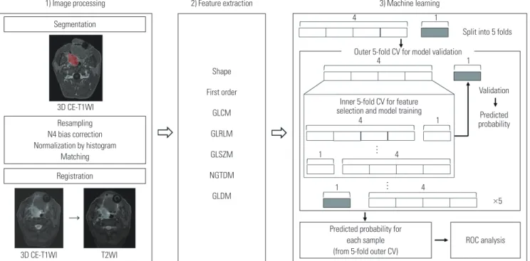
ables that do not follow normal distribution. Elastic net regu-larization, a regularization method used to reduce the variance in linear regression models, has been reported to be useful when the number of predictors is considerably greater than that of the observations or when predictors are correlated as in radiomics data. Analyses were performed with nested validation to avoid overfitting. The details of the nested cross-validation are presented in Supplementary Material 2 (only online). The elastic-net regularized GLM was performed us-ing the “glmnet” package in R software. A schematic diagram of the workflow (from image processing to machine learning) is depicted in Fig. 2.
A total of 102 radiomic features were selected at least once during cross-validation, with an average of 27.6 features se-lected for each fold. Among these, 19 features consisting of 10 first-order features, and 9 texture features were selected at least twice. The generated heatmap of the 19 radiomic features se-lected for the differentiation between SCC and lymphoma yielded different distributions between two pathologies (Fig. 3). The details, including the number of times each feature was selected and the mean coefficient values of the selected features during cross-validation, are summarized in Supple-mentary Table 1 (only online).
The mean AUC of the radiomics classifier was 0.750 [95% confidence interval (CI), 0.613–0.887], with a sensitivity of 84.2%, a specificity of 60.3%, and an accuracy of 65.5% (Fig. 4). Human reader 1 yielded an AUC of 0.613 (95% CI, 0.467–0.759), with a sensitivity of 61.8%, a specificity of 52.6%, and an accura-cy of 59.8%. Reader 2 yielded an AUC of 0.663 (95% CI, 0.531–
0.795), with a sensitivity of 60.3%, a specificity of 63.2%, and an accuracy of 60.9%.
In this study, we assessed the diagnostic value of radiomic features from MR imaging in differentiating between SCC and lymphoma of the oropharynx. In our study, we used conven-tional MR imaging sequences, which are generally employed for head and neck MR imaging in daily clinical practice, and a radiomics-based machine learning model exhibited fair per-formance. In addition, we used open-source tools, such as py-thon and R, instead of in-house software, to increase the avail-ability and reproducibility of the radiomics-based machine learning analyses in external centers.
Although SCC and lymphoma are the most common malig-nancies in the oropharynx, their conventional imaging find-ings are sometimes indistinguishable, as reported previously.4 In the sinonasal cavity, lymphomas are thought to yield larger tumor volume with more homogeneous signal intensity and less intratumoral necrosis than SCC.14,15 These findings may be associated with a prolonged hypoxic microenvironment in SCC and consequent tumor necrosis.16 In addition, several studies have discriminated between SCC and lymphoma with advanced MR imaging techniques, such as diffusion and per-fusion MR imaging. Several investigators reported that lym-phoma yields lower apparent diffusion coefficient values than SCC due to higher degrees of cellularity.2-4 A dynamic perfu-sion study indicated that lymphomas of the oropharynx have lower Ktrans and lower v
e values than SCC, which means that lymphoma is associated with lower vascular permeability and less extravascular extracellular space.6 Therefore, we
hypothe-Segmentation
Registration
1) Image processing 2) Feature extraction 3) Machine learning 4 4 4 4 4 ×5 1
Split into 5 folds Outer 5-fold CV for model validation
Validation Predicted probability 1 1 1 1 3D CE-T1WI 3D CE-T1WI T2WI Resampling N4 bias correction Normalization by histogram Matching Shape First order GLCM GLRLM GLSZM NGTDM GLDM
Predicted probability for each sample (from 5-fold outer CV)
ROC analysis
Fig. 2. Workflow of imaging processing, feature extraction, and machine learning. T2WI, T2-weighted imaging; 3D, three-dimensional; CE-T1WI, contrast-enhanced T1-weighted imaging; GLCM, gray level co-occurrence matrix; GLRLM, gray level run length matrix; GLSZM, gray level size zone matrix; NGT-DM, neighboring gray tone difference matrix; GLNGT-DM, gray level dependence matrix; CV, cross-validation; ROC, receiver operating characteristic.
Inner 5-fold CV for feature selection and model training
sized that these microscopic differences could be reflected by quantitative radiomic features extracted from conventional MR imaging.
The 19 relevant radiomic features that were selected by the nested cross-validation in our study consisted of 10 first-order features and 9 texture features. First-order features represent voxel intensity, which is acquired from histogram-based meth-ods,9 and homogeneous enhancement and low T2 signal inten-sity in lymphomas may mainly contribute to first-order features in radiomics analyses.17 Meanwhile, texture features describe
the statistical interrelationships between voxels and reflect in-tratumoral heterogeneity.9 The increased heterogeneity of the tumor signal intensity with more frequent intratumoral ne-crosis and hemorrhage in SCC may be the main contributor to the texture features.14,15 However, although radiomic features have been shown to capture variable microscopic characteris-tics of the tumor, there are limitations in the understanding of their underlying biologic mechanism and in the identification of the value of individual features.9,18
In fact, in daily clinical practice, radiologic diagnosis is usu-ally based on the integration of various information obtained from imaging studies. Radiologists obtain the information from intrinsic tumor characteristics and from other associated im-aging features, such as tumor growth patterns, adjacent struc-tural changes, and metastatic lymph node appearance. For ex-ample, adjacent bone destruction is more prominent in SCC than in lymphoma,14 and central necrosis is one of the charac-teristic findings of metastatic lymph nodes from SCC.15 There-fore, additional radiomics information obtained from the intrin-sic tumor appearance would be useful to improve the diagnostic performance of radiologists.
A previous study assessed stability and prognostic perfor-mance in various combinations of feature selection and ma-chine learning models in head and neck cancers.19 They sug-gested three classifiers; Bayesian, random forest, and nearest neighbor, as the preferred methods in radiomics-based prog-nostic analyses, owing to their high performances and stabili-ties. The GLM model that was used in our study yielded high-prognostic power but low stability, while the best classifier model could vary according to the clinical outcomes, imaging modalities, and radiomic cohorts.19,20 Large-scale comparative Fig. 4. Receiver operating characteristic curve of the radiomics
elastic-net regularized generalized linear model analysis. The mean area under the curve for the radiomics classifier was 0.750.
Fig. 3. Heatmap of the 19 radiomic features that were selected for the radiomic classifier at least twice during nested cross-validation to differentiate between squamous cell carcinoma and lymphoma. Each column represents individual patients, and each row represents the color-coded z-score of 19 normalized radiomic features respectively.
CE-T1WI: GLDM. Dependence Varianced CE-T1WI: GLDM. Large Dependence Emphasis T2WI: First order. Mean
T2WI: First order. Median
T2WI: First order. Root Mean Squared
T2WI: GLDM. Large Dependence High Gray Level Emphasis T2WI: GLSZM. Large Area Emphasis
T2WI: GLSZM. Large Area High Gray Level Emphasis T2WI: GLSZM. Zone Variance
CE-T1WI: First order. 10 percentile CE-T1WI: First order. 90 percentile CE-T1WI: First order. Mean CE-T1WI: First order. Median CE-T1WI: First order. Minimum CE-T1WI: First order. Root Mean Squared
CE-T1WI: GLSZM. Size Zone Non Uniformity Normalized CE-T1WI: GLSZM. Small Area Emphasis
CE-T1WI: GLSZM. Zone Percentage T2WI: First order. Skewness
Lymphoma Squamous cell carcinoma
Color Key -2 -1 0 1 2 Value 1.0 0.8 0.6 0.4 0.2 0.0 1.0 0.8 0.6 0.4 0.2 0.0 Specificity Sensitivity
studies dealing with various feature selection and machine learning methods across independent cohorts are required to identify the optimal and most reliable estimator with which to differentiate between SCC and lymphoma of the oropharynx.
Our study has several limitations. First, the dataset was small and imbalanced with 68 SCCs and 19 lymphomas. Overfitting is an important issue in machine learning when dealing with high-dimensional features and small populations. We at-tempted to mitigate overfitting by nested cross-validation. Also, data imbalances can have a negative effect on the fitting of the classification algorithm because learning algorithms tend to be biased toward the majority class. We adopted a resampling technique, the synthetic minority over-sampling technique (SMOTE), to overcome the data imbalance in this study. How-ever, this technique does not always guarantee higher perfor-mance: sometimes it even results in lower performance ac-cording to the classifier used, compared with methods with no resampling.21-23 Second, although the model in the present study exhibited fair performance in differentiating between SCC and lymphoma of the oropharynx, it cannot obviate the need for bi-opsy at this point. Further study with a larger population and more elaborate algorithms could support the potential of ra-diomics in noninvasive diagnosis of oropharyngeal tumors. Third, advanced MR imaging techniques, such as diffusion-weighted imaging and perfusion MR imaging, were not includ-ed in this study. Although multiparametric analysis would in-crease diagnostic accuracy in differentiating between SCC and lymphoma, advanced techniques require additional scans and their protocols vary among institutions. Fourth, tumors were semi-automatically segmented, which is user-dependent and time-consuming. Automated tumor segmentation based on deep learning would allow further automation of the workflow, minimize user bias, and enable larger-scale studies. Last, we did not test the model in independent validation cohorts. Fur-ther studies with larger multicenter datasets, such as the Can-cer Genome Atlas dataset, would be required to assess the feasibility and allow the utilization of this technology in clinical practice.
In conclusion, the radiomics-based machine learning mod-el can be useful for differentiating SCC from lymphoma of the oropharynx, although it cannot obviate the need for invasive biopsy procedures presently. Future investigations with larger datasets and more elaborate algorithms are necessary for aug-mentation of the diagnostic performance of radiologists.
ACKNOWLEDGEMENTS
This work was supported by a National Research Foundation of Korea (NRF) grant funded by the Korean Government (Min-istry of Science and ICT) (No. 2019R1A2C1008409).
AUTHOR CONTRIBUTIONS
Conceptualization: Sohi Bae and Jinna Kim. Data curation: Sohi Bae,
Beomseok Sohn, and Jinna Kim. Formal analysis: Yoon Seong Choi and Beomseok Sohn. Funding acquisition: Sung Soo Ahn, Seung-Koo Lee, Jaemoon Yang, and Jinna Kim. Investigation: Sohi Bae, Beomseok Sohn, and Jinna Kim. Methodology: Sohi Bae, Yoon Seong Choi, Beomseok Sohn, Sung Soo Ahn, and Jinna Kim. Project
admin-istration: Sung Soo Ahn, Seung-Koo Lee, Jaemoon Yang, and Jinna
Kim. Resources: Sohi Bae, Beomseok Sohn, Seung-Koo Lee, and Jinna Kim. Software: Yoon Seong Choi, Beomseok Sohn, Jaemoon Yang, and Sung Soo Ahn. Supervision: Sung Soo Ahn, Seung-Koo Lee, and Jinna Kim. Visualization: Yoon Seong Choi, Beomseok Sohn, and Sung Soo Ahn. Writing—original draft: Sohi Bae, Yoon Seong Choi, and Beomseok Sohn. Writing—review & editing: Sung Soo Ahn, Seung-Koo Lee, Jaemoon Yang, and Jinna Kim. Approval of final
man-uscript: all authors.
ORCID iDs
Sohi Bae https://orcid.org/0000-0002-5048-4627 Yoon Seong Choi https://orcid.org/0000-0002-6034-9912 Beomseok Sohn https://orcid.org/0000-0002-6765-8056 Sung Soo Ahn https://orcid.org/0000-0002-0503-5558 Seung-Koo Lee https://orcid.org/0000-0001-5646-4072 Jaemoon Yang https://orcid.org/0000-0001-7365-0395 Jinna Kim https://orcid.org/0000-0002-9978-4356
REFERENCES
1. Harnsberger HR, Bragg DG, Osborn AG, Smoker WR, Dillon WP, Davis RK, et al. Non-Hodgkin’s lymphoma of the head and neck: CT evaluation of nodal and extranodal sites. AJR Am J Roentgenol 1987;149:785-91.
2. Ichikawa Y, Sumi M, Sasaki M, Sumi T, Nakamura T. Efficacy of diffusion-weighted imaging for the differentiation between lym-phomas and carcinomas of the nasopharynx and oropharynx: cor-relations of apparent diffusion coefficients and histologic features. AJNR Am J Neuroradiol 2012;33:761-6.
3. Maeda M, Kato H, Sakuma H, Maier SE, Takeda K. Usefulness of the apparent diffusion coefficient in line scan diffusion-weighted imaging for distinguishing between squamous cell carcinomas and malignant lymphomas of the head and neck. AJNR Am J Neurora-diol 2005;26:1186-92.
4. Sumi M, Ichikawa Y, Nakamura T. Diagnostic ability of apparent diffusion coefficients for lymphomas and carcinomas in the phar-ynx. Eur Radiol 2007;17:2631-7.
5. Abdel Razek AAK. Arterial spin labelling and diffusion-weighted magnetic resonance imaging in differentiation of recurrent head and neck cancer from post-radiation changes. J Laryngol Otol 2018; 132:923-8.
6. Park M, Kim J, Choi YS, Lee SK, Koh YW, Kim SH, et al. Applica-tion of dynamic contrast-enhanced mri parameters for differenti-ating squamous cell carcinoma and malignant lymphoma of the oropharynx. AJR Am J Roentgenol 2016;206:401-7.
7. Bernstein JM, Homer JJ, West CM. Dynamic contrast-enhanced magnetic resonance imaging biomarkers in head and neck can-cer: potential to guide treatment? A systematic review. Oral Oncol 2014;50:963-70.
8. Asaumi J, Yanagi Y, Konouchi H, Hisatomi M, Matsuzaki H, Kishi K. Application of dynamic contrast-enhanced MRI to differentiate
malignant lymphoma from squamous cell carcinoma in the head and neck. Oral Oncol 2004;40:579-84.
9. Gillies RJ, Kinahan PE, Hricak H. Radiomics: images are more than pictures, they are data. Radiology 2016;278:563-77.
10. Lambin P, Leijenaar RTH, Deist TM, Peerlings J, de Jong EEC, van Timmeren J, et al. Radiomics: the bridge between medical imaging and personalized medicine. Nat Rev Clin Oncol 2017;14:749-62. 11. Dang M, Lysack JT, Wu T, Matthews TW, Chandarana SP,
Brock-ton NT, et al. MRI texture analysis predicts p53 status in head and neck squamous cell carcinoma. AJNR Am J Neuroradiol 2015;36: 166-70.
12. Ramkumar S, Ranjbar S, Ning S, Lal D, Zwart CM, Wood CP, et al. MRI-based texture analysis to differentiate sinonasal squamous cell carcinoma from inverted papilloma. AJNR Am J Neuroradiol 2017;38:1019-25.
13. Ger RB, Zhou S, Elgohari B, Elhalawani H, Mackin DM, Meier JG, et al. Radiomics features of the primary tumor fail to improve pre-diction of overall survival in large cohorts of CT- and PET-imaged head and neck cancer patients. PLoS One 2019;14:e0222509. 14. Kim SH, Mun SJ, Kim HJ, Kim SL, Kim SD, Cho KS. Differential
di-agnosis of sinonasal lymphoma and squamous cell carcinoma on CT, MRI, and PET/CT. Otolaryngol Head Neck Surg 2018;159:494-500.
15. Kato H, Kanematsu M, Watanabe H, Kawaguchi S, Mizuta K, Aoki M. Differentiation of extranodal non-Hodgkins lymphoma from squamous cell carcinoma of the maxillary sinus: a multimodality imaging approach. Springerplus 2015;4:228.
16. Li JZ, Gao W, Chan JY, Ho WK, Wong TS. Hypoxia in head and neck
squamous cell carcinoma. ISRN Otolaryngol 2012;2012:708974. 17. Derinkuyu BE, Boyunag˘a Ö, Öztunalı Ç, Tekkes¸in F, Damar Ç,
Alımlı AG, et al. Imaging features of Burkitt lymphoma in pediat-ric patients. Diagn Interv Radiol 2016;22:95-100.
18. Lambin P, Rios-Velazquez E, Leijenaar R, Carvalho S, van Sti-phout RG, Granton P, et al. Radiomics: extracting more informa-tion from medical images using advanced feature analysis. Eur J Cancer 2012;48:441-6.
19. Parmar C, Grossmann P, Rietveld D, Rietbergen MM, Lambin P, Aerts HJ. Radiomic machine-learning classifiers for prognostic biomarkers of head and neck cancer. Front Oncol 2015;5:272. 20. Mackin D, Fave X, Zhang L, Fried D, Yang J, Taylor B, et al.
Mea-suring computed tomography scanner variability of radiomics features. Invest Radiol 2015;50:757-65.
21. Xie C, Du R, Ho JW, Pang HH, Chiu KW, Lee EY, et al. Effect of machine learning re-sampling techniques for imbalanced datas-ets in 18F-FDG PET-based radiomics model on prognostication performance in cohorts of head and neck cancer patients. Eur J Nucl Med Mol Imaging 2020 Apr 6 [Epub]. Available at: https:// doi.org/10.1007/s00259-020-04756-4.
22. Park YW, Oh J, You SC, Han K, Ahn SS, Choi YS, et al. Radiomics and machine learning may accurately predict the grade and his-tological subtype in meningiomas using conventional and diffu-sion tensor imaging. Eur Radiol 2019;29:4068-76.
23. Park YW, Choi YS, Ahn SS, Chang JH, Kim SH, Lee SK. Radiomics MRI phenotyping with machine learning to predict the grade of lower-grade gliomas: a study focused on nonenhancing tumors. Korean J Radiol 2019;20:1381-9.