Clear cell chondrosarcoma (CCCS) is a very rare malignant bone tumor that can behave clinically as a benign or low-grade malignant bone tumor. It was first reported as a clear cell variant of chondro-sarcoma by Unni et al. in 1976.1) CCCS has a strong predilection for the epiphysis or metaphysis of long bones, especially the proximal end of the femur and humerus. However, there are only three cases of CCCS in the diaphysis of long bones2,3) and furthermore, this tumor has never been reported to affect the diaphysis of the tibia. We report an extremely rare case of CCCS that involved the middle shaft of the tibia.
Case Report
A 42-year-old male was referred from the primary hospital due to a 2-month history of mild pain of the right shin and abnormal find-ings on radiographs and MRI. Physical examination revealed a slight swelling of his right shin but there was no tenderness, warmth, or a mass-like lesion.
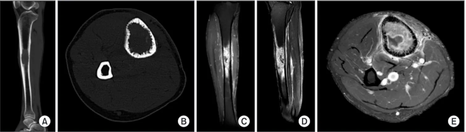
On radiographic examination, an intramedullary oval-shaped ra-diolucent lesion was notable in the middle of the right tibia. A thin
but relatively distinct sclerotic border was identified at the anterior and distal endosteal portions of the lesion. In contrast, the posterior endosteal portion of the lesion had an ill-defined margin with a slightly infiltrative pattern. There was no periosteal reaction evident (Fig. 1A, B).
MRI performed at the primary hospital had revealed a well-delineated lesion 4.5×2.5×2.0 cm in the medullary cavity of the tibial shaft. T1-weighted images gave intermediate signal intensity and T2-weighted images gave intermediate to high signal intensity with a relatively homogeneous appearance. The lesion was slightly expansile and had a sclerotic border with low signal intensity and adjacent marrow edema. Slight edema was also noted in the sub-cutaneous fat layer of the anterior tibia (Fig. 1C, D). A bone scan revealed highly increased uptake in the middle shaft of the tibia but no other abnormalities (Fig. 1E).
Given the location of the tumor and its radiologic features, we as-sumed it to be fibrous dysplasia and performed an incisional biopsy, which identified it as a CCCS.
To re-evaluate the tumor, we performed CT and gadolinium-enhanced MRI (the previous MRI had included only limited images). CT showed an intramedullary lesion with permeative endosteal bone destruction but incomplete disruption of the cortex (Fig. 2A, B). MRI showed similar findings with enhancement and without any muscu-lar involvement, except a small area of hemorrhagic necrosis, which was thought to be due to previous biopsy (Fig. 2C, D, E).
Chest CT, abdominal US and PET-CT showed no local or distant
Copyrights © 2014 by The Korean Bone and Joint Tumor Society
“This is an Open Access article distributed under the terms of the Creative Commons Attribution Non-Commercial License (http://creativecommons.org/licenses/by-nc/3.0/) which permits unrestricted non-commercial use, distribution, and reproduction in any medium, provided the original work is properly cited.”
대한골관절종양학회지:제20권 제2호 2014
Clear Cell Chondrosarcoma of the Tibia Diaphysis: A
Case Report
Chang Min Kang, Chung Soo Han, Gwang Young Jung, Ho Yeon Jeong, and Young Jun Kim
Department of Orthopedic Surgery, School of Medicine, Kyung Hee University, Seoul, Korea
Clear cell chondrosarcoma is a very rare malignant bone tumor that shows a strong predilection for the epiphysis or metaphysis of long bones. Many studies have reported that the proximal end of the femur is the most commonly affected site, followed by the proximal end of the humerus. Histopathologically, tumor cells of this type have centrally located round nucleoli with clear cytoplasm and a distinct cytoplas-mic membrane. Generally, clear cell chondrosarcomas is not confused with conventional chondrosarcomas. However, when it involves the diaphysis in long bones, diagnosis can be hindered, as only three reports of this exist in the literature. We report herein an unusual case of clear cell chondrosarcoma of the tibial diaphysis in a 42-year-old male.
Key words: tibia, diaphysis, clear cell chondrosarcoma
Received October 20, 2014 Revised November 24, 2014 Accepted November 26, 2014
Correspondence to: Chung Soo Han
Department of Orthopedic Surgery, School of Medicine, Kyung Hee University, 1, Hoegi-dong, Dongdaemun-gu, Seoul 130-702, Korea
90
Chang Min Kang, et al
metastasis.
We planned wide resection of the tumor and structural allogenic bone graft of the site. Using wide margins, we resected the tibia
shaft including the tumor and transplanted a piece of structural al-logenic bone the same length of the tibia in its place. There was no muscular involvement, so we were able to save the muscles of the
Figure 1. (A, B) The radiographs showed an intramedullary oval-shaped radiolucent lesion of the right tibia diaphysis. A distinct sclerotic border was identified, but the posterior endosteal portion of the lesion showed an ill-defined margin with a slightly infiltrative pattern. There was no periosteal reaction. (C) T1-weighted coronal images showed a well-marginated lesion with intermediate signal intensity in the medullary cavity of the tibia shaft. (D) T2-weighted sagittal images showed intermediate to high signal intensity with a relatively homogeneous appearance without soft tissue expansion. (E) A bone scan revealed highly increased uptake in the middle shaft of the tibia, and showed no other abnormalities elsewhere.
A B C D E
Figure 2. (A, B) CT showed an intramedullary lesion with permeative endosteal bone destruction. Complete cortical disruption was not noted. (C−E) MRI repeated after surgical biopsy showed a similar appearance with enhancement.
A B C D E
Figure 3. (A−F) Resection of the tibia shaft including the tumor was performed and structural allogenic bone was transplanted into the empty space. The allo-bone was fixed with an intramedullary nail and plate.
F A
B
C
lower leg. The allogenic bone was fixed using an intramedullary nail and a one-third tubular plate on each end of the allogenic bone was added for rigid plate fixation. Finally, autogenic cancellous bone was harvested from the patient’s iliac crest and transferred into the gap between the allogenic bone and remaining tibia (Fig. 3).
Histopathology revealed cells with large, round nuclei and cen-trally located nucleoli. The cells had pale, clear or slightly eosino-philic cytoplasm with a distinct cytoplasmic membrane. Numerous osteoclastic giant cells were present. Many lesions also contained zones of low-grade chondrosarcoma with focally calcified and ossi-fied hyaline cartilage and mildly atypical nuclei (Fig. 4).
The patient has been disease-free for 7 months. Bony union is under process between allogenic bone and remaining tibia with 3 months of a long leg cast and a patella tendon bearing brace after-ward.
Discussion
One of the subtypes of chondrosarcoma, CCCS, is a very rare malig-nant bone tumor that comprises approximately 2% of chondrosar-comas.2,4) A recent US study analyzed 2890 cases of chondrosarcoma, 13 of which were CCCS (0.4%).5)
In most cases, diagnosis can be made microscopically. Histo-pathologic findings are characteristic and diagnostic. The tumor cells have a centrally located, round nucleus surrounded by clear, abun-dant cytoplasm and a well-defined cytoplasmic border. Most CCCS lesions contain variable amounts of hyaline cartilage, calcification and osteoid production that can be seen in chondroblastomas.1-3)
On conventional radiographs, CCCS typically presents as a radio-lucent lesion with a distinct sclerotic border. The degree of radiolu-cency can be affected by calcified chondroid or osseous components, which can be confusing to physicians. Cortical involvement is com-mon, but extensive cortical destruction or expansion to soft tissue is unusual. Computed tomography can more reliably detect cortical involvement and calcification, especially in flat bone and vertebrae. MRI is the most useful imaging modality for detecting expansion to soft tissue. However, all radiologic features are nonspecific.1,6,7) While conventional chondrosarcoma usually arises in the me-taphysis or diaphysis, CCCS has a strong predilection for the epiphysis and metaphysis of long bones. Very few cases have been reported with expansion to the diaphysis or arising from the diaphy-sis itself.1,2,3) The most common site of CCCS is the proximal femur followed by the proximal humerus.1,2,4,6) Some reports have described other unusual sites including the scapula, rib, phalanx, etc.2,6,8) In our case, radiographs revealed a simple radiolucent lesion with slight endosteal erosion but no periosteal reaction in the middle of the tibia. We consider fibrous dysplasia or low-grade osteosarcoma initially based on these findings. In fact, CCCS in the proximal tibia has been reported in only a few cases and only three cases in the diaphysis of long bones. Bjornsson et al.2) reported one case of CCCS which had occurred in the diaphysis of the femur among 32 cases of long bone CCCS, but they omitted radiologic findings of the case. Memis et al.3) reported two unusual CCCS cases. One was an ex-pansile lytic lesion involving the proximal two-thirds of the humeral shaft, and the other was a lesion in the medial cortex of the femoral shaft, which showed a highly destructive pattern. All diagnoses could Figure 4. Histopathologic appearance. The cells had pale, clear cytoplasm with a distinct cytoplasmic membrane and centrally located large, round nuclei, which are typical features of CCCS (H&E, ×160, ×250).
92
Chang Min Kang, et al
be made only on histopathology.
When tumorous appearances are shown in the diaphysis of long bone, we have to consider Ewing sarcoma, osteomyelitis, osteoid osteoma, osteoblastoma, histiocytosis, lymphoma, fibrous dysplasia, and adamantinoma. Among them, the differential diagnosis of low-grade central osteosarcoma is difficult. Unni et al.9) described a low-grade central osteosarcoma shows a mixture of sclerosis and lysis with poor margination, but without a highly destructive appearance. Andresen et al.10) described four radiographic patterns of low-grade central osteosarcoma: (1) lytic with varying amounts of thick and coarse trabeculation (31%); (2) predominantly lytic with few thin, incomplete trabecula (30%); (3) densely sclerotic (24%); (4) mixed lytic and sclerotic (14%).
Many treatment modalities have been reported.1,2,7,8) Simple exci-sion or intraleexci-sional curettage resulted in recurrence and metastasis in more than 80%; thus, wide excision with sufficient surgical margins is recommended, as in our case.
Conclusion
To our knowledge, this is the first report of CCCS in the tibia di-aphysis. Due to the tendency of this tumor to affect the epiphysis, it was nearly impossible to diagnose CCCS prior to biopsy when the diaphysis showed a radiographically benign or low-grade malignant bony lesion. When a non-aggressive bone tumor of the diaphysis is diagnosed in a long bone, we must always approach diagnosis using the basic principles for these tumors and CCCS must not be exclud-ed from the differential diagnoses.
References
1. Unni KK, Dahlin DC, Beabout JW, Sim FH. Chondrosarcoma: clear-cell variant. A report of sixteen cases. J Bone Joint Surg Am. 1976;58:676-83.
2. Bjornsson J, Unni KK, Dahlin DC, Beabout JW, Sim FH. Clear cell chondrosarcoma of bone. Observations in 47 cases. Am J Surg Pathol. 1984;8:223-30.
3. Memis A, Arkun R, Basdemir G, Sabah D, Ustün EE. Clear cell chondrosarcoma: unusual radiologic appearances with histologic correlation. Eur Radiol. 2002;12:427-30.
4. Itälä A, Leerapun T, Inwards C, Collins M, Scully SP. An in-stitutional review of clear cell chondrosarcoma. Clin Orthop Relat Res. 2005;440:209-12.
5. Giuffrida AY, Burgueno JE, Koniaris LG, Gutierrez JC, Dun-can R, Scully SP. Chondrosarcoma in the United States (1973 to 2003): an analysis of 2890 cases from the SEER database. J Bone Joint Surg Am. 2009;91:1063-72.
6. Collins MS, Koyama T, Swee RG, Inwards CY. Clear cell chondrosarcoma: radiographic, computed tomographic, and magnetic resonance findings in 34 patients with pathologic correlation. Skeletal Radiol. 2003;32:687-94.
7. Ayoub KS, Grimer RJ, Carter SR, Mangham DC, Davies AM, Tillman RM. Clear cell chondrosarcoma of bone. Sarcoma. 1999;3:115-9.
8. Present D, Bacchini P, Pignatti G, Picci P, Bertoni F, Cam-panacci M. Clear cell chondrosarcoma of bone. A report of 8 cases. Skeletal Radiol. 1991;20:187-91.
9. Unni KK, Dahlin DC, McLeod RA, Pritchard DJ. Intraosseous well-differentiated osteosarcoma. Cancer. 1977;40:1337-47. 10. Andresen KJ, Sundaram M, Unni KK, Sim FH. Imaging
fea-tures of low-grade central osteosarcoma of the long bones and pelvis. Skeletal Radiol. 2004;33:373-9.
경골 간부에 발생한 투명세포연골육종: 증례 보고
강창민 • 한정수 • 정광영 • 정호연 • 김영준
경희대학교 의과대학 정형외과학교실 투명세포연골육종은 매우 드문 골종양으로 장골의 골단이나 골간단에 발생한다. 많은 연구에서 대퇴골의 근위부를 가장 흔하게 발생 하는 부위로 보고하였고, 상완골의 근위부가 그 다음으로많이 보고되었다. 조직학적으로 이 종류의 종양 세포들은 투명한 세포질과 뚜 렷한 세포질막과 함께 중심성으로 위치하는 둥근 핵소체를 가지고 있다. 일반적으로 투명세포연골육종은 고식적인 연골육종과 혼동되 지 않는다. 하지만, 방사선학적으로 장골의 간부에 발생한 경우 진단이 어려우며, 이전 문헌을 통하여 단 3례만이 보고된 바 있다. 저자 들은 이전에 보고된 바 없는 42세 남자 환자의 경골 간부에서 발생된 투명세포연골육종을 보고하고자 한다. 색인단어: 경골, 간부, 투명세포연골육종 접수일 2014년 10월 20일 심사수정일 2014년 11월 24일 게재확정일 2014년 11월 26일 교신저자 한정수 서울시 동대문구 회기동 1번지, 경희대학교 의과대학 정형외과학교실 TEL 02-958-8369, FAX 02-964-3865, E-mail cshan1129@yahoo.co.krCopyrights © 2014 by The Korean Bone and Joint Tumor Society
“This is an Open Access article distributed under the terms of the Creative Commons Attribution Non-Commercial License (http://creativecommons.org/licenses/by-nc/3.0/) which permits unrestricted non-commercial use, distribution, and reproduction in any medium, provided the original work is properly cited.”