The functional motifs for nucleocytoplasmic shuttling of
C-terminal fragment of c-Met: A novel “pH-dependent”
nuclear localization signal
by
Shubhash Chandra Chaudhary
Major in Cancer Biology
Department of Biomedical Sciences
The Graduate School, Ajou University
The functional motifs for nucleocytoplasmic shuttling of
C-terminal fragment of c-Met: A novel “pH-dependent”
nuclear localization signal
by
Shubhash Chandra Chaudhary
A Dissertation Submitted to The Graduate School of
Ajou University in Partial Fulfillment of the Requirements for the
Degree of Ph.D. in Cancer Biology
Supervised by
Jae-Ho Lee, M.D., Ph.D.
Major in Cancer Biology
Department of Biomedical Sciences
The Graduate School, Ajou University
This certifies that the dissertation
of Shubhash Chandra Chaudhary is approved.
SUPERVISORY COMMITTEE
Gyesoon Yoon
Sooyoul Kim
Chang-Dae Bae
Hyeseong Cho
Jae-Ho Lee
The Graduate School, Ajou University
December 19th, 2014
ACKNOWLEDGEMENTS
First and foremost I would like to designate my deepest gratitude to my honorable supervisor, Professor Dr. Jae-Ho Lee, for giving me the great opportunity to join his beautiful laboratory of Biochemistry and Molecular Biology Department with a pleasant environment and to work on exciting and challenging projects on Cancer Biology. I would like to express my gratitude for providing me with continuing support and trust, as well as guidance, advice and gentle encouragement throughout my whole Ph.D. research and making my dream of Ph.D. true, fruitful and stimulating. I immensely appreciate his enthusiasm and devotion to help and support me unconditionally and providing inspirational facts and thoughtful criticism to grow me as a biomedical research scientist.
To this dissertation, I would like to thank to my advisory committee members, Professor Dr. Gyesoon Yoon, Professor Dr. Sooyoul Kim, Professor Dr. Chang-Dae Bae, and Professor Dr. Hyeseong Cho for their brilliant comments and enormous valuable suggestions and for their precious time and effort on my thesis.
I would like to thank to all work colleagues at the Biochemistry and Molecular Biology Department of Ajou University for their support and guidance throughout my studies and experimental research. I will never forget to appreciate my lab buddy Seung Hoon Jang whom I call brother and Dr. Bok Soon Lee who helped and guided me and taught a lot for my basic experimental knowledge in the laboratory. I would like to acknowledge Min-Guk Cho for his significant contribution to the experimental parts of my project and technical support. I would also like to thank Dr. In Jeong Lee, Mihyang Do, Yun-Yeon Park and Ju Hyun Ahn not only for their help in laboratory works but also for their innovating suggestions, encouragement ,exciting discussion on variety of subjects and sharing unconventional ideas, culture, customs, socio-economic status , family life-style of people in Korea and many more.
members and well-known and dedicated Professors in the department. I am immensely grateful to our department’s administrative staffs-Hee Yong Kim and Yu Jin Lee for their memorable help in various activities and programmes of the department and particularly to In-Hae Kwak for her invaluable contribution not only in making the experimental instruments update but also for her kind co-operation in some particular experiments. I have had very pleasant moment and experiences for playing base ball game and mountain climbing with group members during membership trainings and memorable trips and picnic into the mountains and jungles. My time at Ajou University Medical School was enjoyable due to having very kind, friendly and funny friends and groups that became a part of my life.
This thesis is dedicated to my most lovely family who has supported me during this period since I was born and for their unconditional love, sympathy and encouragement for allowing me to pursue Ph.D. degree in Korea coming from my home town. I would like to thank my father for all the sacrifices that he has made for making me happy by supporting me tirelessly to my every plan in the journey of my higher education and my late mother who loved and supported me a lot in my all pursuits during her life time. They have taught me about hard work and self-respect, and help others in hard situations. I am really grateful to them for emotional, moral and financial support. Thank you to my entire family members, brother-sisters, friends and relatives for their unconditional love and sympathy.
I would like to express my heartfelt thanks to Professor Dr. Devinder Thapa for his guidance, encouragement and enthusiastic words for my motivation. I would also like to thank to my friend Prashamsa Pant, or as I call her, my dear friend Prashu, for her valuable suggestions, motivation, support and encouragement and for her nice talks and emotional feelings during my hard time of study making my life enjoyable and more content. Moreover, I will definitely not forget Dr. Dilli Poudel and all my friends of Sungkyunkwan University particularly to Bhushan Shrestha and Prashanta Dhoj Adhikari for their valuable suggestions, moral support, and guidance and more especially for their invitation during feast and festivals making the environment festive gathering together, laughing together and playing games together around Sungkyunkwan University.
Last but certainly not the least, I specifically dedicate this piece of work to my lovely sister Aasha Chaudhary not only for making phone calls and messaging to me time to time to know about my health condition, progress of my study but also for her strong emotional and moral support for my happiness, even during tough times in the Ph.D. pursuit and unconditional help by sending me delicious food items and necessary thing from home town, Kathmandu Nepal
i
-ABSTRACT-
The functional motifs for nucleocytoplasmic shuttling of C-terminal
fragment of c-Met: A novel “pH-dependent” nuclear localization
signal
The C-terminal fragment of the c-Met receptor tyrosine kinase is present in the nuclei of various cells irrespective of ligand stimulation, but neither the specific amino acid motif nor the responsible nuclear localization signal (NLS) has not been previously reported. Here, we report that two histidine residues separated by a 10-amino-acid spacer (H1068-H1079) located in the juxtamembrane region of c-Met function as a putative novel NLS. Immunocytochemistry and cellular fractionation assays revealed that deletion of these sequences significantly abolished the nuclear translocation of c-Met in HeLa cells, as did substitution of the histidines with alanines. This substitution also decreased the nuclear translocation of the c-Met fragment and its association with importin β, the carrier protein. The putative NLS of c-Met is unique in that it relies on histidines, whose positive charge changes depending on pH, rather than the lysines or arginines, commonly found in classical bipartite NLSs, suggesting the possible ‘pH-dependency’ of this NLS. Indeed, decreasing the cytosolic pH either with nigericin, a Na+/H+ exchanger or low pH KRB (pH=6.5) buffer significantly increased the level of nuclear c-Met and the interaction of the c-Met fragment with importin β, indicating that low cytosolic pH itself enhanced nuclear translocation of cytosolic fragment of c-Met. Consistent with this, nigericin treatment also enhanced the nuclear accumulation of endogenous c-Met in HeLa cells. Moreover, replacement of histidines either with lysines or arginines abolished the “pH-dependency” at the same time. To the best of our knowledge, the putative aberrant bipartite NLS of c-Met seems to be the first example of what we call a “pH-dependent” NLS. Furthermore, we also report here a putative leucine-rich nuclear export signal (NES) in c-Met which plays a decisive role for nucleocytoplasmic shuttling of the receptor. Taken together, these results suggest that the
ii
shuttling of the C-terminal fragment of c-Met between the cytosol and nucleus appears to be due to the presence of both a putative NLS and an NES in the juxtamembrane region.
Key words: c-Met, proto-oncogene, monopartite, NLS; nuclear localization signal, NES; nuclear export signal, receptor tyrosine kinase, hepatocyte growth factor (HGF), pH-dependent, Low pH buffer, Exportin-1
iii
TABLE OF CONTENTS
ABSTRACT ... i
TABLE OF CONTENTS... iii
LIST OF TABLES ... vi
LIST OF ABBREVIATIONS ... vii
I. INTRODUCTION ... 1
II. MATERIALS AND METHODS ... 19
A. Reagent and antibody ... 19
B. Plasmid construction ... 19
C. Cell culture and transfection ... 20
D. Cell fractionation ... 20
E. Western bloting ... 20
F. Immunocytochemistry ... 21
G. Decrease of cytosolic pH ... 21
H. Cytosolic pH measurement ... 21
I. Importin β binding assay ... 22
J. Time-lapse analysis ... 22
K. Leptomycin B treatment ... 23
iv
M. Statistical analysis ... 23
III. RESULTS ... 25
Part. I. Nuclear translocation of C-terminal fragment of c-Met... 25
A. The c-Met fragment localizes to the nuclei of HeLa cells ... 25
B. Serial deletion of juxtamembrane domain shows that c-Met contains an NLS at H1068-H1079 ... 29
C. Two histidine residues are important for the nuclear translocation of c-Met fragment ... 34
D. The NLS-directed nuclear accumulation of the c-Met fragment is pH-dependent .. 41
E. Low pH KRB buffer solution enhances the nuclear accumulation of c-Met fragment ... 48
F. Decrease of cytosolic pH enhances nuclear accumulation of ~ 60 kDa Met. ... 51
Part. II. Nuclear export of C-terminal fragment of c-Met ... 53
A. c-Met harbors a putative leucine-rich nuclear export signal (LR-NES) ... 53
IV. DISCUSSION ... 59
V. CONCLUSION ... 64
VI. REFFERENCES ... 65
v
LIST OF FIGURES
Fig. 1. Structure of Met receptor tyrosine kinase ... 2
Fig. 2. HGF- Met Signaling Pathways ... 5
Fig. 3. Schematic representation of nuclear pore complex ... 14
Fig. 4. Shematic diagram representing nuclear import and export process ... 16
Fig. 5. Schematic representation of interaction between importin α and cargo protein containing NLS ... 17
Fig. 6. The C-terminal fragment of c-Met localizes in the nuclei of HeLa cells ... 28
Fig. 7. Serial deletion of juxtamembrane domain of c-Met shows that c-Met harbors NLS motif at aaH1068-H1079 ... 33
Fig. 8. Two histidine residues are crucial for the nuclear translocation of c-Met fragment ... 37
Fig. 9. Substitution and /or deletion of hydrophobic amino acids with alanines or insertion of alanines within NLS did not abolish the nuclear translocation of F-3 ... 40
Fig. 10. Nuclear accumulation of c-Met fragment is cytosolic pH-dependent ... 45
Fig. 11. Jxtm1 fragment show essentially the same results with F-3 fragment ... 47
Fig. 12. Low pH KRB buffer solution enhances the nuclear accumulation of c-Met fragment ... 50
Fig. 13. Decrease of cytosolic pH enhances nuclear translocation of 60kDa endogenous c-Met fragment ... 52
Fig. 14. Nuclear export signal (NES) is present in juxtamembrane domain of c-Met .. 55
vi
LIST OF TABLES
Table 1. Amino acid alignment of known bipartite nuclear localization signals ... 15 Table 2.Primer sequences ... 24
vii
LIST OF ABBREVIATIONS
NLS : Nuclear localization signal NES : Nuclear export signal NPC : Nuclear pore complex CRM1 : Chromosome maintenance 1 HGF : Hepatocyte growth factor EGF : Epidermal growth factor RTK : Receptor tyrosine kinase
KRB : Krebs-Ringer Bicarbonate Buffer NPL : Nucleoplasmin
RB : Retinoblastoma
1
I. INTRODUCTION A. Structure of Met receptor tyrosine kinase
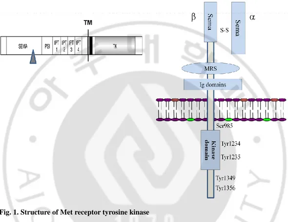
Historically, Met; the cell surface receptor tyrosine kinase was identified as the protein product of a transforming oncogege (Cooper and Park et al., 1984). The proto-oncogene was thus initially known as a transforming gene from chemically induced human osteogenic sarcoma cell line (Park et al., 1987). The product of this gene, c-Met, is synthesized as a 170 kDa single-chain precursor, which undergoes intracellular proteolytic cleavage producing a matured form of Met, the heterodimer (Comoglio et al.2001; Birchmeier et al.2003; Christensen et al. 2005 and Ma et al., 2003). This mature Met heterodimer is composed of an extracellular α- chain (50 kDa) and a membrane spanning β- chain (145 kDa), linked together by disulphide bonds (Figure 1).
The subunit is heavily glycosylated, extracellular and heterodimerized to the amino terminal portion of β- chain. The β subunit composed of an extracellular portion of Met contains a SEMA domain forming the major ligand-HGF binding site and receptor dimerization and a membrane-spanning segment with a transmembrane domain. Its intracellular region consists of juxtamembrane region followed by a tyrosine kinase catalytic domain and a C-terminal regulatory tail with multifunctional docking sites that binds to various signaling molecules for intracellular downstream signaling cascades (Ponzetto et al., 1994, Perschard et al., 2004).
2
A B
Fig. 1. Structure of Met receptor tyrosine kinase
(A) Met proto-oncogene is synthesized as a single-chain precursor about 170 kDa, which is proteolytically cleaved (shown by triangle) at SEMA domain producing the mature form of Met. (B) Mature Met heterodimer is composed of an extracellular α chain (50 kDa) and a membrane- spanning β chain (145 kDa), linked together by disulphide bonds. The SEMA domain lying at extracellular portion is crucial for HGF binding and receptor dimerization. (Cancer and Metastasis Reviews 22: 309–325, 2003).
3
B. HGF-Met Signaling
Hepatocyte growth factor (HGF), also known as scatter factor (SF) is the one and only one ligand of Met receptor (Vigna and Comoglio et al., 1999). Met, after binding with HGF gets dimerized and activated, subsequently undergoes autophosphorylation in the intracellular domain and upregulation of Met kinase activity followed by substrate phosphorylation leading to various cellular responses such as cell proliferation, motility, cell survival, cell adhesion, differentiation, angiogenesis and more related phenomena. Such complex biological activities are termed ‘‘as invasive growth’’ (Trusolino L. and Comoglio PM. 2002; Boccaccio C. and Commoglio PM. 2006)that occurs in normal cellular development.
During development, Met plays a crucial role for proper growth and development of placenta, kidney, liver, neuronal, and skeletal muscles. In adults, it is involved in haematopoiesis (Ikera et al., 1996) and is upregulated during tissue injury as in wound healing.
Activation of Met through the HGF binding leads to receptor dimerization and phosphorylation of selected tyrosine kinases within the cytoplasmic domains of the receptor, causing conformational changes. The “three tyrosine” motif Y1230,
Y1234 and Y1235 present within the kinase domain of c-Met are conserved in other kinase receptors. The tyrosines Y1234, and Y1235 including Y1230 in the activation loop of kinase domain is associated with full activation of enzymes and the enzymatic activity of Met is robustly up-regulated upon phosphorylation of these residues while the phosphorylation of Y1349 and Y1356 at C-terminal tail creates multisubstrate docking sites to recruit intracellular signal transducers including SOS, RAS, PI3K, STAT3, PLC-γ, and Src via several adaptor protein such as Grb2, SHC, Crk/CRKL and large adaptor protein GAB1 (growth- factor-receptor bound protein 2 (Grb2)- associated binder 1 ) containing -SH2 and –SH3 biding domains. Through these different components of signaling complex including adaptor proteins and signal tranducers, activated Met relays the different downstream signal cascades including RAS-MAPK and PI3K-AKT pathways (Figure 2).
Juxtamembrane domain of c-Met harbors a regulatory motif (Vigna et al., 1999; Lee et al., 20000) consisting of two distinct phosphorylation sites, Ser975 and Tyr1003.
4
Both of which are involved in inhibitory phenomena i.e. down-regulation and/or internalization of MET. Phosphorylation of S975 in the juxtamembrane domain is involved in MET internalization while phosphorylation of Y1003 induces to bind with CBL, the E3 ubiquitin ligase. Binding of CBL also leads to the recruitment of the endophilin-CIN85 complex, causing MET internalization and degradation (Ma and Maulik et al., 2003). The internalization of MET is necessary for ERK signaling (Kermorgant et al., 2005).The inhibitory activity of S975 is mainly controlled by Ca2+ / calmodulin-dependent kinase.
5
Fig. 2. HGF- Met Signaling PathwaysUpon HGF binding to c-Met, it gets activated , dimerized and activated Met transmits downstream signaling cascades inducing a wide variety of biological phenomena, including cell proliferation, survival, motility, scattering, branching morphogenesis and anti-apoptosis. (Paolo M. Comoglio, 2001).
6
C. Dysregulation of Met signaling and cancer
Despite the HGF-Met signaling plays a pivotal role in normal cellular processes, dysregulation of HGF-Met pathway either by autocrine manner or paracrine manner leads to malignant cell transformation, tumor progression causing to tumor metastasis (Jeffer et al.,1996; Birchmeier et al.,2003;Trusolino and comoglio et al.,2002, Danilkovitch-Miagkova and Berton Zbar et al.2002).The aberrant activation of Met is generally influenced either by Met over expression or Met gene amplification, missense point mutations in kinase domain and/or juxtamembrane domain or alternative splicing. Interestingly, Met expression level is associated with the progression of many different types of tumors, the elevated level of Met was observed in later stage or in metastatic tumors (Ma, PC.et al., 2003).
Met receptor predominantly found to be expressed in epithelial derived cells while HGF is expressed in surrounding mesenchyme or stromal cells. Thus expression of HGF in Met positive (but HGF- negative) epithelial cells or expression of Met receptor in HGF positive (but Met-negative) stroma cells leads to the autocrine activation of the Met receptor, that has the transforming potential (Renzo et al.,1991; Lyer et al.,1990). Therefore, autocrine activation of Met receptor is one of the mechanisms involved in aberrant activation of HGF Met signaling, leading to tumor formation and progression as frequently observed various type of tumor cells and tissues.
Met receptor has found to be over-expressed in different tumor cells and tissues as in carcinomas and lymphomas (Jiang et al., 1999).The Met gene amplification or over-expression of Met receptor tyrosine kinase is associated with transcriptional activation as observed in primary tumors ((Di Renzo et al., 1995).Tumor progression and development is also linked with the expression level of Met receptor as observed in metastatic tumors.
The missense point mutation is also one of the main causes of dysregulation of Met receptor tyrosine kinase. Most of the point mutations found to be located in tyrosine kinase domain of c-Met receptor are associated with the enhancement of tyrosine kinase activity (Jeffers et al., 1997) as observed in papillary renal carcinoma (Schmidt et al., 1997).
7
Alternative splicing of Met is also associated with dysregulation of Met receptor. It had been demonstrated that the splicing variant of c-Met with the deletion of exon-14 showed aberrant activation of Met which led to the tumor development as shown in nude mouse (Lee et al., 2006)
8
D. Nuclear Met and its implication
The cytosolic fragments, the truncated form of different cell surface proteins, or the receptor tyrosine kinases including FGFR family members and FGFR have been reported in the nucleus of the cell. These intracellular fragments or the full form of the receptors have been shown to regulate various essential functions in the nuclei of cells. Among them, nuclear EGFR was found to induce the transcription of various genes that are essential for cell proliferation (Low et al., 2005), cell cycle regulation, DNA repair and cell survival (Dittmann et al., 2010; Wang et al., 2006; Bandyopadhyay et al., 1998) and has been correlated with treatment resistance and poor prognosis in cancer (Hoshino et al., 2007; Laimer et al., 2007 and Hung et al., 2008). Furthermore, a truncated fragment of ErbB-4 found in the nucleus has been shown to be involved in activating or regulating the transcription of target genes (Maher et al., 1996 and Komuro et al., 2003).
It had been previously demonstrated that c-Met was found to present in the cells nuclei. (Dawidson et al., 2008). Recently, it has been reported c- terminal fragment of c-Met ~ 60 kDa in the nuclear fractions of several cancerous and noncancerous cell lines including HEK293; A431 cell lines (Pozner-Moulis et al., 2006) and in MDAMB231 breast carcinoma cells (Matteucci et al., 2009) even irrespective of ligand stimulation. The functional role and biological relevance of nuclear c-Met is still not well understood. However, some authors suggested that C-terminal fragment of nuclear c-Met may play a substantial role as a transcription factor in enhancing the c-Met signaling pathway by activating certain genes which may be associated with either cell proliferation (Rodrigues et al.,2007), or cell migration and motility which leads to invasive phenomenon in MDA-MB231 breast carcinoma cells (Matteucci et al.,2009).
Various RTKs, such as epidermal growth factor receptor (EGFR), ErbB-2, ErbB-4 and fibroblast growth factor receptor 1, reportedly translocate into the nuclei of cells as either intact or truncated forms (Maher et al., 1996 and Komuro et al., 2003). A cytosolic fragment may be generated from a transmembrane protein in two different ways: cleavage within the transmembrane domain by γ-secretase (Brown et al., 2000) or cleavage within the
9
cytoplasmic domain by proteases such as the proteasome (Hoppe et al., 2000) or caspases (Baess et al., 2001). However, the underlying mechanism(s) for the cleavage of cytosolic portion of Met and its translocation to the cell nuclei is still unclear.
10
E. Mechanism of nuclear import and export process
The nucleus of eukaryotic cells is separated from the cytoplasm by the nuclear envelop and the transport systems have established between the two compartments for the movement of biomolecules including proteins and nucleic acids in and out selectively and efficiently. The traffic between nucleus and cytoplasm occurs through a specialized, cylindrical, supramolecular structure called nuclear pore complex (NPC) forming a central aqueous channel with approximately 145 nm in diameter and 80 nm length in vertebrates (Figure 3).The nuclear pore complex acts as a filter running through a nuclear membrane. The NPC is made of ~30 different types of proteins called nucleoporins (Suntharalingam et al., 2003, Alber, F. et al., 2007) and is anchored to the nuclear envelope. The NPC allows to small molecules, metabolites and ions that do not bind to nucleoporins to diffuse passively between the nucleus and cytoplasm, but acts as a highly efficient molecular filter for macromolecules. Transport of almost all macromolecules into and out of the nucleus is achieved through a common energy dependent, active mechanistic process that requires soluble nuclear transport factors (NTFs) and specific transport signals (Köhler et al., 2007; Stewart et al., 2007, Görlich et al., 1997).
The translocation of protein molecules larger than 30~40 kDa (Yoneda et al., 200 and Nigga et al.) from the cytoplasm to the nucleus and vice versa requires a certain specific amino acid sequence called a nuclear localization signal (NLS) and nuclear export signal (NES) ( Kiseleva et al.,200 and Sorokin et al.,2007). During both import and export processes of most of the proteins, including membrane proteins (receptors) and nucleic acids a large evolutionary conserved families of karyopherins-β are involved. Most karyopherins-β participates either in nuclear trafficking called importins or nuclear export and is referred as exportins. In general, the nuclear import of proteins occurs via the formation of a ternary complex composed of the carrier proteins, i.e., importin α and importin β, and the NLS-containing cargo protein. Importin α acts as an adaptor that recognizes and binds the NLS of the cargo protein, while importin β enables the formation of a trimeric complex by mediating the binding of importin α at importing β binding domain (IBB) of importin α along with the NLS at cytoplasm, docks at the NPC through the FG repeats nucleoporins (Gorlich et al.,
11
1994 and 1995) and thereafter the complex is transported into the nucleus. Binding of Ran-GTP to the import receptor (importin β) in the nucleus promotes the separation of the importin α-cargo complex, discharging the cargo protein in the nucleus. Importin α and β are then recycled to the cytoplasm by different routes for further cycle of nuclear import. Therefore, the major nucleocytoplasmic trafficking process is governed by a small Ran GTPase. It is believed that importin α and importin β1 dimer is unstable in the presence of Ran GTP and therefore, there is rapid hydrolysis of Ran GTP during the docking of importin α/importin β/cargo with NLS at NPC to maintain the stable state of the trimer complex (Sorokin at al., 2007).
Many classical and non-classical NLSs have been identified to date. The classical monopartite NLS generally consists of a short stretches ~ 4-6 predominantly basic amino acids, such as lysine and arginine; a characteristic example is found in the Simian Virus 40 Large T-antigen (SV40-T-ag) as represented by PKKKRKV, specifying its import into the nucleus (Kalderon et al., 1984). A second type of classical NLS is the bipartite NLS, which is characterized by the presence of two basic amino acid clusters separated by a 10-to 12- amino-acid spacer, as seen and best studied in nucleoplasmin (KRPAATKKAGQAKKKK) (Robbins et al., 1991 and Dingwall et al., 1982). Numerous bipartite NLSs have been reported in various proteins with more than 12-amino-acid spacers (Table 1), still they migrate to the nucleus through the similar nuclear import process. Astonishingly, precise amino-acid sequence of the of the linker was found not to be important, and certain much longer linker segments could be tolerated and still promote efficient nuclear import (Robbins et al., 1991; Makkerh et al., 1996).
The proteins, on the other hand, containing nuclear export signals (NESs) exit out of the nucleus through the same aqueous channel of NPC located in nuclear envelope. The NES is the certain amino acid stretches (~3-4 amino acids) enriched in hydrophobic residues, specifically leucine-rich sequences of amino acids recognized by a soluble export receptor, known as Exportin1 or Crm1 (chromosome region maintenance 1) (Fried at el., 2003). The nuclear export signal in protein leads the protein to exit out of the nucleus. A possible consensus nuclear export sequence is denoted by ‘LX 1-3 LX 2-3 LXL’, where L =
12
leucine and X = amino acid; the last leucine can be replaced by conservative substitutions (isoleucine, valine, etc) (Bogerd et al., 1996 and Ikuta et al., 1998). The leucine rich NESs has been reported in various protein including HIV-1 Rev, PKI and HTLV-1 REX proteins (Figure14). Nuclear export of the protein via leucine-rich nuclear export signal takes place in a same manner as nuclear import, by the formation of a trimeric complex between protein carrying the NES, exportin 1, the export receptor and RanGTP in the nucleus. This trimeric complex is then transported out of the nucleus into the cytoplasm, where it dissociates and releases the protein in the cytosol where RanGTP gets hydrolyzed to RanGDP which is then utilized for the further process (Mattaj et al., 1998) (Figure 4).
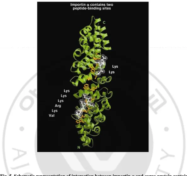
The crystal structure of importin α in an extended beta-strand conformation has two distinct sites on the concave surface of its superhelix (Figure 5). The larger site is at the amino-terminal end of the receptor while the smaller site is located at the carboxy-terminal end. At each site, the NLS peptide binds in an antiparallel direction via interactions with conserved tryptophan and asparagine pairs in repeat helix 3.
The binding of the NLS peptide occurs through two distinct types of interactions; hydrophobic interaction and electrostatic interaction respectively. The hydrophobic interactions occur between the aliphatic portions of the peptide side chains and the conserved receptor tryptophan residues while the electrostatic interactions occur between the basic groups of the NLS peptide and conserved negatively charged residues at the ends of the hydrophobic grooves of importin α at the carboxy-terminal smaller site, specifically only two residues in the bound NLS peptide are recognized. In the case of bipartite NLS cargo, in similar fashion, the smaller peptide-binding site allows optimal recognition of only two basic amino acids, whereas the larger site allows for the optimal recognition of five lysine or arginine residues (Figure 5) provided that in bipartite NLSs, the larger cluster of basic residues of NLS must be carboxy-terminal to the smaller cluster.
Therefore, during the nuclear import process, importin α binds with the NLS-containing cargo protein through ionic interactions that form between the basic amino acids of the NLS and acidic amino acids of importin α (Lange et al.,2007). Nevertheless,
13
there are some previous reports that some NLS-bearing proteins are imported to the nucleus by direct binding with importin β without the involvement of importin α as in Rex protein of HTLV-1(Palmeri et al.,1999; Truant et al., 1999), Smad 3 (Xio et al.,2000), ribosomal protein L23a (Jackel and Gorlich et al.,1998 ) and histones (Muhlhausser et al.,2001).
14
Fig. 3. Schematic representation of nuclear pore complexNuclear pore complex (NPC) is a cylindrical, supramolecular structure forming a central aqueous channel showing major components including cytoplasmic filament, cytoplasmic ring, nuclear envelop and nuclear baskets. Nuclear import and export of biomolecules takes place through the central channel by simple diffusion or NLS and/ or NES mediated active transport process. (Sorokin AV, Kim ER, Ovchinnikov LP, Biochemistry (Mosc). 2007 Dec;72(13):1439-57).
15
Table 1. Amino acid alignment of known bipartite nuclear localization signals
Protein Name (Source)
A possible consensus of bipartite NLSs
Spacer length K R – X (10-12) –K (K/R) (K/R) or K R – X (10-12) –K (K/R) –X – (K/R) (K/R) (K/R) – X (10–12 ) – (K/R) K R – X(10-12) – K R R K Npl K R P A A T K K A G Q A K K K K L D 10 NIN2 R K K R K T E E E S P L K D K A K K S K 13 SWI5 K K Y E N V V I K R S P R K R G R P R K 11 Rb K R S A E G S N P P K P L K K L R 12 PB2 K R K R D S S I L T D S Q T A T K R I RM A 13 CBP80 S R R R H S D E N D G G O P H K R R K T S 13 c-Met Q H V V I G P S S L I V H 10 Npl, nucleoplasmin; NIN2, Xenopus phosphoprotein NIN2; SWI5, Saccharomyces cerevisiae protein SWI5; Rb, retinoblastoma protein; Pb2, influenza virus polymerase Pb2; CBP80, human cap-binding protein 80; c-Met, human receptor tyrosine kinase protein c-Met.
Ref. (Molecular basis for specificity of nuclear import and prediction of nuclear localization. (Biochim Biophys Acta. 2011 Sep; 1813 (9):1562-77.)
16
Fig. 4. Shematic diagram representing nuclear import and export process
Carrier proteins importin α and importin-β bind to cargo protein containing NLS forming trimer complex in the cytoplasm, docks at the NPC and is translocated to the nucleus where RanGTP dissociates the cargo–carrier complex. The carrier–RanGTP complex is then exported to the cytoplasm where RanGAP activates the RanGTPase activity and dissociates the carrier–Ran complex, thus freeing the carrier for a further cycle of nuclear import. Similar process occurs for nuclear export process where carrier protein is exportin-1 with cargo containing NES.
17
Fig. 5. Schematic representation of interaction between importin α and cargo protein containing NLS
Two peptide-binding sites in the NLS receptor importin α (also known as karyopherin α) show the association with cargo proteins. The figure shows a ribbon diagram of the importin α polypeptide, highlighting the small and large peptide-binding sites. The conserved residues in repeat helix 3 are shown in yellow, and the amino acids of the NLS peptides that make specific contacts are shown in white.
18
F.
Nuclear localization signal and nuclear export signal of c- Met: A putative novel “pH-dependent” nuclear localization signal in juxtamembrane regionThe nuclear translocation or nuclear export of C-terminal fragment of the c-Met should require the presence of specific amino acid sequences, the NLS or NES respectively, but neither relevant sequence was found by the available nuclear localization search engine nor had been reported in previous studies. Here, we introduced that C- Met harbors a NLS possessing two histidines with 10 amino acid spacers in juxtamembrane domain between 1067aa-1080aa and may function as a bipartite NLS which can be characterized by 1068HVVIGPSSLIVH1079. Although this is similar to the known bipartite NLSs, the NLS found in c-Met contains histidines rather than lysines or arginines. The positivity of these individual histidine residues are weaker than those of the two or three lysine/arginine residues commonly present in classical bipartite NLSs. The positivity of histidine residues was expected to be increased with the decrease of intracellular pH. Thus, we speculated that a small change in intracellular pH could change the strength of the interaction between importin and the histidine-containing NLS of the c-Met fragment, thereby affecting its nuclear translocation and thus c-Met harbors a putative pH- dependent nuclear localization signal in juxtamembrane region.
c-Met, in addition to putative ‘pH-dependent’ NLS harbors conserved leucine-rich nuclear export signal similar to other proteins with classical NESs lying before the putative NLS in its juxtamembrane segment. The consensus sequence of putative NES c-Met is L-SA-L-NPE-L-V.
19
II. MATERIALS AND METHODS A. Reagent and antibody
F12 (Dulbecco’s Modified Eagle’s Medium/Nutrient Mixture F-12 Ham), PEI (Polyethylenimine), 2-deoxyglucose, NaN3, leptomycin B and nigericin were purchased from
Sigma-Alderich Chemical Co. (St. Louis, MO). DMEM (Dulbecco’s Modified Eagle’s Medium) and Lipofectamine™ 2000 were obtained from Invitrogen Corporation (CA, USA). BCECF-AM was from Molecular Probes (Eugene, OR). Mouse monoclonal anti-c-Met antibody (25H2) was from Cell Signaling Technology (Beverly, MA). Rabbit polyclonal anti-c-Met (C-28) (sc-161), Mouse monoclonal anti-GFP (sc-9996), anti-α-tubulin (sc-23948) and anti-lamin B (sc-374015) were purchased from Santa Cruz Biotechnology (Santa Cruz, CA,). Anti-importin β antibody (3E9) was from Abcam (MA, USA).
B. Plasmid construction
Plasmids for Jxtm1,-2,-3 green fluorescent protein (GFP) were from Dr. Rimm (Yale University, New Haven, CT). For the generation of GFP fusion constructs Fragment number-2 (F-number-2) and Fragment number-3 (F-3), truncating in the juxtamembrane region D97number-2 and Y1026, P1027 and I1084 of c-Met and cloned into the pEGFP-C3 fusion vector. We amplified using the primers containing XhoI and BamHI sites, respectively in a PCR using the template DNA of human c-Met. The DNA products were cloned into pEGFP-C3 fusion vector in the similar manner. A series of mutant plasmids were generated by PCR mutagenesis using either QuickChange Multi Site-Directed Mutagenesis kit (Stratagene, #200531, Agilent Technologies, USA) for amino acid substitution or QuikChange II XL kit (Stratagene #200521) for deletion and /or Muta-DirectTM Site-Directed Mutagenesis Kit, iNtRON Biotechnology (Gyeeonggi-do, Korea) for insertion or substitution of amino acids according to manufacturer’s instructions. Primer sequences used are given in (Table 2). All PCR products were checked by sequencing to confirm the presence of mutation and to verify the absence of secondary point mutations.
20
C. Cell culture and transfection
HeLa and Chang cell lines have been purchased from ATCC, and passaged and used in our laboratory for fewer than 6 months after resuscitation. ATCC performs cell line authentication by using STR profiling. HeLa cells were grown in F12 and Chang cells were grown in DMEM respectively supplemented with 10% fetal bovine serum (FBS) and 1% antibiotic-antimycotic (Gibco, Life technology) at 37°C under 5% CO2 in air and 95% humidity. Cells were transiently transfected with each GFP-fusion plasmids or si-RNA using PEI or Lipofectamine™ 2000, respectively according to the manufacturer’s instructions. D. Cell fractionation
HeLa and Chang cells were grown to confluence and the cells pre-treated with MG132 (10µM) for 2 hr were harvested and washed with PBS, resuspended in 200µl of TM-2 buffer (20 mM Tris-Cl, pH 7.5, 5 mM MgCl2 , 0.3 M sucrose containing freshly added protease
inhibitors) and incubated on ice for 5 min, subsequently added with 10 µl 10% Triton X-100 and again incubated on ice for 5 min. Cells were sheared by needle (22-guage) and then centrifuged (1,200xg, 4°C, 15 min). The supernatant was used as the cytoplasmic fraction. The pellet was resuspended in the TM-2 buffer, sheared again using a needle, and then centrifuged (1,200xg, 4°C, and 15 min). Pellet was washed three times , and resuspended in the mixture of 85µl of TM-2 buffer containing 10µl of 3M KCl, 5µl of 10% Triton X-100 and incubated for 10 min, and then centrifuged at 12,000xg for 2 min. Supernatant was used as the nuclear fraction.
E. Western bloting
Cells were harvested and lysed in radioimmunoprecipitation assay buffer (RIPA) buffer (50 mM Tris-HCl, pH 7.4, 1% NP40, 0.1%SDS, 150 mM NaCl,5Mm EDTA) containing freshly added protease inhibitor (lmg/ml aprotinin, l mg/ml leupeptin) from Amresco (Life Science Research Products and Biochemicals) and/or phosphatase inhibitors (1 mM sodium fluoride,1mM sodium orthovanadate).Lysed cells were centrifuged at 12,000xg for 15 min, Lysate was used for protein quantification using Bio-Rad Protein Assay (Hercules, CA) reagent. Proteins were resolved by SDS-PAGE (polyacrylamide gel electrophoresis) and
21
transferred to a nitrocellulose transfer membrane (WhatmanR , PROTRANR ). Membranes were blocked in 1% BSA in PBST, (BSA/PBS/1%Tween) for 1 hour. Primary antibodies at a 1:1,000 dilution were added to BSA/PBS/ Tween and incubated for overnight at 40 C tempera ture. Membranes were washed with PBST and then incubated with horseradish peroxidase conjugated goat anti-mouse or goat anti-rabbit secondary antibody (Invitrogen) at a dilution of 1:5000 for 1 hour at room temperature and subjected to immunoblotting using ECL detection kit reagent.
F. Immunocytochemistry Cells (1Χ105
in number)grown on coverslips were transfected with plasmids or si-RNAs. At 24 hr after transfection, cells were supplied with complete media. At 48 hr after transfection, cells were washed with PBS and fixed with 1:1 ice cold methanol-acetone for 15 min. Cells were then permeabilized with 0.2% (V/V) Triton Χ-100 in PBS for 5 min, and then washed 3 times with PBS and then blocked with 1% (W/V) BSA for 1 hr. Cells were then incubated with primary antibodies at 4°C for overnight and then washed with PBS. Cells were incubated with goat anti-mouse or goat anti-rabbit Alexa 448 for 1 hr at room temperature. After washing three times with PBS, cells were stained with diamidino-2-phenylindole (DAPI) for 10 min and washed three times with PBS. Coverslips were then mounted with mounting medium (Biomeda corp., Foster city, CA). Images were taken under fluorescence or confocal microscope (LSM 710, Zeiss).
G. Decrease of cytosolic pH
HeLa cells transfected with GFP-fusion constructs were treated either with nigericin at different concentrations as indicated in the text or with pH 6.5 Krebs-Ringer bicarbonate (KRB) solution (140 mM NaCl, 3.6 mM KCl, 0.5 mM NaH2PO4, 0.5 mM MgSO4, 1.5 mM
CaCl2, 10 mM HEPES, 2 mM NaHCO3, 5.5 mM glucose). Cells were then incubated at 37°C
under a humidified atmosphere of 5% CO2 for different durations. Cells were then harvested
for further assays.
H. Cytosolic pH measurement
22
fluorescence dye, BCECF-AM (2.5 M) for 30 min at room temperature. After wash-out, nigericin (1 M) or acidic pH (6.5) was applied for the indicated time (0~60 min) in KRB solution containing sulfinpyrazone (100 M). Fluorescence imaging of BCECF was performed by using an inverted microscope (IX-70, Olympus, Tokyo, Japan) equipped with a cooled charge-coupled device (CCD) camera (Cascade 512B, Photometrics, Tucson, AZ). Cells were alternately excited at 440 nm and 490 nm by a light source (DG-4, Sutter, Novato, CA) and acquired images at 540 nm were analysed using Metafluor 6.3 software (Universal Imaging, Molecular Devices). Titration of pH was carried out by clamping the cytosolic pH with high K+ buffers (125mM KCl, 5mM NaCl, 1mM NaH2PO4, 1mM MgSO4, 10mM
HEPES) of defined pH containing nigericin (5 μM) and monensin (5 μM). I. Importin β binding assay
Lysate (500 µg) of HeLa cells transfected with various constructs was incubated with 1µg of anti-importin β (3E9) antibody for 16 hr at 4°C with gentle rotation. Twenty five l of 50% slurry of rProtein G Agarose were added and the mixture was incubated for additional 1 hr to pull down the immune complex. After three successive wash with RIPA buffer containing freshly added protease inhibitors (l mg/ml aprotinin, l mg/ml leupeptin) and phosphatase inhibitors (1mM sodium fluoride, 1mM NaVO4), bound proteins were subjected to Western blotting.
J. Time-lapse analysis
HeLa cells were seeded onto a 6-well plate and transfected with GFP-fusion constructs. Twenty-four hour after transfection, 2-channel time-lapse video microscopy was performed using a fully motorized Axiovert 200M microscope (Carl Zeiss), equipped with AxiocamHRm. Temperature and CO2 control was maintained using the Incubator S-M and
heating insert M06 controlled by Tempcontrol 37-2 and CTI-Controller 3700. Both phase contrast and GFP fluorescence images were acquired for 48 h with a lapse time of 5 min using AxioVision 4.3 software. For routine and quantitative analyses, images were acquired using a 20x objective lens (LD Plan-Neofluar 20x/0.4 Corr Ph2, Carl Zeiss).
23
K. Leptomycin B treatment
HeLa cells transiently transfected with GFP-Jxtm1 fusion construct were treated with leptomycin B at different concentrations. Cells were then incubated at 37°C under a humidified atmosphere of 5% CO2 for four hour. Cells were then harvested for further assays.
L. ATP depletion
HeLa cells, transiently transfected with indicated plasmids were subjected to ATP depletion by the treatment with 2-deoxyglucose (2-DG) and sodium azide (NaN3) for indicated time
points. Western blotting was carried out with anti-GFP antibody after cellular fractionation. C,cytoplasmic fraction; N, nuclear fraction
M. Statistical analysis
All data were presented as mean ± standard deviation (SD). Each experiment was performed in triplicates. Statistical differences were analyzed by Student’s t-test or ANOVA (Analysis of variance) test, and p values <0.05 were regarded as statistically significant.
24
Table 2.Primer sequences25
III. RESULTS
Part. I. Nuclear translocation of C-terminal fragment of c-Met A. The c-Met fragment localizes to the nuclei of HeLa cells
Previous reports showed the presence of cystolic fragment ~ 60 kDa of c-Met in the nuclei of both cancerous and non-cancerous cell lines in spite of the diffusion limit of its size through the nuclear pore complex. The nuclear localization of the c-Met C-terminal fragment should require the presence of an NLS, but neither relevant sequence was found by the available nuclear localization search engine nor had been reported in previous studies. So, the aim of present study was to find out the motif or amino acid residues responsible for nuclear import of such large size of c-Met cytosolic fragment.
To search for the NLS of c-Met, we first selected HeLa cell line as it found to contain all the molecular machineries required for nuclear translocation of the c-Met fragment. For this, we first investigated the presence of the c-Met fragment in the nuclei of HeLa cells. Cells were stained with an antibody specific for the C-terminus of c-Met (C-28) and observed by confocal microscopy, which revealed a fine and distinct granular pattern of staining in the nuclei as shown by arrow (Figure 6A). We next performed cellular fractionation to confirm the immunocytochemical data. Immunoblotting using another antibody specific to the terminus of c-Met (25H2) clearly detected the ~ 60 kDa C-terminal fragment of c-Met in the nuclear fraction. This is consistent with previous reports on the nuclear C-terminal fragment of c-Met in various cells (Pozner-Moulis et al., 2006; Matteucci et al., 2009).
However, the nuclear fraction from Chang liver cells did not show the nuclear c-Met band despite the presence of wild-type c-Met in the non-nuclear fraction (Figure 6B), indicating that the nuclear translocation of the c-Met fragment is cell-type-specific. The observed expression of the c-Met fragment in the nuclear fraction of HeLa cells was clearly abolished by the c-Met-siRNA (Figure 6C), confirming the presence of the nuclear c-Met fragment in the nuclei of HeLa cells. To confirm the specificity of this signal, we carried out RNAi-mediated gene knockdown experiments, and found that the distinct
26
punctate pattern was significantly and dose-dependently abrogated by a c-Met-specific siRNA (Figure 6D) supporting the specificity of antibody. Based on these findings, we used the HeLa cell line as the model for our investigations.
28
Fig. 6. The C-terminal fragment of c-Met localizes in the nuclei of HeLa cells
(A) Sub-confluent HeLa cells were fixed and stained with C-terminal antibody for c-Met (C-28, green) and DAPI for nucleus (blue). Representative images show the nuclear patches of c-Met. Size bar;10µm. (B) Cell lysates from the indicated cells were fractionated as described in Materials and Methods, and then subjected to Western blot analysis by using antibody against C-terminus of c-Met (25H2). Notice the c-Met receptor tyrosine kinase (145 kDa) in cytosolic fraction and a 60kDa fragment of c-Met (triangle) in nuclear fraction only in HeLa cells. Purity of cellular fractionation was checked by Lamin B (nuclear marker) and tubulin (cytoplasmic marker). (C) Cell lysates from HeLa cells transfected with indicated siRNAs were fractionated and subjected to Western blot analysis as in (B), triangle; 60kDa nuclear c-Met fragment. (D) HeLa cells were transfected with control or c-Met siRNAs at the indicated concentrations and fixed at 24 hr after transfection. Immunocytochemistry was performed using C-28 antibody, and DAPI staining was used to visualize nuclei. Representative images show disappearance of nuclear c-Met signal in the presence of c-Met siRNA. Met (green) and nuclei (blue). Size bar; 10µm. Each experiment was performed in triplicate with similar results.
29
B. Serial deletion of juxtamembrane domain shows that c-Met contains an NLS at H1068-H1079 The nuclear c-Met fragment is not the spliced variant of Met as reported
previously rather it clearly originates from the cytosolic portion of full-size Met, no previous study had clearly identified the precise mechanism of nuclear translocation or the specific NLS involved. A previous report found that c-Met does not contain a classical NLS (Pozner-Moulis et al., 2006). Therefore, our main goal was to identify one or more motifs essential for the nuclear translocation of the c-Met fragment. We generated various GFP fusion constructs of the cytosolic portion of c-Met (Figure 7A), and transiently transfected these constructs into HeLa cells to analyze their subcellular distribution. Immunocytochemistry, and Western blotting after subcellular fractionation of Jxtm1 , GFP-F-2 and GFP-F-3 transfected cells enabled us to position of the putative NLS localizing between P1027 and I1084 in the juxtamembrane domain of c-Met (7B-E); this particular motif ‘P1027-I1084’ lies on the GFP-F-3 fragment shown in Figure 7A.
The overall size of EGFP-Met constructs namely F-2 and F-3 fragment is ~36.5 kDa, might be small enough to passively diffuse through the NPC. However, EGFP (~30 kDa) neither contains NLSs nor NESs and thus, it is a neutral protein which localizes ubiquitously (nucleus and cytoplasm of the cells. Nonetheless, practically it is predominantly found to localize in cytoplasm (more than 75 ~ 80% of the cells) and also not detected in nuclear fraction (Figure 7B and D). These results convinced us that observed nuclear localization of GFP-F-3 fragment might be due to the presence of putative NLSs.
To uncover the exact NLS, we generated a series of deletion mutants of GFP-(F-3) fusion constructs (Figure 7F) and investigated their intracellular distributions using Immunocytochemistry, and Western blotting following subcellular fractionation. Two of the deletion mutants (∆L1035-S1044 and ∆I1053-L1062) showed nuclear localization patterns similar to that of the wild-type F-3 fragment, indicating that the putative NLS was not located in these fragments. In contrast, the ∆H1068-H1079 deletion robustly abolished nuclear localization, as observed by immunocytochemistry (Figure 7G, left panel). The percentage of cells showing ubiquitous (cytosolic and nuclear) localization was significantly decreased by this deletion, while the proportion showing cytosol-only localization was
30
significantly increased compared to the results obtained with the wild-type F-3 fragment (Figure 7G, right panel). Similar results were obtained from a subcellular fractionation assay, wherein the protein level of the ∆H1068-H1079 mutant was substantially increased in the cytosolic fraction but decreased in the nuclear fraction (Figure 7H). Although, the ∆S1043-H1052 deletion mutant also showed decreased nuclear localization in our immunocytochemical analysis (Figure 7G), our subcellular fractionation assay clearly indicated that ∆H1068-H1079 but not ∆S1043-H1052 harbored the major NLS in the juxtamembrane fragment of c-Met (Figure 7F).
33
Fig. 7. Serial deletion of juxtamembrane domain of c-Met shows that c-Met harbors NLS motif at aaH1068-H1079
(A) Schematic representation of full and different truncated forms of Met with different domains fused
to C-terminus of pEGFP plasmid as described in Materials and Methods. (B) HeLa cells were transfected with indicated pEGFP and GFP-Jxtm1, and subjected to immunocytochemistry using GFP antibody. Confocal images were taken at 40X objective. (C) Cellular fractionation of HeLa cells transiently transfected with pEGFP and Jxtm1 and was followed by western bloting with GFP antibody. Purity of cellular fractionation was checked by lamin B (nuclear marker) and tubulin (cytoplasmic marker) respectively C, cytoplasmic fraction; N, nuclear fraction. (D) HeLa cells were transfected with indicated GFP-F2 and GFP-F3, and subjected to immunocytochemistry using GFP antibody. Confocal images were taken at 40X objective. The percentages of cells depicting subcellular distribution constructs are shown in the bar graph on the right (E) Cellular fractionation of HeLa cells transiently transfected with fusion constructs (GFP-F2 or GFP-F3) was followed by western bloting with GFP antibody as in (C). (F) Amino acid sequences of fragment No.3 (F-3) and schematic diagrams of deletion mutant constructs of F-3 fragment.GFP (green) and Juxtamembrane fragment (black solid line) with red solid verticalline indicating two histidine residues. (G) HeLa cells were transfected with indicated deletion mutant constructs of GFP-F3, and subjected to immunocytochemistry using GFP antibody. Confocal images were taken at 40X objective. Size bar; 10µm. The percentages of cells depicting subcellular distribution of deletion mutant constructs are shown in the bar graph on the right. Error bars; standard deviations, Three independent experiments were performed in triplicates. **, p<0.01 by Student’s t-test, n=200~270 (H) Cellular fractionation and Western blot analysis by using anti-GFP was performed after transfection as in (C). Purity of cellular fractionation was checked as in C. Each experiment was performed in triplicates with equivalent results. The densitometry graph on the right shows the nucleo-cytoplasmic ratio (nuclear GFP intensity divided by lamin intensity/ cytosolic GFP intensity divided by tubulin intensity). **, p<0.01 by Student’s t-test.
34
C. Two histidine residues are important for the nuclear translocation of c-Met fragment Classical bipartite NLSs comprise two stretches of basic amino acids (lysines or arginines) separated by a 10- to 12-amino-acid spacer, as previously described for many nuclear proteins, including nucleoplasmin( Robbins et al., 1991;Dingwall et al.,1982) ( Table 1). In bipartite NLSs, the positive charges provided by lysines or arginines are essential because they enable the electrostatic interaction with negatively charged amino acid residues on importin α, resulting in binding between the importin and the cargo protein (Lange et al.,2010;Kohler et al.,1997) We hypothesized that the putative NLS found in c-Met (H1068-H1079) might be a variant of this classical bipartite NLS, with two histidines replacing the lysines or arginines found in classical bipartite NLSs. To test this, we created point mutants of the F-3 protein wherein one or both histidines of the putative NLS motif were substituted with alanine (H1068A, H1079A and H1068A_H1079A), and examined the importance of the histidine residues for the nuclear localization of c-Met. Indeed, the mutant constructs all yielded significantly higher fractions of cells showing cytoplasm-onlylocalization (from ~10% to 70~80%), indicating that nuclear translocation was blocked in the mutant-expressing cells (Figure 8A). Next, we examined whether the histidine residues provided the positive charge required for the association of the c-Met fragment with importin α. Since mammalian cells may express six different forms of importin α but only one form of importin β (Kohler et al.;1997,Gabriel at al.,2011) and both receptors form ( β-α-cargo ) ternary complex with cargo containing NLS for nuclear import, we detected importin β. As expected, the wild-type F-3 fragment clearly co-immunoprecipitated with importin β, and substitution of both histidines with alanine significantly decreased this protein-protein interaction (Figure 8B). These results are consistent with our hypothesis that these two histidines are essential for the function of the NLS of c-Met, where they provide the positive charges required for the electrostatic interaction with importin α.
When we replaced either or both of the histidine residues with more basic lysine the nucleo-cytoplasmic ratios of the lysine-substituted mutants were higher than that of the wild-type F-3 construct (Figure 8C, lower right panel). In this case, we measured nucleo-cytoplasmic intensity ratios. Simple counting of the cells showing different subcellular localizations failed to reveal any difference (Figure 8C, right upper panel)
35
because the category of cells showing both nuclear and cytosolic localization (Figure 8C, right upper panel) contained cells with varied proportions of nuclear and cytosolic signals. A representative line scan analysis also revealed a similar tendency (Figure 8C, middle panel). Together, these findings support our belief that the two histidine residues in the NLS of c-Met confer the positive charges needed for the complex formation with importin molecules and the subsequent nuclear translocation of the c-Met fragment.
Astonishingly, precise amino-acid sequence of the of the linker was found not to be important, and certain much longer linker segments could be tolerated and still promote efficient nuclear import (Robbins et al., 1991; Makkerh et al., 1996).However, recently some reports have indicated that the linker sequences of an NLS, particularly hydrophobic amino acids, are involved in importin-cargo binding (Lange at al., 2010, Fontes et al., 2003 and Jang et al., 2012). So, we thought that whether the substitution of hydrophobic amino acids with alanines or deletion of two or four hydrophobic amino acids or insertion of two or four alanines within the linker of nuclear localization signal affect nuclear translocation. But neither substitution of hydrophobic amino acid with alanines ( Figure 9A) nor insertion of 2-4 alanines (Figure 9B) nor the deletion of 2-2-4 hydrophobic amino acids (1069VVI1071 and 1076LIV1078) within the H1068-H1079 linker of c-Met abolished the nuclear import rather showing similar subcellular distribution to that of F-3 wild type construct indicating that two histidines residues are enough to provide interacting site for the cargo-importin binding and are sufficient for the association .
37
Fig. 8. Two histidine residues are crucial for the nuclear translocation of c-Met fragment
(A) HeLa cells were transfected with wild type F-3, indicated histidine-to-alanine mutant constructs or pEGFP as a vector control. Immunocytochemistry by using anti-GFP antibody was performed as in Figure 7. Representative images were taken from fluorescence microscopy at 40X objective. Bar graph on right panel shows the percentage of subcellular distribution of cells (n=120~225). ***, p<0.001 by Student’s t-test (B) Cell lysates from HeLa cells transfected with F-3(WT) or H1068A_H1079A mutant were subjected to immunoprecipitation by using anti-GFP antibody, which was followed by immunoblotting with importin beta antibody. Bar graph in right shows the relative binding tendency of importin β with cargo protein compared to F-3(WT). (C) HeLa cells were transfected with wild type F-3 or indicated histidine-to-lysine mutant constructs, and subjected to immunocytochemistry by using anti-GFP antibodies. Bar graph displaying on right upper panel shows the percentage of subcellular distribution of cells (n=102~156) and on the right lower panel shows the relative nucleo-cytoplasmic ratio of green fluorescence intensity. Middle panel shows the intensity changes revealed by line scan analysis performed following the arrow. Error bars; standard deviations of three independent experiments.*, P< 0.05, **, P< 0.01 by Student’s t-test,n=25
38
0 20 40 60 80 100 120 C C+ N N C C+ N N C C+ N N C C+ N N C C+ N N C C+ N NGFP F-3 (WT) HA3 A3H HA3_A3H 2A
%
o
f C
40
Fig. 9. Substitution and /or deletion of hydrophobic amino acids with alanines or insertion of alanines within NLS did not abolish the nuclear translocation of F-3
(A) HeLa cells were transfected with wild type F-3, indicated hydrophobic amino acids substituted to alanine mutant constructs or pEGFP as a vector control. Immunocytochemistry by using anti-GFP antibody was performed as in Figure 8A. Representative images were taken from fluorescence microscopy at 40X objective. Bar graph displaying on lower panel shows the percentage of subcellular distribution of cells (n=120~150). (B-C) HeLa cells were transfected with wild type F-3 or alanines inserted or hydrophobic amino acids deleted as indicated histidine mutant constructs, and subjected to immunocytochemistry by using anti-GFP antibodies. Bar graph displaying lower upper panel shows the percentage of subcellular distribution of cells (n=105~125).
41
D. The NLS-directed nuclear accumulation of the c-Met fragment is pH-dependent
The imidazole group of histidine has a pKa of 6.0~6.2, which is fairly close to physiological pH and thus, it can be protonated or deprotonated depending on the intracellular pH of the cell (Li et al., 2011, Rötzschke et al., 2002 and Kampmann et al., 2006). Therefore, under physiological conditions, a relatively small shift in the environmental pH (especially to the acidic side) will readily change the average positive charge of a histidine residue. Thus, we speculated that a small change in intracellular pH could change the strength of the interaction between importin α and the histidine-containing NLS of the c-Met fragment, thereby affecting its nuclear translocation.
To explore the effect of decreased cytosolic pH on the nuclear translocation of the c-Met fragment, we used nigericin, which reduces cytosolic pH via the exchange of Na+ and H+ from vesicular organelles (Nilsson et al., 2003; Lin et al., 2003).We measured the actual cytosolic pH change with the pH-sensitive dye, BCECF-AM (Corvini et al., 2000; Weinlich et al., 1998). As shown in Figure 10A, treatment of HeLa cells with 1 uM nigericin significantly decreased the cytosolic pH as early as 30 min post-treatment, and this decrease of pH remained continued on time-dependent manner thereafter. We then explored the effect of nigericin on the nuclear translocation of the c-Met fragment. As expected, decreasing the cytosolic pH significantly increased the nuclear accumulation of the fusion protein both time- and dose-dependently (Figure 10B and C). The increase in the nuclear protein level was observed as early as 15 min after nigericin treatment and reached up to 10-fold at 1 hr post-treatment; the protein level in the cytosolic fraction decreased only slightly because most of the fusion proteins were localized in the cytosol (Figure 7C and E). We obtained essentially the same result using the Jxtm1 fragment of c-Met protein (Figure 10D and Figure 11A-D), which has similar size to the ~ 60 kDa C-terminal fragment of c-Met, ruling out the possibility that this result was an artifact due to our use of a small fragment of c-Met (Figure 7A). Since there was some discrepancy in the timings of the pH decrease and the nuclear translocation, we used time-lapse microscopy for real-time measurement of nuclear translocation (Figure 10E). When the fluorescence intensities of two circular areas inside the cytosol and nucleus, respectively, were measured from the time-lapse images (Figure 10E, right panel), we clearly saw that the nucleo-cytoplasmic ratio of the fusion protein increased
42
significantly at ~30 min after nigericin treatment, which was consistent with the cytosolic pH change measured using BCECF-AM (Figure 10A). These data collectively indicated that the novel NLS of c-Met appears to be sensitive to changes in pH (hereinafter called “pH-dependent”).
Next, we examined whether this pH dependency could be altered by replacing the critical histidines with other amino acids. Indeed, the increase of nuclear localization by nigericin treatment was abolished when histidine residues were substituted with either lysines or alanines (Figure 10F). Unexpectedly, nigericin treatment decreased the protein levels of these mutants in the nuclear fraction, which we don't understand the reason yet. Collectively, however, these data provide additional evidence that the putative bipartite NLS is pH-dependent, and this pH dependency stems from the critical histidine residues.
We then examined whether a decrease in cytosolic pH could enhance the binding of the c-Met fragment to importin α and β. Indeed, we found that a decrease in cytosolic pH enhanced the interaction between importin β and the F-3 fragment in immunoprecipitation assays run after 30 min of nigericin treatment (Figure 10G). This is consistent with the time point at which the decrease in cytosolic pH became significant (Figure 10A). In lysine- or alanine-mutant-transfected cells, in contrast, a decrease in cytosolic pH did not alter the level of cargo-associated importin β (Figure 10H), indicating that the pH-dependent change in the association of the c-Met fragment with importin is governed by these histidine residues.