ICCAS2005 June 2-5, KINTEX, Gyeonggi-Do, Korea
1. INTRODUCTION
The recent extension of the average lifespan of human has led to a huge increase in the population of the elderly and, in result, the occurrence of the traffic accidents. Due to the increase in the traffic accidents, there has been increasing number of reports on the illnesses related to the central nerve system such as cerebral apoplexy, brain damage, cerebral palsy, and regressive brain illnesses. These illnesses deteriorate the activities, the sensory perceptions, and the cognition processes of the patients, causing great difficulties in walking and everyday activities of them.
Recently, a research on postural control by Latt [1] realized that the stimulus for positional response of vestibular organ in normal state is in continuous sine waves. Seidler [2] studied the effects of short-term balance training for postural control of the elderly. Henry [3] studied postural control during A/P surface translation. From these previous studies, we can determine that three major senses are used in postural control. The three senses are vision, proprioception, and vestibular senses. The training of the three senses should be the basis for the balance training before walking. In maintaining balance, at least two of these three factors should function correctly. Therefore, a multi-dimensional rehabilitation method is needed in the rehabilitation of the balance control capability. However, the conventional researches on the rehabilitation have been insignificant due to the nonchalance of the society. Even most of the simple rehabilitation equipment is old and insufficient, and the methods used in the equipments are quite ancient.
The present study introduces a new rehabilitation training system for balance control of the patients in bed using a tilting bed, a visual display, and a force plate providing sense of balance and the sensation of walking to the patient. The tilting bed is automatically controlled by a computer, and can tilt up and down and move sideways in uniform velocity enabling the training for balance control of the patient in bed. The sense of
balance can be provided by the visual display and the force plate as well as tilting of the bed. In addition to providing the training for balance control, an assessment of active and passive balance control can be also carried out with the system.
2. SYSTEM CONFIGURATION
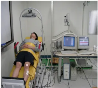
The present study proposes a new training device which can overcome the disadvantages of monotonous training environment of the existing devices by stimulating various senses increasing the efficiency of the training for balance control. Figure 1 shows the training system developed in this study. As shown in the figure, the system consists of a tilting bed, a visual display, and a force plate.
Fig. 1 Configuration of the early rehabilitation training system for posture control
Development of an Advanced Early Rehabilitation Training System for
Postural Control Using a Tilting Bed
Chang-Ho Yu
*, Kyung Kim
*, Yong-Yook Kim
**, Tae-Kyu Kwon
***, Chul-Un Hong
***,
and Nam-Gyun Kim
****Department of Biomedical Engineering, Chonbuk National University, Jeonju, Korea (Tel : +82-63-270-2246; E-mail: combo418@hanmail.net, kkruddy@hotmial.com)
**Center for Healthcare Technology Development, Jeonju, Korea (Tel : +82-63-270-4063, Fax: +82-63-270-2247; E-mail: yykim@chonbuk.ac.kr) ***Division of Bionics and Bioinformatics, Chonbuk National University, Jeonju, Korea
(Tel : +82-63-270-2247; E-mail: kwon10, cuhong, ngkim@chonbuk.ac.kr)
Abstract: We propose a new early rehabilitation training system for postural control using a tilting bed and a force plate. The
conventional rehabilitation systems for postural control cannot be applied to the patients lying in bed because the rehabilitation training using those systems is possible only when the patient can stand up by himself or herself. Moreover, there has not existed any device that could provide the sense of balance or the sensation of walking to the patients in bed. By using a tilting bed, a visual display, and a force plate, we have developed a new rehabilitation training system for balance control of the patients in bed providing sense of balance and the sensation of walking to the patient. Through the experiments with real people, we verified the effectiveness of the new early rehabilitation training system. The results showed that this system is an effective system for the early rehabilitation training and that our system might be useful as clinical equipment.
Keywords: Somatic sensation, Postural Control, Early rehabilitation training
ICCAS2005 June 2-5, KINTEX, Gyeonggi-Do, Korea
Figure 2 shows the newly developed early rehabilitation
training system. The figure shows the bed in tilted angle. The system also includes numerous sensors including the load cells in force plate, and the sensors for electromyogram. The signals from the sensors are fed to a computer through an A/D board. Potentiometer resistive transducers detect the tilt angle, which is a degree sensor. The height and the sideway rotation adjustments of the bed are enabled by two motors. The system is designed to help faster recovery of the patients and the effects of rehabilitation by stimulating various sensory organs simultaneously.
Fig. 2 The early rehabilitation training system A force plate was used for the lower body rehabilitation training. The force plate was in rectangular shape with the width of 400 mm and the depth of 300mm. Four circular shaped load cells (50kg weight limit) were placed in the plate with vertical spacing of 180 mm and horizontal spacing of 280mm. The signals from the load cells were amplified since the actual voltages from each load cell was only a few mill volts (mV). To make the amplifier to output 10V for 50kg of weight, the magnitude of the amplification was set to be 250 times. The amplified signal was fed to an A/D converter to be used in the computer analysis. In addition, a low-pass filter was installed between the amplifier and the A/D board so that only the signals below 300Hz can be passed to the A/D converter. When the bed is tilted, the weight of the subject is applied to the force plate creating a distribution of pressure on the force plate. By analyzing the four signals from the four load cells, the location of the center of the pressure (COP) can be determined. Then, this information on the location of the COP is fed back to the subject using a visual display so that the subject can know where the COP is, and he or she can control the location of COP using his or her low limb muscles.
3. METHODS
3.1 Subjects
The subjects were young males in their twenties. Before the test took place, the subjects were all informed of the entire procedures of the test and signed a consent form.
3.2
Experimental procedureIn the training system, the subjects are instructed to move
the COP shown in the visual display to follow certain paths by using the lower limb muscles when the bed is tilted and pressure is applied to the force plate. The training using the system consisted of COP linear tracking training and COP sine wave tracking training: the COP linear tracking training was done by moving the COP following linear paths in upward, downward, right, left, upper right, upper left, lower right, and lower left directions shown in Figure 3; and the COP sine wave tracking was done by moving the COP following the sine wave shaped paths in horizontal and vertical directions.
Fig. 3 Directions of COP maintaining training
3.2 Data acquisition
Experiments were carried out while the subject was lying on the tilting bed with upper part of the body tied to the bed. The signals from the load cells were used in determining the location of COP and calculating the deviation from the desired path. The EMG signals were measured from four kinds of muscles: rectus femoris of the left foot, biceps femoris, gastrocnemius, and anterior tibial muscle. The EMG signals were examined to find out which muscle was mostly used.
To evaluate the effectiveness of the postural control training using the system, we measured and tracked various parameters such as COP moving time and COP maintaining time: COP moving time means the time it takes for a subject to move the COP to a desired location, and COP maintaining time means the duration of time a subject maintains the location of COP inside a desired area
.
4. RESULTS AND DISCUSSIONS
To examine the serviceability of the system and effects of the training, the performance of the training was assessed. Major factors related to balancing sensors and data were analyzed.
4.1 The EMG changes in according to degree shifts
Figure 3 displays the EMG signals of the tibialis anterior according to degree shifts. In this experiment, the degree variations were given using the tilting bed at 0°, 15°, 30°, and 45° up and down. As a result, as tilting bed angles increases, the uses of muscles decrease. In EMG signal changes between 0° and 45° in tibialis anterior and gastrocnemius, EMG signal at 0° was larger than at 45°, as shown in Fig. 3. This is due to the fact that EMG signal was large because at 0° there was no
ICCAS2005 June 2-5, KINTEX, Gyeonggi-Do, Korea
weight exerted on the plate, training by just lower body
muscles, but at 45° EMG signals lessened due to the fact that the training was carried out with both the weight exerted on the plate and the lower body muscles.
0 10 20 30 -0.5 0.0 0.5 1.0 EM G S ign al (V ) T im e(S ) EM G S ign al (V ) 0(degree) -0.5 0.0 0.5 1.0 45(degrees)
Fig. 4 EMG signal of tibialis anterior on the tilting angle
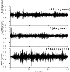
0 1 0 2 0 3 0 - 0 . 2 - 0 . 1 0 . 0 0 . 1 0 . 2 0 . 3 EM G Signa l( V ) EM G Signal( V ) T i m e ( s ) EM G Signa l( V) + 1 5 ( d e g r e e s ) - 0 . 2 - 0 . 1 0 . 0 0 . 1 0 . 2 0 . 3 0 ( d e g r e e ) - 0 . 2 - 0 . 1 0 . 0 0 . 1 0 . 2 0 . 3 - 1 5 ( d e g r e e s )
Fig. 5 EMG signal of gastrocnemius on the tilting angle of left-right 0.E+00 1.E-04 2.E-04 3.E-04 4.E-04 5.E-04
Area of power spectrum
(Vrms*Hz)
Anterior Posterior Left Right
Position 15 degrees 30 degrees 45 degrees 0.E+00 1.E-04 2.E-04 3.E-04 4.E-04 5.E-04 A rea of p ow er s p ectru m (Vrms*Hz)
Anterior-Left Anterior-RightPoeterior-Left Posterior-Right
Position
15 degrees 30 degrees 45 degrees
Fig. 6 Area of power spectrum of Tibialis anterior
0.E+00 1.E-04 2.E-04 3.E-04 4.E-04 5.E-04 A rea of p ow er s p ectru m (Vrms*Hz)
Anterior Posterior Left Right
Position 15 degrees 30 degrees 45 degrees 0.E+00 1.E-04 2.E-04 3.E-04 4.E-04 5.E-04 A rea of pow er spectrum (Vrms*Hz) Anterior-Lef t Anterior-Right Post erior-Lef t Post erior-Right Position 15 degrees 30 degrees 45 degrees
Fig. 7 Area of power spectrum of Rectus femoris Figure 4 shows the muscular conductivity signal of the gastrocnemius according to sideways rotation in the tilting bed. When we changed to 15° both to the left and the right, the changes in gastrocnemius showed changes in EMG signals, as shown in Fig. 4. The measured muscle is on the left foot, therefore EMG signal at +15° tended to be larger than 0° and -15° due to the fact that the signals put force in the direction of the weight. Also in -15° the muscle movement was smaller due to the training executed in the opposite direction of the weight. Therefore at -15° the EMG signal tended to be smaller.
To know activated muscles related to the COP training, we analyzed EMG signal into frequency-domain according to the COP training in all directions. During analyzing on my experiment, the area of power spectrum is related to the activated quantity of the muscular conductivity signal. Figure 5 and 6 display the analytic area of power spectrum according to degree shifts in tilting bed and these are about rectus femoris and tibialis anterior. Figure 5 and 6 also displayed the
ICCAS2005 June 2-5, KINTEX, Gyeonggi-Do, Korea
graphs of the eight training positions which consist of
anterior, posterior, left, right, anterior-left, anterior-right, posterior-left and posterior-right. According to Fig. 5 and 6, the larger of the degrees, the shorter of the area of power spectrum and the activated quantity of muscles in all directions.
The lower body muscle used in COP tracking training and sine wave tracking training were generally the muscles below the knees such as tibialis anterior and gastrocnemius, rather than those above, such as rectus femoris and biceps femoris. In this experiment the EMG signals were measured only on the left foot.
Therefore in experiments where the positions were shifted toward the right, the left foot was hardly used, which caused almost insignificant amount of EMG signal in the left foot muscles. On the other hand, in experiments positioned toward the left, EMG signals were measured. In sine wave tracking training, EMG signals of the tibialis anterior and gastrocnemius were mainly measured. In horizontal tracking the EMG signals of the gastrocnemius exterior was mainly measured, which takes major control of the sideways movements, and in vertical tracking tibialis anterior as well as gastrocnemius movements were measured, which takes major control of going forward and backward.
Let us now take a look at the movements of the lower body muscles according to degree shifts. At 0° the experiment is executed without any weight exerted on the plate, forcing a lot of strength from the lower body. Therefore at 0° the EMG signals of tibialis anterior and gastrocnemius exterior are large. As the degree is increased, the exerted weight increases, so it becomes easier to execute the experiment. Therefore the EMG signals of tibialis anterior and gastrocnemius exterior gradually decrease.
4.2 Evaluation of COP Training
Fig. 8 The COP moving time
Fig. 9 The COP maintaining time
The lower body rehabilitation training system was executed in a basic loading training by pressing the legs
against the plate at 0° and 45°, COP, a measuring of weight shift, a muscle training program doing weight transfer tracing the sine wave, and rehabilitation training evaluation through games.
Figure 7 displays the COP measurements and training results of the tibialis anterior and gastrocnemius due to degree shifts (at 0° and 45°). According to Fig. 7 and 8, the larger of the degrees, the shorter the COP movement time in all directions, and the longer the COP maintenance time. This is due to the fact that the weight exerted on the plate was increased as the degree became larger. Therefore as the degree became larger, the COP movement and maintenance became easier.
5. CONCLUSIONS
In this study, the early rehabilitation training system was devised for the patients to start the rehabilitation process right after hospitalization, which in the past was only possible after a certain time of recovery, leading to the following conclusions:
1. The present study developed an automatic tilting bed controlled by computers, tilting up, down and sideways in uniform velocity and slopes, to enable training of improving postural balance.
2. When the force plate was shifted sideways, it was confirmed by EMG signals that tibialis anterior, gastrocnemius, biceps femoris and rectus femoris were used simultaneously or one by one.
3. In general, COP movement time was long due to difficulties in postural balancing control in the rear, left, and rear left, and it was discovered that the times in these positions could be shortened by training.
ACKNOWLEDGMENTS
This research was supported by the Korean Ministry of Education and Human Resources Development through the Center for Healthcare Technology Development.
REFERRENCES
[1] L. D. Latt, P. J. Sparto, J. M. Furman, and M. S. Redfern, “The steady-stady postural response to continuous sinusoidal galvanic vestibular stimulation,” J. of Gait
Posture, Vol. 18, pp. 64-72, 2003.
[2] R. D. Seidler, and P. E. Martin, “The effects of short term balance training on the postural control of older adults,”
Gait and Posture, Vol. 6, pp. 224-236, 1997.
[3] S. M. Henery, and J. Fung, “Control of stance during lateral and anterior/posterior surface translations,” IEEE
Trans. on Rehabilitation Engineering, Vol. 6, No. 1, pp.
32-42, 1998.
[4] J. Massion, “Movement, posture and equilibrium: interaction and coordination,” Prog. Neurobiol., Vol. 38, pp. 35-56, 1992
[5] R. A. Speer, A. D. Kuo, and F. B Horak, “Contributions of altered sensation and feedback responses to changes in coordination of postural control due to aging.” Gait and
Posture, Vol. 16, pp. 20-30, 2002
[6] C. A. Laughton, M. Slavin, K. Katdare, L. Nolan, J. F. Bean, D. C. Kerrigan, E. Philips, L. A. Lipsite, and J. J. Collins, “Aging, muscle activity and balance control: physiologic changes associated with balance impairment,”
Gait and Posture, Vol. 18, pp. 101-108, 2003