Lesion of subthalamic nucleus in Parkinsonian
rats: Effects of dopamine D1 and D2 receptor
agonists on the neuronal activities of the
substantia nigra pars reticulata
Park Yong Sook
Department of Medicine
Lesion of subthalamic nucleus in Parkinsonian
rats: Effects of dopamine D1 and D2 receptor
agonists on the neuronal activities of the
substantia nigra pars reticulata
Directed by Professor Park Yong Gou
The Doctoral Dissertation
submitted to the Department of Medicine,
the Graduate School of Yonsei University
in partial fulfillment of the requirements
for the degree of Doctor of Philosophy
Park Yong Sook
June 2006
This certifies that the Doctoral Dissertation of
Park Yong Sook is approved.
---
Thesis Supervisor : Park Yong Gou
---
Thesis Committee Member#1 : Chang Jin Woo
---
Thesis Committee Member#2 : Chang Jong Hee
---
Thesis Committee Member#3 : Kim Dong Goo
---
Thesis Committee Member#4 : Ahn Duck Sun
The Graduate School
Yonsei University
ACKNOWLEDGEMENT
I would like to thank my teachers, who have given support and encouragement, Professor Park Yong Gou, Professor Chang Jin Woo and Professor Chang Jong Hee. I would also like to thank for the help to committee members, Professor Kim Dong Goo and Professor Ahn Duck Sun. I am particularly grateful to Professor Chung Sang Sup, Professor Choi Joong Uhn and Professor Kim Dong Seok for allowing an opportunity of learning and practice. I am obliged to Professor Jeon Byung Chan for careful concern. Special thanks to Jeon Mi Fa, Cho Yoon Hee and all members of a laboratory for their assistance and significant help all through the completion of this process. Also special thanks to Mrs. Ann Kye Soon for her kindness and thoughtful consideration.
June 2006 Park Yong Sook
i
CONTENTS
ABSTRACT···vii
I. INTRODUCTION ··· 1
II. MATERIALS AND METHODS ··· 3
1. Surgical procedures for MFB and STN lesions··· 3
2. Extracellular microrecordings ··· 4
3. Histology and immunohistochemistry ··· 5
4. Data analysis ··· 6
III. RESULTS ··· 7
1. Histological findings of rat parkinsonian models··· 7
2. Effects of STN lesions on firing rate and firing patterns··· 7
3. Effects of SKF38393 on the firing rates and firing patterns ··· 12
4. Effects of Quinpirole on the firing rate and firing patterns ··· 14
IV. DISCUSSION··· 15
1. Effects of STN lesions on the firing rates and firing patterns of SNpr in rat parkinsonian model··· 15
2. Effects of SKF38393 and Quinpirole on the firing rates and firing patterns of SNpr in rat parkinsonian model with STN lesion··· 16
V. CONCLUSION ··· 19
REFERENCES ··· 20
ii
LIST OF FIGURES
Figure 1. Surgical procedure for the MFB ··· 4 Figure 2. Immunohistochemistry of striatum, substantia nigra and subthalamic nucleus ··· 9 Figure 3. SNpr discharge pattern recorded in 6-OHDA-lesioned rats ··· 11 Figure 4. The effects of D1 agonist (SKF38393) and D2 agonist (Quinpirole) on the firing
pattern of SNpr neurons in rat parkinsonian models ··· 13
LIST OF TABLES
Table 1. Spontaneous activity of SNpr single units recorded from 6-OHDA lesioned rats with a kainic acid lesion of the STN and intrastriatal selective D1, D2 agonist microinjection. ··· 10
iii
ABSTRACT
Lesion of subthalamic nucleus in Parkinsonian rats: Effects of dopamine D1
and D2 receptor agonists on the neuronal activities of the substantia nigra
pars reticulata
Park Yong Sook
Department of Medicine
The Graduate School, Yonsei University
(Directed by Professor Park Yong Gou)
Object. It was hypothesized that dopamine agonist administration and subthalamic nucleus (STN) lesion in the rat might have a synergistic effect on the neuronal activities of substantia nigra pars reticulata (SNpr) as observed in patients with Parkinson’s disease. The effects of SKF38393 (a D1 receptor agonist) and Quinpirole (a D2 receptor agonist) were compared in parkinsonian rat models with 6- hydroxydopamine (6-OHDA) after STN lesion. The changes of the firing rates and firing patterns of SNpr neurons were analyzed.
Methods. SKF38393 and Quinpirole were consecutively injected intrastriatally. SNpr was microrecorded to ascertain the activity of the basal ganglia output structure. The effect of SKF38393 or Quinpirole injection on the firing rate and firing patterns of SNpr was investigated in medial forebrain bundle (MFB) lesioned rats and in MFB + STN lesioned rats.
iv
Results. The administration of SKF38393 decreased SNpr neuronal firing rates in the lesioned rats, but did not alter the mean neuronal firing rate in the SNpr neurons of MFB+ STN lesioned rats, nor induce significant changes in the percentage of burst neurons. In rats prepared from 6-OHDA, Quinpirole significantly decreased the spontaneous firing rate. However, after an additional STN lesion, it induced a significant change in the percentage of burst neurons.
Conclusions. The results demonstrated that STN lesion decreases SNpr hyperactivity and the proportion of burst neurons among total neurons, but that dopamine receptor agonists, such as SKF38393 and Quinpirole, did not change the firing pattern.
--- Key words : 6-hydroxydopamine; Substantia nigra pars reticulata; Kainic acid;
1
Lesion of subthalamic nucleus in Parkinsonian rats: Effects of dopamine D1
and D2 receptor agonists on the neuronal activities of the substantia nigra
pars reticulata
Park Yong Sook
Department of Medicine
The Graduate School, Yonsei University (Directed by Professor Park Yong Gou)
I. Introduction
Parkinson’s disease (PD) is a neurodegenerative movement disorder induced by a progressing deficit of dopamine cells in the pars compacta of the substantia nigra (SNpc), leading to dopamine depletion in the striatum.1 Since the discovery of the dopamine precursor, levodopa, it has remained as the most effective medical treatment for PD. However, after several years of exposure to levodopa, the majority of patients develop on-off motor fluctuations, painful dystonia, wearing-off phenomena and levodopa-induced dyskinesia.2 The mechanisms underlying the motor complications in PD remain poorly understood. As well, it is now well known that deep brain stimulation (DBS) of the subthalamic nucleus (STN) is reported to decrease motor fluctuations and dyskinesia in patients with PD despite the fact that most patients continue to take levodopa.3 The STN lesion in the rat also normalizes the decreased activity of globus pallidus internus (Gpi) in the rat with a previous medial forebrain bundle (MFB) lesion.4 Thus both strategies, dopamine agonist administration and STN lesion in the rat, might have a
2
synergistic effect on the neuronal activities of substantia nigra pars reticulata (SNpr) as observed in PD patients treated with bilateral DBS.
In this study, the effects of SKF38393 (a D1 agonist) and Quinpirole (a D2 agonist) were compared in parkinsonian rat models with 6-hydroxydopamine (6-OHDA) after STN lesion. To investigate these effects, the changes of the firing rates and firing patterns of SNpr neurons were analyzed.
3
II. Materials and Methods
1. Surgical procedures for MFB and STN lesions
Male adult Sprague-Dawley rats weighing 200-250 g were used for the first surgical procedure. Rats were divided into 4 groups: (i) control group, 7 rats without lesions; (ii) 6-OHDA lesioned group, 7 rats that had an MFB lesion induced by 6-OHDA; (ⅲ) 6-OHDA + STN lesioned group, 7 rats with a 6-OHDA lesion of MFB plus a STN lesion, induced by kainic acid; and (ⅳ) 6-OHDA + STN sham group, 7 rats treated with saline instead of kainic acid in STN. Five animals per group were housed in a temperature-controlled room on a 12hr-light/12hr-dark schedule with free access to food and water. Rats were anesthetized with a mixture of ketamine (75 mg/kg), acepromazine (0.75 mg/kg) and rompun (4 mg/kg) and mounted in a stereotaxic apparatus. Six-OHDA hydrobromide (Sigma, St Louis, MO, 8 µg free base in 0.2 % ascorbic acid) was injected into MFB according to the following stereotaxic coordinates: AP –4.4 mm, ML 1.2 mm relative to bregma, and DV -7.5 mm from the dura. The injection was made at a rate of 0.5 µl/min using a cannula, and was controlled using a Hamilton microsyringe. A polyethylene tube connected the cannula and the microsyringe (Fig. 1). To prevent the noradrenergic neurons being destroyed, desipramine (12.5 mg/kg, i.p.) was administered 30 min prior to the 6-OHDA infusion. The STN lesion was achieved by injecting 1µg of kainic acid (Sigma, St Louis, MO) dissolved in 0.5 µl of saline into the right STN (coordinates: AP -3.8 mm, ML 2.5 mm relative to bregma, and DV –8.0 mm from the dura) at the rate of 0.25 µl/min. In the group of rats with combined lesions (6-OHDA + STN lesion), the STN lesion was performed 3 weeks after the 6-OHDA lesion. Sham lesion was performed by using the same protocols as used for the combined lesions, but saline was injected instead of kainic acid.
4
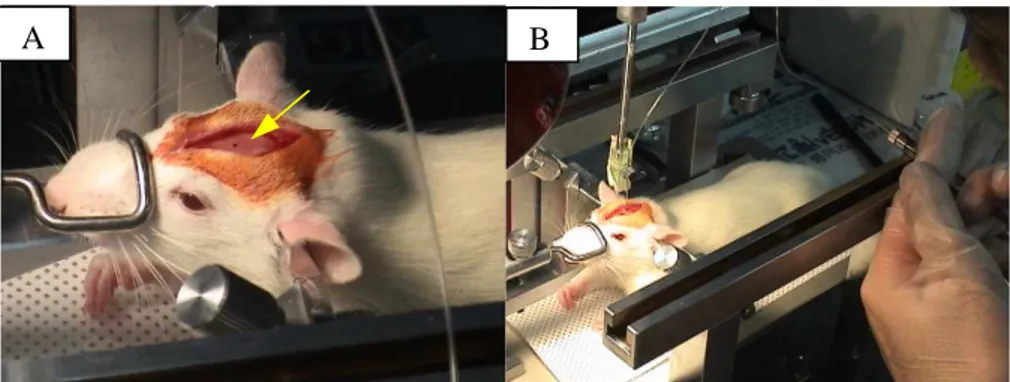
Fig. 1. Surgical procedure for the MFB. A: Mounted in a stereotactic apparatus. Arrow indicated the dot for bregma. B: Parkinsonian model by lesioning of medial forebrain bundle with 6-hydroxydopamine using a cannula controlled with a Hamilton microsyringe.
2. Extracellular microrecordings
Extracellular, single unit recordings were undertaken in rats anesthetized with urethane (1.3 mg/kg i.p.). A glass microelectrode (impedance, 7-10 Mohm) filled with 2.5% Pontamine sky blue in 0.5 M sodium acetate buffer (pH 7.6) was used to investigate the single recordings. Microelectrodes were stereotaxically guided through a drilled skullburr hole to the target coordinates (SNpr: AP –5.3 mm, ML 2.4 mm relative to bregma, and DV 7.5-8.0 mm from the dura). Electrical signals were amplified using a DAM80 preamplifier (WPI, UK) in bridge mode, displayed on a storage oscilloscope and monitored with an audio amplifier. Single unit activity was isolated with a window discriminator, and firing rate data were collected on a computer equipped with Spike 2 software (version 2.18, Cambridge Electronic Design, UK). Visual inspection of digital neuronal activity and raster displays were useful complements to the computer-based analysis of the discharge patterns of these units. The isolated units were monitored for at least 10 min to
5
ensure the stability of their firing rate, firing pattern and spike morphology, and then 5-10 min of spontaneous activity was recorded. The selective D1-class dopamine agonist SKF38393 (Sigma, St Louis, MO, 10 nmol/0.5 µl), or the selective D2-class agonist Quinpirole (Sigma, St Louis, MO, 10 µmol/0.5 µl) was injected in the striatum [coordinates AP –0.8 mm, ML 3.0 mm relative to bregma, and DV 1.3 mm from the dura]. This procedure required less than 2 min, and the needle was left in place until the end of the recording, when the location of the tip of the recording microelectrode was marked, at –15 uA for 20-30 min, by an iontophoretic deposit of Pontamine Sky Blue.
The mean firing rate, the mean interspike interval (ISI), autocorrelogram and discharge pattern were investigated for each neuron. The ISIs allowed an evaluation of the neurons’ degree of burst frequency, following an algorithm described by Hutchinson et al.5,6 Bursting cells had a degree of burstiness score of more than 10, which was calculated from the reciprocal of the modal interval divided by the mean firing rate.
3. Histology and immunohistochemistry
After the extracellular single unit recording, rats were anesthetized and transcardially perfused with 125 ml of normal saline followed by 250 ml of ice-cold 4% paraformaldehyde. Brains were removed, postfixed for 10 hours, and transferred to 30% sucrose until equilibrated. Twenty micrometer sections were cut frozen and then immunoreacted with a primary, polyclonal antibody against rat tyrosine hydroxylase (TH, Pel-freeze, Rogers, AK) at a dilution of 1:750, and then with a biotinylated goat anti-rabbit IgG (Vector Labs, Burlingame, CA) secondary antibody. The signal was amplified using avidin and biotinylated horseradish peroxidase using an Elite ABC Vectastain Kit (Vector, Burlingame, CA). 3,3'-Diaminobenzidine tetrachloride dehydrate was used as a chromogen and cobalt
6
chloride/nickel ammonium was used to intensify color changes. This immunostaining allowed the determination of the extent of dopaminergic cell degeneration. Only rats with a total loss of TH immunoreactivity were used for the electrophysiological analysis. The STN lesions and the localization of the recorded basal ganglia nuclei were studied in 20-µm sections stained with cresyl violet.
4. Data analysis
Statistical analysis was performed with the SPSS version 11.0 statistical software packages (SPSS Inc., Chicago, IL). Comparisons of the firing rates from different rats in each group were performed using analysis of variance (ANOVA). Results showing significant differences between groups were compared using Kruskal-Wallis one-way ANOVA and then the Mann-Whitney U-test. Statistical significance was set at p < 0.05.
7
III. Results
1. Histological findings of rat parkinsonian models
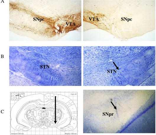
The extent and location of the lesions induced by 6-OHDA were confirmed by assessing the loss of TH-immunoreactive cells in SNpc in a rat parkinsonian model with 6-OHDA (Fig. 2A). The STN lesions were also evaluated after conducting the experiments and they revealed local gliosis at the STN level (Fig 2B). Those rats in which the STN lesion extended to the nearby basal ganglia nuclei or missed the STN localization were excluded from the data analysis. The localizations of the recorded SNpr were also confirmed by cresyl violet staining (Fig. 2C).
2. Effects of STN lesions on firing rate and firing patterns
In each group, the mean firing rates and the total number of cells recorded are shown in Table 1. The number of cells recorded per track was similar for each group. The firing patterns in SNpr were classified into a regular non-burst pattern and a burst pattern (Fig. 3). In normal unlesioned rats (n = 35), the mean firing rates of neurons in SNpr were 20 ± 1.9 spikes/s (Table 1). Compared with the normal control rats, parkinsonian rat models with 6-OHDA exhibited significantly increased mean firing rates in SNpr (28 ± 1.5 spikes/s, p < 0.05). Following STN lesion in parkinsonian rats, the mean firing rate in SNpr was reduced compared to that of parkinsonian rats (21 ± 1.8 vs. 28 ± 1.5 spikes/s, respectively, p < 0.05). No statistically significant difference was observed between the mean firing rates of MFB lesioned and sham STN lesioned parkinsonian rats. Regular neurons represented 76% of the neurons in normal rats, and burst neurons, 24% (Fig. 4). In 6-OHDA lesioned rats, the number of burst neurons increased from 24% to 35%.
8
The STN lesions in the 6-OHDA lesioned rats increased the percentage of regular neurons to 87% in the group of STN lesioned rats versus 76% in the normal rats.
9
A
B
C
Fig. 2. A: Immunohistochemistry of tyrosine hydroxylase (TH) showing the total degeneration of dopamine fibers in the striatum, and dopamine cell bodies in the SNpc on the 6-OHDA injected side (right) compared to the normal side (left). B: Cresyl violet-stained sections illustrating a unilateral kainic acid lesion in the subthalamic nucleus. Arrow indicates the location of the lesion. C: Photomicrograph showing the Pontamine Sky Blue mark corresponding to a neuron recorded at the end of a track of pars reticulata of substantia nigra (right) and its corresponding atlas. Magnification, ×40
SNpc SNpc VTA VTA SNpr STN STN
10
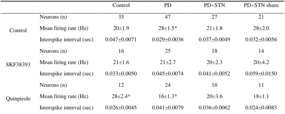
Table 1. Spontaneous activity of SNpr single units recorded from 6-OHDA lesioned rats with a kainic acid lesion of the STN and intrastriatal selective D1, D2 agonist microinjection.
The values mean ±SEM. * P < 0.05 in comparison with values from control subjects
Control PD PD+STN PD+STN sham
Neurons (n) 35 47 27 21
Mean firing rate (Hz) 20±1.9 28±1.5* 21±1.8 28±2.0 Control
Interspike interval (sec) 0.047±0.0071 0.029±0.0036 0.037±0.0049 0.032±0.0056
Neurons (n) 16 25 18 14
Mean firing rate (Hz) 21±1.6 21±2.7 20±2.3 20±4.2 SKF38393
Interspike interval (sec) 0.033±0.0050 0.045±0.0074 0.041±0.0052 0.059±0.0150
Neurons (n) 12 24 16 11
Mean firing rate (Hz) 28±2.4* 16±1.3* 20±3.6 18±1.1 Quinpirole
11
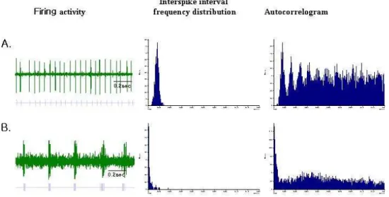
Fig. 3. SNpr discharge pattern recorded in 6-OHDA-lesioned rats. Left: Neuronal activity, each dot corresponds to a spike (Scale bar 1.4 s) raster display. Middle: Interspike interval histograms. Right: autocorrelogram. A: regular non-burst pattern, B: burst pattern
12
3. Effects of SKF38393 on the firing rates and firing patterns
The effect of the D1 agonist, SKF38393, applied by intrastriatal injection, on the firing rate of SNpr units is shown in Table 1. The administration of SKF38393 decreased SNpr neuronal firing rates in 6-OHDA lesioned rats (from 28 ± 1.5 to 21 ± 2.7 spikes/s, n = 25), but did not significantly alter the mean firing rate of SNpr neurons in the intact rat (21 ± 1.6 vs. the control at 20 ± 1.9 spikes/s, n = 16). SKF38393 did not alter the mean neuronal firing rate in the SNpr neurons of MFB+ STN lesioned rats (20 ± 2.3 vs. 21 ± 1.8 spikes/s, respectively, n = 18). In rats with 6-OHDA lesions of MFB, SKF38393 induced a decrease in the percentage of burst neurons (from 35% to 21%). Additional SKF38393 injection in STN lesioned and MFB lesioned rats increased the percentage of burst neurons (from 13% to 15%) but the change was not significant.
13
A
B
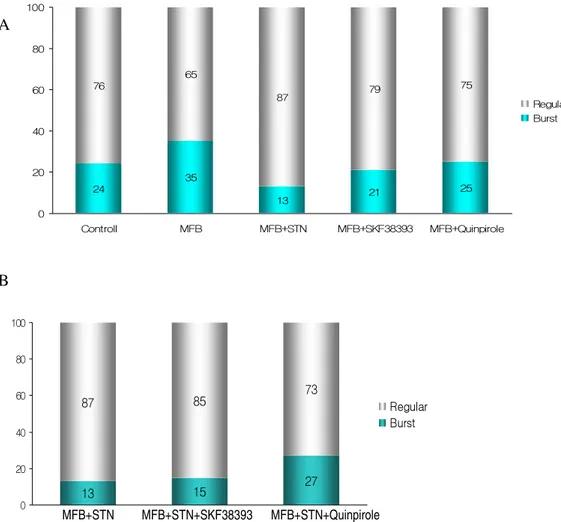
Fig. 4. The effects of D1 agonist (SKF38393) and D2 agonist (Quinpirole) on the firing pattern of SNpr neurons in rat parkinsonian models. A: The proportion of burst neurons in the SNpr of each group. B: The effects of SKF38393 and Quinpirole on the firing pattern of SNpr neurons in rat parkinsonian models after the STN lesion. 13 15 27 87 85 73 0 20 40 60 80 100 SNc+STN SNc+STN+SKF38393 SNc+STN+Quinpirole Regular Burst MFB+STN MFB+STN+SKF38393 MFB+STN+Quinpirole 24 35 21 25 76 65 87 79 75 13 0 20 40 60 80 100 Controll MFB MFB+STN MFB+SKF38393 MFB+Quinpirole Regular Burst
14
4. Effects of Quinpirole on the firing rate and firing patterns
The excitatory effect of dopamine on the neuronal activity of STN appeared to be largely mimicked by the dopamine D2-like receptor agonist. Quinpirole increased the mean firing rate of SNpr neurons in normal rats from 20 ± 1.9 to 28 ± 2.4 spikes/s (p < 0.01). However, in neurons prepared from 6-OHDA lesioned rats, Quinpirole decreased the spontaneous firing rate from 28 ± 1.5 to 16 ± 1.3 spikes/s (p < 0.01). In rats with 6-OHDA lesions of MFB, Quinpirole induced a significant change in the percentage of burst neurons (from 35% to 25%). However, Quinpirole after the additional STN lesion in rats with 6-OHDA lesions of MFB induced a significant increase in the percentage of burst neurons (from 13% to 27%).
15
IV. Discussion
1. Effects of STN lesions on the firing rates and firing patterns of SNpr in rat parkinsonian model
In accordance with predictions, our data demonstrated that SNpr neurons recorded in rat parkinsonian models also exhibited basal firing rates of SNpr neurons that were faster in MFB lesioned rats (28±1.5Hz) than those observed in SNpr of normal rats (20±1.9Hz). We observed that rat PD models had more burst neurons (35%) in SNpr than the control rats (24%). Following STN lesioning for the rat PD models, the percentage of burst neurons decreased by 13% (p<0.05). This finding suggested that STN lesion decreased the percentage of SNpr burst neurons.7
However, many reports have shown controversial results. Kreiss et al.8 reported that rat PD models had more regular neurons in STN than the control rats did. Rohlfs et al.9 reported that SNpr neuronal activity in rat parkinsonian models was significantly lower than the respective activity in the control groups, and that, in contrast, contralateral SNpr neuronal activity in rat parkinsonian models was significantly higher than the respective activity in the contralateral SNpr neuronal activity of control rats. In addition to the significant reduction in the mean firing rate, they also reported that many neurons in SNpr on the lesioned side exhibited a burst pattern. MacLeod et al.10 suggested that the continuous burst type of firing could be an indication of a compensatory physiological mechanism for the loss of spontaneous firing, and that the decreased mean firing rate of ipsilateral SNpr neurons, observed in rats within 2 weeks post-lesion, can be explained by assuming a postsynaptic inhibitory action of dopamine in the striatum. However, our data did not seem to support these suggestions. All of our rat parkinsonian models with 6-OHDA clearly demonstrated an increase in the mean firing rates in SNpr. In
16
addition, the results of our rat PD models with STN lesions further supports the theory that the mean firing rates in SNpr are reduced by the presence of STN lesions.
2. Effects of SKF38393 and Quinpirole on the firing rates and firing patterns of SNpr in rat parkinsonian model with STN lesion
The STN hyperactivity is based on the hypothesis that the loss of dopamine in the striatum reduces the activity of the inhibitory GABAergic pallido-subthalamic pathway.11,12 Our results also demonstrate that SKF38393 and Quinpirole decreased the mean firing rate in SNpr of rats with MFB lesion with 6-OHDA. However, we did not find a further decrease in the SNpr firing rate after the administration of SKF38393 or Quinpirole in additional STN lesions of MFB lesioned rats with 6-OHDA. This may suggest that D2 agonist has a stronger influence on the firing rates of SNpr because it could affect the function of STN and pedunculopontine nucleus7, the existence of interaction between indirect pathway and direct pathway, or some role for STN in the genesis or maintenance of SNpr neural activity.
Under the balanced condition between direct and indirect pathways, namely, normal control group or under the corrected state of striatopallidal disinhibition, namely, STN and MFB lesioned group, augmentation of D1 receptor hardly affected the firing rate and firing pattern of SNpr, however, augmentation of D2 receptor resulted in increasing firing rate (control group) and increasing the proportion of burst neurons (MFB + STN group). This means D1 or D2 dopamine agonist alone is not sufficient to affect the SNpr neural activity. Other reports support the consistent results.8,13 Kreiss et al. indicated that the integration of dopamine D1 and D2 receptors rather than predominant action of dopamine D2 receptors was considered to regulate STN neuronal activity.8 Calabresi et al.
17
reported that stimulation of both D1 and D2 dopamine receptors is required for the expression of long term depression at the single cell level in striatal slices.14 Other in vitro studies have shown that D1 and D2 dopamine receptors expressed in the same cell were able to interact synergistically.15,16 Gerfen et al17 have demonstrated that D1 agonists are able to modify the expression of an immediate early gene mRNA exclusively in striatal enkephalin-negative neurons and that D2 agonists have an opposite effect exclusively on enkephalin-positive neurons. The coadministration of D1 and D2 agonists produced a potentiated effect in enkephalin-negative neurons. These results also suggest that this synergistic action seen at the single cell level results from interneuronal interactions between separate populations of striatal neurons sensitive either to D1 or D2 receptor agonists. While Other reports have shown that microiontophoretically administered D1 and D2 dopamine receptor agonists do not interact synergistically at the single cell level within the striatum,18,19 suggesting that the D1–D2 receptor interaction could also involve other mechanisms. An alternative mechanism could be an interplay between the direct striatonigral and the indirect striato-pallido-subthalamo-nigral pathways at the SNr level, resulting in a greater number of responding units in the SNr when both pathways are activated by the appropriate dopamine receptor agonists. Alternatively, burst activity could originate in other structures projecting to STN, such as the sensorimotor cortex or the intralaminar thalamic nuclei.9
Recent studies have provided evidence that an irregular or burst firing pattern might correlate better than firing rate with signs of PD.20,21 In our experiment, the proportion of burst neurons in SNpr of 6-OHDA lesioned rats was significantly reduced by STN lesions. These results suggest that the interruption of the indirect pathway by a STN lesion regularizes the SNpr neuron discharge pattern in 6-OHDA lesioned rats. From recent evidence, it appears that STN participates in the genesis of the burst pattern activity of globus pallidus (GP) and SNpr neurons in
18
rats with 6-OHDA lesions, and that STN lesions can reverse this abnormal spontaneous pattern.13,22,23
19
V. Conclusions
This study demonstrated that dopamine agonists and STN lesion decreased the hyperactive firing rate of SNpr neurons in 6-OHDA lesioned rats. Concerning the firing pattern, the STN lesion, but not SKF38393 or Quinpirole, reduced the percentage of burst neurons in 6-OHDA lesioned rats. However, Quinpirole with STN lesion demonstrated a higher percentage of burst neurons than did STN lesion in 6-OHDA lesioned rats. D1–D2 receptor interactions may be considered. To clear the exact interactive mechanism of D1 and D2 agonist and the corresponding location, it should be followed a study using a nonselective dopamine agonist and D1, D2 selective antagonist.
20
References
1. DeLong MR: Primate models of movement disorders of basal ganglia origin. Trends Neurosci 1990;13:281-285.
2. Obeso JA, Olanow CW, Nutt JG: Levodopa motor complications in Parkinson's disease. Trends Neurosci 2000;23:S2-7.
3. Limousin P, Pollak P, Benazzouz A, et al: Effect of parkinsonian signs and symptoms of bilateral subthalamic nucleus stimulation. Lancet 1995;345:91-95.
4. Perier C, Marin C, Jimenez A, et al: Effect of subthalamic nucleus or entopeduncular nucleus lesion on levodopa-induced neurochemical changes within the basal ganglia and on levodopa-induced motor alterations in 6-hydroxydopamine-lesioned rats. J Neurochem 2003;86:1328-1337.
5. Hutchinson WD, Levy R, Dostrovsky JO, et al: Effects of apomorphine on globus pallidus neurons in parkinsonian patients. Ann Neurol 1997;42:767-775.
6. Hutchison WD, Allan RJ, Opitz H, et al: Neurophysiological identification of the subthalamic nucleus in surgery for Parkinson's disease. Ann Neurol 1998;44:622-628.
7. Jeon MF, Ha Y, Cho YH, et al: Effect of ipsilateral subthalamic nucleus lesioning in a rat parkinsonian model: study of behavior correlated with neuronal activity in the pedunculopontine nucleus. J Neurosurg 2003;99:762-767.
8. Kreiss DS, Mastropietro CW, Rawji SS, et al: The response of subthalamic nucleus neurons to dopamine receptor stimulation in a rodent model of Parkinson's disease. J Neurosci 1997;17:6807-6819.
9. Rohlfs A, Nikkhah G, Rosenthal C, et al: Hemispheric asymmetries in spontaneous firing characteristics of substantia nigra pars reticulata neurons
21
following a unilateral 6-hydroxydopamine lesion of the rat nigrostriatal pathway. Brain Res 1997;761:352-356.
10. MacLeod NK, Ryman A, Arbuthnott GW: Electrophysiological properties of nigrothalamic neurons after 6-hydroxydopamine lesions in the rat. Neuroscience 1990;38:447-456.
11. Albin RL, Young AB, Penney JB: The functional anatomy of basal ganglia disorders. Trends Neurosci 1989;12:366-375.
12. Miller WC, DeLong, M.R.: Altered tonic activity of neurons in the globus pallidus and subthalamic nucleus in the primate MPTP model of Parkinsonism. The basal ganglia. II. New York Plenum; Vol 32 pp 415-427, 1987
13. Murer MG, Riquelme LA, Tseng KY, et al: D1-D2 dopamine receptor interaction: an in vivo single unit electrophysiological study. Neuroreport 1997;8:783-787.
14. Calabresi P, Maj R, Mercuri NB, et al: Coactivation of D1 and D2 dopamine receptors is required for long-term synaptic depression in the striatum. Neurosci Lett 1992;142:95-99.
15. Bertorello AM, Hopfield JF, Aperia A, et al: Inhibition by dopamine of (Na(+)+K+)ATPase activity in neostriatal neurons through D1 and D2 dopamine receptor synergism. Nature 1990;347:386-388.
16. Piomelli D, Pilon C, Giros B, et al: Dopamine activation of the arachidonic acid cascade as a basis for D1/D2 receptor synergism. Nature 1991;353:164-167.
17. Gerfen CR, Keefe KA, Gauda EB: D1 and D2 dopamine receptor function in the striatum: coactivation of D1- and D2-dopamine receptors on separate populations of neurons results in potentiated immediate early gene response in D1-containing neurons.J Neurosci 1995;15:8167-8176.
22
18. Hu XT, Wang RY: Comparison of effects of D-1 and D-2 dopamine receptor agonists on neurons in the rat caudate putamen: an electrophysiological study. J Neurosci 1988;8:4340-4348.
19. Shen RY, Asdourian D, Chiodo LA: Microiontophoretic studies of the effects of D-1 and D-2 receptor agonists on type I caudate nucleus neurons: lack of synergistic interaction. Synapse 1992;11:319-329.
20. Bergman H, Wichmann T, Karmon B, et al: The primate subthalamic nucleus. II. Neuronal activity in the MPTP model of parkinsonism. J Neurophysiol 1994;72:507-520.
21. Hollerman JR, Grace AA: Subthalamic nucleus cell firing in the 6-OHDA-treated rat: basal activity and response to haloperidol. Brain Res 1992;590:291-299.
22. Burbaud P, Gross C, Benazzouz A, et al: Reduction of apomorphine-induced rotational behaviour by subthalamic lesion in 6-OHDA lesioned rats is associated with a normalization of firing rate and discharge pattern of pars reticulata neurons. Exp Brain Res 1995;105:48-58.
23. Ni ZG, Bouali-Benazzouz R, Gao DM, et al: Time-course of changes in firing rates and firing patterns of subthalamic nucleus neuronal activity after 6-OHDA-induced dopamine depletion in rats. Brain Res 2001;899:142-147.