The Protective Effects of
Epigallocatechin Gallate (EGCG)
against Ischemia/Reperfusion Injury
in the Mouse Hepatocyte
Jee Hoon Hwang
Department of Medicine
The Protective Effects of
Epigallocatechin Gallate (EGCG)
against Ischemia/Reperfusion Injury
in the Mouse Hepatocyte
Directed by Professor Dong Kyun Rah
The Master's Thesis
submitted to the Department of Medicine
the Graduate School of Yonsei University
in partial fulfillment of the requirements for the degree of
Master of Medical Science
Jee Hoon Hwang
This certifies that the Master's Thesis of
Jee Hoon Hwang is approved.
---
Thesis Supervisor : Dong Kyun Rah
---
Hoguen Kim
---
Jong-Chul Park
The Graduate School
Yonsei University
June 2008
ACKNOWLEDGEMENTS
First, I thank my supervisor Dong Kyun Rah, for his continuous support in the master program. Professor Rah was always there to listen and to give advice. He taught me how to ask my questions and express my ideas. He showed me different ways to approach a research problem and rhe need to persistent to accomplish my goal.
A special thanks goes to my co-supervisor, Hogeun Kim, who is most responsible for helping me complete the writing of this article as well as the challenging research that lies behind it. Professor Kim has been a friend and mentor. He taught me how to write academic papers, made me a better doctor, had confidence in me when I doubted myself, and brought out the good ideas in me.
Let me also say ‘thank you’ to my another co-supervisor Jong-Chul Park. Without his encouragement and constant guidance, I could not have finished this article. He always there to meet and talk about my ideas, to proofread and mark up ,my papers and chapters, and ask me good questions to help me think through my problems.
I am deeply grateful to my friend, Young Woo Cheon, for his encouraging help and bright ideas through all the years of progress. Despite his many obligations he has always found time for a prompt response to my questions and need for advice.
Last, but not least, I thank my family: my wife So Hyun Kwack, amd my sons Hyun Jun and Hyun Bin
<TABLE OF CONTENTS>
ABSTRACT……… 1
I. INTRODUCTION……… 4
II. MATERIALS AND METHODS……… 7
1. Mouse hepatocytes culture ……… 7
2. Dilution test to determine cytotoxicity of EGCG ……… 7
3. Dilution test to determine adequate amount of oxidative stress …… 8
4. EGCG treatment ……… 8
5. Oxidative stress induction ……… 9
6. Cell viability measurements by MTT assay ……… 9
7. Statistical analysis ……… 10
III. RESULTS……… 10
1. Dilution test to determine cytotoxicity of EGCG ……… 10
2. Protective effects of EGCG against H
2O
2-induced oxidative stress … 13
3. Protective effects of EGCG against
Xanhine(Xn)/Xanthine-oxidase(XO)-induced oxidative stress ……… 17
IV. DISCUSSION……… 21
V. CONCLUSION……… 23
REFERENCES……… 24
LIST OF FIGURES
Figure 1. Morphological changes of mouse hepatocytes with EGCG
treatment. ···
12
Figure 2. H
2O
2-induced cytotoxicity in mouse
hepatocytes··· 14
Figure 3. Morphological changes in mouse hepatocytes treated with
H
2O
2··· 15
Figure 4. Morphological changes in mouse hepatocytes treated with
xanthine oxidase ··· 19
LIST OF TABLES
Table 1. EGCG-induced cytotoxicity in mouse hepatocytes
··· 11
Table 2. Effect of EGCG (1 h treatment) with H
2O
2-induced
cytotoxicity of mouse hepatocytes ··· 16
Table 3. Effect of EGCG (24 h treatment) with H
2O
2-induced
cytotoxicity of mouse hepatocytes ···16
Table 4. Xanthine oxidase -induced cytotoxicity in mouse
hepatocytes ··· 18
Table 5. Effect of EGCG (1 Hour treated) with Xanthine oxidase
-induced cytotoxicity of mouse hepatocytes ···20
Table 6. Effect of EGCG (24 Hours treated) with Xanthine oxidase
Abstract
The Protective Effects of Epigallocatechin Gallate (EGCG)
against Ischemia/Reperfusion Injury in the Mouse Hepatocytes
Jee Hoon Hwang
Department of Medicine
The Graduate School, Yonsei University
(Directed by Professor Dong Kyun Rah)
Reactive oxygen species have been implicated in the pathogenesis of hepatic injury after ischemia/reperfusion (I/R). Recently, green tea polyphenol (GTP) has been found to protect the myocardium and kidney cells against I/R injury. Green tea consists mainly of polyphenols (catechins) which constitute about 40% of the dry weight of solids in brewed green tea, of which (–)-epigallocatechin gallate (EGCG) is the most abundant and the most extensively studied catechin.
Less attention, however, has been paid to the protective effects of EGCG with respect to hepatocytes. This study was designed to investigate the potential protective
roles played by EGCG against the injurious effects of reactive oxygen species in mouse hepatocytes.
Mouse hepatocytes were cultured in Dulbecco's modified Eagle's medium supplemented with 5% fetal bovine serum. Then to determine the maximal concentration of EGCG without cellular toxicity, dilution test was used. In dilution test, mouse hepatocytes were treated with 0.2, 0.4, 0.6, 0.8and 1.0 mM of EGCG. After 24 hours of EGCG treatment, the viability of the hepatocytes was assessed by MTT assay. The oxidative stress was induced by two exogenous methods: (1) H2O2
addition and (2) an enzymatic system, xanthine oxidase (XO) and its substrate xanthine (Xn, 250 µM). To determine an adequate amount of oxidative stress, different concentrations of H2O2 (20, 10, 1, 0.1 mM) were added to mouse hepatocyte.
After 24 hours of incubation, the cell viability and morphology were respectively measured. Similarly, different concentrations of xanthine oxidase (10, 1, 0.1 U/L) were added to mouse hepatocyte. After 24 hours of incubation, the cell viability and morphology were measured. In order to examine the ability of EGCG to protect the mouse hepatocytes against ROS-mediated oxidative stress, the cells were pre-incubated for 1 h and 24 h in the presence of EGCG at a final concentration of 0.2mM in the medium, which was added to the cell suspension. After the oxidative stress was induced by H2O2 or XO, the cell viability and morphology were evaluated.
in viability of mouse hepatocytes. In the microscopic observations, the morphological changes and necrotic detachment were appreciably induced by both treatments. The H2O2-induced alterations were near completely prevented by pre-incubating the
mouse hepatocyte with 0.2 mM EGCG for 1 h and 24 h (p<0.05). When the oxidative stress was induced by XO, the XO-induced alterations were also prevented by pre-incubating the mouse hepatocyte with 0.2 mM EGCG for 1 h and 24 h (p<0.05). But no significant difference was observered between 1hr EGCG treatment group and 24hr EGCG treatment group.
The results of this study suggest that EGCG can reduce hepatocyte cellular injury by preventing the oxidative stress dependent injuries on I/R and may be used in liver transplantation as an antioxidant.
--- Key words : green tea polyphenol, epigallocatechin gallate, ischemia/reperfusion injury, anti-oxidants, mouse hepatocyte
The Protective Effects of Epigallocatechin Gallate (EGCG)
against Ischemia/Reperfusion Injury in the Mouse Hepatocytes
Jee Hoon Hwang
Department of Medicine
The Graduate School, Yonsei University
(Directed by Professor Dong Kyun Rah)
I. INTRODUCTION
Liver ischemia initiates a complex and interrelated sequence of events, resulting in the injury and death of hepatocyte.1 Reperfusion, although essential for the survival of ischemic liver tissue, causes additional damage (reperfusion injury).2 Together, ischemia/reperfusion (I/R) of the liver contribute to the hepatocyte dysfunction and injury associated with hepatic failure.3,4 Although the exact mechanisms involved in the pathogenesis of ischemic/reperfusion liver injury have not been fully elucidated, it is generally believed that reactive oxygen species (ROS) and reactive nitrogen species are the key mediators of I/R-induced damage to the liver. Oxidative and nitrosative
stress cause lipid peroxidation of cell membranes, 5,6 protein and enzyme oxidation,7 and some irreversible DNA changes,8, 9 collectively leading to the loss of cell viability, either via necrotic or apoptotic pathways. One possible way of preventing ROS-mediated cellular injury is to augment the endogenous oxidative defenses by the dietary intake of antioxidants such as vitamins A, C and E. Recently, a great attention has been focused on a variety of non-vitamin antioxidants such as phenolic compounds, which may also contribute to the cellular antioxidative defense mechanisms, and which can be found in many plant species, such as green tea, fruits and vegetables.
One of the approaches to limit apoptotic or necrotic cell death in response to I/R injury may be antioxidant therapy. Some antioxidants such as vitamins C and E have been shown to have protective properties against ischemia-induced tissue damage.10, 11 Recently, considerable attention has been focused on a variety of non-vitamin antioxidants such as phenolic compounds, including quercetin, curcumin, resveratrol etc., which might contribute to liver protection.11
Hyon performed one of the first studies suggesting that non-vitamin antioxidants may offer tissue preservation; he observed that green tea polyphenol (GTP) may contribute to the physiological preservation of tissues or organs, particularly in rat pancreatic islets.12 Since then, little evidence has been accumulated showing these beneficial preservative effects of GTP compared to other effects such as
anticarcinogenic and anti-inflammatory activities. The extension of Hyon’s observation regarding the preservation of tissues for transplantation will make it possible to store tissues or organs for longer periods by a concentration-adjusted GTP treatment. Previous studies have demonstrated that GTP can reduce I/R-induced injuries in the livers, hearts, intestines and nerves of rats or mice.13,14,15,16 Only a few reports, however, have focused on its protective activities against I/R injury in mouse hepatocyte.13, 17 Green tea consists mainly of polyphenols (catechins) which constitute about 40% of the dry weight of solids in brewed green tea, of which (–)-epigallocatechin gallate (EGCG) is the most potent antioxidant and the most extensively studied catechin.18
Consequently, we sought to determine whether EGCG pretreatment would protect mouse hepatocytes from cellular damage after I/R injury. I/R hepatocytes represent a suitable and well-characterized model for studying free radical-induced oxidative stress and inflammatory response, which can contribute significantly to tissue injury and the functional damage of liver tissues. Our principal hypothesis was that the feasible protective mode of action of EGCG in vitro model of mouse hepatocyte I/R injury would be through prevention of oxidative stress dependent injuries by its anti-oxidant effects.
II. MATERIALS AND METHODS
1. Mouse hepatocytes culture
The mouse hepatocytes were purchased from Young Science (Seoul, Korea) and used between passages 5 and 10. The cells were routinely maintained in Dulbecco's modified Eagle's medium (Sigma, St. Louis, MO, USA) supplemented with 5% fetal bovine serum (Sigma) and a 1% antibiotic antimycotic solution (including 10,000 units penicillin, 10 mg streptomycin and 25 µg amphotericin B per ml, Sigma) at 37 °C in a humidified atmosphere of 5% CO2 in air.
2. Dilution test to determine cytotoxicity of EGCG
The mouse hepatocytes were treated with 0.2, 0.4, 0.6, 0.8and 1.0 mM of EGCG. (kindly supplied by PFI Inc., Kyoto, Japan) After 24 hours of EGCG treatment the viability of the hepatocytes was assessed by MTT assay.[reduction of 3-(4,5-dimethylthiazol-2-yl)-2,5-diphenyltetrazolium bromide to a purple formazan product]. Non-treated control group was used to compare viability with same time of incubation.
3. Dilution test to determine adequate amount of oxidative stress
The oxidative stress was induced by two exogenous methods: (1) H2O2 addition
and (2) an enzymatic system, xanthine oxidase (XO) and its substrate xanthine (Xn, 250 µM). To determine an adequate amount of oxidative stress, different concentrations of H2O2 (20, 10, 1, 0.1 mM) were added to mouse hepatocyte. After
24 hours of incubation, the cell viability and morphology were measured. Similarly, different concentrations of xanthine oxidase (10, 1, 0.1 U/L) were added to mouse hepatocytes. After 24 hours of incubation, the cell viability and morphology were respectively measured.
4. EGCG treatment
The EGCG compounds extracted from green tea were kindly supplied by PFI Inc., Kyoto, Japan. Its purity exceeded 90%. In order to examine the ability of EGCG to protect the mouse hepatocytes against ROS-mediated oxidative stress, the cells were pre-incubated for 1 h and 24 h in the presence of EGCG at a final concentration of 0.2mM in the medium, which was added to the cell suspension. After pre-incubation, the excess EGCG was completely removed and the medium was exchanged before
adding the oxidative stress-inducing agents. This avoided a direct reaction between the EGCG and the oxidant source in the medium.
5. Oxidative stress induction
According to the results from the dilution test of H2O2 and xanthine oxidase,
oxidative stress was induced after pre-incuation with EGCG. The oxidative stress was induced by two exogenous methods: (1) H2O2 (10, 1 mM) addition and (2) an
enzymatic system, xanthine oxidase (10, 1 U/L) and its substrate xanthine (Xn, 250 µM). Finally, the cell viability and morphology were evaluated.
6. Cell viability measurements by MTT assay
The toxic effect of ROS on mouse hepatocytes and the protective effect of EGCG were assessed by using a viability test with MTT assay. [reduction of 3-(4,5-dimethylthiazol-2-yl)-2,5-diphenyltetrazolium bromide to a purple formazan product].
7. Statistical analysis
Results are expressed as means ± standard deviation. A p value of less than 0.05 was considered statistically significant. The effects of increasing H2O2 and XO
concentrations on mouse hepatocytes viability were analyzed by Student's t-test.
III. RESULTS
1. Dilution test to determine cytotoxicity of EGCG
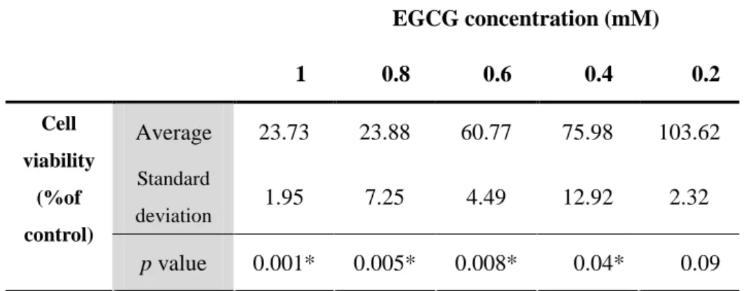
The mouse hepatocytes viability was same as control group when EGCG
concentration was 0.2 mM (table 1). The significant differences on cell viability were observed between EGCG treated group and non treated control group except when EGCG concentration was 0.2 mM . Optical microscopic examination showed a significant EGCG dose-dependent decrease in hepatocyte viability (fig. 1).
Table 1. EGCG-induced cytotoxicity in mouse hepatocytes. The cell viability was measured by MTT analysis. (* p < 0.05 vs. the non-treated at the same time, analyzed by a Student’s t-test, n = 6) EGCG concentration (mM) 1 0.8 0.6 0.4 0.2 Average 23.73 23.88 60.77 75.98 103.62 Standard deviation 1.95 7.25 4.49 12.92 2.32 Cell viability (%of control) p value 0.001* 0.005* 0.008* 0.04* 0.09
Figure 1. Morphological changes of mouse hepatocytes with EGCG treatment. Optical microscopic examination showed a significant EGCG dose-dependent morphological changes of hepatocytes. (A) Normal hepatocyte (B) 0.2mM EGCG treatment (C) 0.4mM EGCG treatment (D) 0.6mM EGCG treatment (E) 0.8mM EGCG treatment (F) 1 mM EGCG treatment
2. Protective effects of EGCG against H2O2-induced oxidative stress
To investigate the ROS-induced cytotoxic effects on the mouse hepatocytes, increasing H2O2 concentrations were added to the medium and after incubation the
cellular and morphological alterations were examined. To characterize in greater detail the overall cellular injury by the agent, a MTT assay in microplate format was performed. Incubating the cells in the presence of millimolar H2O2 concentrations
resulted in a significant (p<0.05) dose-dependent decrease in hepatocyte viability (Fig. 2 ). After 24 hours of treatment with 1 mM H2O2, an approximate 32% loss of cell
viability was observed (Fig. 2, 3).
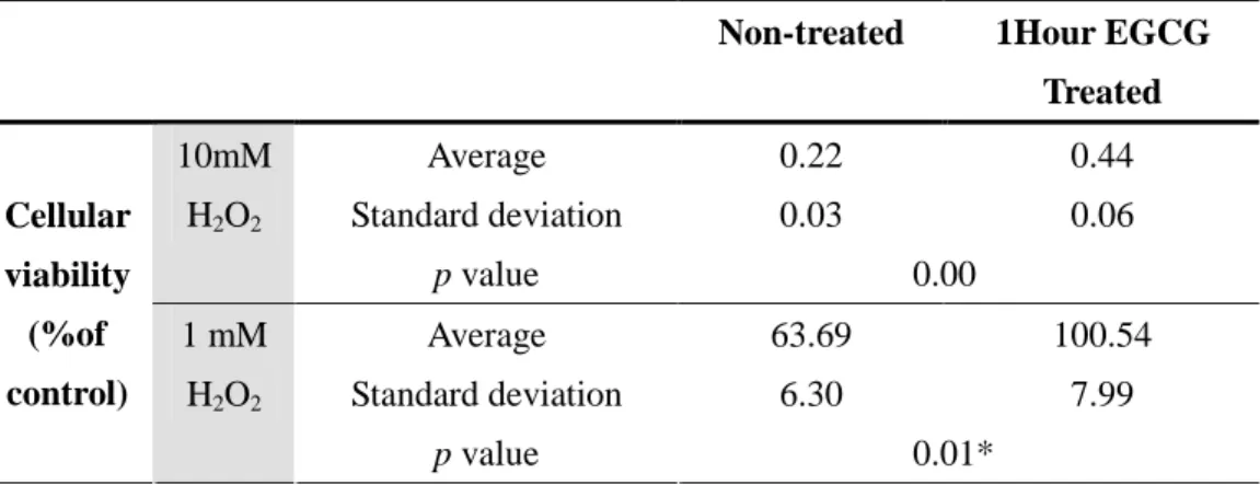
These markers were then used to verify the protective effect of EGCG against H2O2
-induced oxidative stress to the mouse hepatocytes. When the cells were pretreated with 0.2mM EGCG prior to being challenged with 1 mM H2O2, in conditions similar
to those used in the experiments mentioned above, a complete protection of cell viability was observed, suggesting that the EGCG suppresses the H2O2-induced
cytotoxicity attributed to its biological activity. Both 1hr-treated group and 24hrs-treated group had significant effect compared with the non 24hrs-treated group (Table 2 and 3). But no significant difference was observed between 1hr EGCG treatment group and 24hr EGCG treatment group .
0.00 20.00 40.00 60.00 80.00 100.00 120.00 20 10 1 0.1 H2O2 concentration (mM) H2O2 concentration (mM) H2O2 concentration (mM) H2O2 concentration (mM) % o f c o n tr o l % o f c o n tr o l % o f c o n tr o l % o f c o n tr o l 20 10 1 0.1
Figure. 2. H2O2-induced cytotoxicity in mouse hepatocytes (* p < 0.05 vs. the
non-treated at the same time, analyzed by a Student’s t-test, n = 6)
Figure. 3. Morphological changes in mouse hepatocytes treated with H2O2 . Optical
microscopic examination showed a significant H2O2 dependent morphological change
and increase of necrotic debri.
Table 2. Effect of EGCG (1 h treatment) on H2O2-induced cytotoxicity of mouse
hepatocytes. The cell viability was measured by MTT analysis. (* p < 0.05 vs. the non-treated at the same time, analyzed by a Student’s t-test, n = 6).
Non-treated 1Hour EGCG
Treated Average 0.22 0.44 Standard deviation 0.03 0.06 10mM H2O2 p value 0.00 Average 63.69 100.54 Standard deviation 6.30 7.99 Cellular viability (%of control) 1 mM H2O2 p value 0.01*
Table 3. Effect of EGCG (24h treatment) on H2O2-induced cytotoxicity of mouse
hepatocytes. The cell viability was measured by MTT analysis. (* p < 0.05 vs. the non-treated at the same time, analyzed by a Student’s t-test, n = 6).
Non-treated 24Hour EGCG
Treated Average 0.55 0.60 Standard deviation 0.06 0.17 10mM H2O2 p value 0.65 Average 73.57 97.23 Standard deviation 9.34 3.81 Cellular viability (%of control) 1 mM H2O2 p value 0.003*
3. Protective effects of EGCG against Xanhine(Xn)/Xanthine-oxidase(XO)-induced oxidative stress
The cytotoxic effects induced in the mouse hepatocytes were then investigated by another ROS-generating system, consisting of XO and its substrate Xn. This system enzymatically generates superoxide radicals and H2O2 during the conversion of Xn to
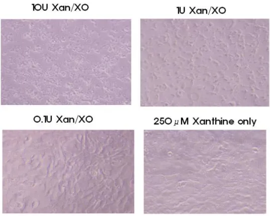
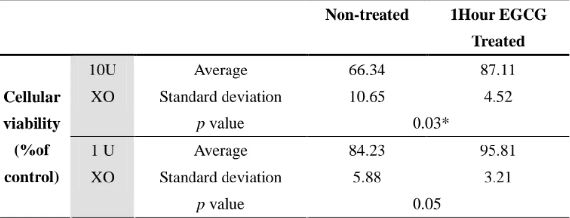
uric acid. Figure. 4 shows the effects of XO activity on mouse hepatocytes viability. When the cells were incubated with increasing XO concentrations in the presence of 250 µM Xn, a marked decrease in viability was observed. About 31% decrease in viability was observed after incubating the cells treated with 10 U/l of the enzyme (Table 4). The addition of XO or Xn alone did not affect the cell viability. Subsequently, the protective effect of EGCG against Xn/XO-induced oxidative stress in the mouse hepatocytes was also investigated under these experimental conditions. Pretreating the cells with 2mM EGCG significantly prevented the Xn/XO-induced loss of the mouse hepatocytes viability, indicating that the EGCG acted as a biological antioxidant. Both 1hr-treated group and 24hrs-treated group had significant ( p< 0.05) effect in comparision with the non treated group (Table 5 and 6). However no significant difference was observed between 1hr EGCG treatment group and 24hr EGCG treatment group.
Table 4. Xanthine oxidase -induced cytotoxicity in mouse hepatocytes. The cell viability was measured by MTT analysis. (* p < 0.05 vs. the non-treated at the same time, analyzed by a Student’s t-test, n = 6).
Xanthine oxidase concentration (mM)
10 1 0.1 Xanthine only Average 71.40 84.47 105.81 98.72 Standard deviation 2.97 0.03 0.85 7.11 Cell viability (%of control) p value 0.001* 0.003* 0.04* 0.79
Figure 4. Morphological changes in mouse hepatocytes treated with xanthine oxidase. Optical microscopic examination showed a significant xanthine oxidase dependent morphological change and increase of necrotic debri
Table 5. Effect of EGCG (1 h treatment) on Xanthine oxidase -induced cytotoxicity of mouse hepatocytes. The cell viability was measured by MTT analysis. (* p < 0.05 vs. the non-treated at the same time, analyzed by a Student’s t-test, n = 6).
Non-treated 1Hour EGCG
Treated Average 66.34 87.11 Standard deviation 10.65 4.52 10U XO p value 0.03* Average 84.23 95.81 Standard deviation 5.88 3.21 Cellular viability (%of control) 1 U XO p value 0.05
Table 6. Effect of EGCG (24 h treatment) on Xanthine oxidase cytotoxicity of mouse hepatocytes. The cell viability was measured by MTT analysis. (* p < 0.05 vs. the non-treated at the same time, analyzed by a Student’s t-test, n = 6).
Non-treated 24Hours EGCG
Treated Average 67.25 88.60 Standard deviation 10.10 3.85 10U XO p value 0.02* Average 87.62 99.69 Standard deviation 3.75 4.41 Cellular viability (%of control) 1 U XO p value 0.002*
IV. DISCUSSION
In an in vitro experimental system, the total ethanol extract from the leaves of
Chromolaena odorata showed significant antioxidant effects on hydrogen peroxide
and hypoxanthine/XO-induced damage to fibroblasts and keratinocytes, which might be related to the wound healing process .19 This prompted us to consider the influence of H2O2 and Xn/XO on the viability and morphology of mouse hepacytes. In the
present study, the injurious effects of ROS, such as H2O2 and Xn/XO, on the viability
and morphology of the mouse hepatocytes during the culture period were examined using MTT analysis and optical microscopy (Fig. 3,4,5 and Fig. 6). The simultaneous use of the MTT analysis in combination with morphological observations enables us to suggest the mechanism of action if the metabolic activity affected by the cytotoxic agents can be detected on the principle of an indirect method. It was shown that the EGCG could act as a biological antioxidant in a cell culture experimental system and protect the mouse hepatocyte from oxidative stress-induced toxicity, which might inhibit ischemic and reperfusion injury under similar pathologic conditions, such as liver transplantation. It was suggested that these results might be related to the intrinsic properties of EGCG, which passes readily through the cell membrane due to its amphipathic properties. The EGCG is easily adsorbed by lipid bilayers, extracellular matrices and various cell membrane receptors. The adsorption of EGCG to such proteins is rapid, and their desorption rates are low. Consequently, the mouse
hepatocytes could be protected from I/R-induced injuries due to the adsorption of EGCG to various membrane proteins and lipids. Moreover, it has been reported that EGCG showed excellent adsorption to the collagen in the extracellular matrix and various receptors on the cell membrane.12,20. In relation to these results, EGCG have already been shown to be significantly effective for protecting rat calvarial osteoblasts from ROS (e.g. H2O2 and Xn/XO)-induced oxidative stress.
21
Previous studies on polyphenolic compounds have demonstrated that polyphenols are potent antioxidant, antiinflammatory and antiproliferative agents, which is thought to have chemopreventive properties with respect to carcinogenesis.
This study was not intended to investigate the in vivo effects of the EGCG by means of oral administration or subcutaneous injection but to examine the protective effects of EGCG against exogenous ROS-induced oxidative stress in mouse hepatocytes as an in vitro experimental model. This will help the future use of EGCG as a commercial product. Although the barrier effect of EGCG treatment could not be fully explained, these observations might have important implications for the successful protection of hepatocytes under surgical conditions.
V. CONCLUSION
In vitro experimental results oxidative stress was induced in cultured hepatocyte, either by adding 1, 10mM H2O2 or by the action of 1, 10 U/l xanthine oxidase (XO) in
the presence of xanthine (250 µM). Both treatments produced a significant reduction in hepatocyte viability.
The H2O2 -induced alterations were completely prevented by pre-incubating the
Hepatocyte with 0.2mM EGCG for 1 h. When the oxidative stress was induced by XO, the cell viability and morphology were also significantly maintained at the 0.2mM EGCG. These results demonstrate that EGCG can act as a biological antioxidant in a cell culture experimental model and prevent oxidative stress-induced cytotoxicity in hepatocytes.
REFERENCES
1.Massip-Salcedo M, Roselló-Catafau J, Prieto J, Avíla MA, Peralta C. The response of the hepatocyte to ischemia. Liver international 2007;27(1):6-16.
2.Luedde T, Trautwein C. Intracellular survival pathways in the liver. Liver international 2006;26(10):1163-74.
3.Zhang W, Wang M, Xie HY, Zhou L, Meng XQ, Shi J, et al. Role of reactive oxygen species in mediating hepatic ischemia-reperfusion injury and its therapeutic applications in liver transplantation. Transplantation proceedings 2007;39(5):1332-7. 4.Mizunuma K, Ohdan H, Tashiro H, Fudaba Y, Ito H, Asahara T. Prevention of ischemia-reperfusion-induced hepatic microcirculatory disruption by inhibiting stellate cell contraction using rock inhibitor. Transplantation 2003;75(5):579-86. 5.Lloberas N, Torras J, Herrero-Fresneda I, Cruzado JM, Riera M, Hurtado I, et al. Postischemic renal oxidative stress induces inflammatory response through PAF and oxidized phospholipids. Prevention by antioxidant treatment. The FASEB journal 2002;16(8):908-10.
6.Fukai M, Hayashi T, Yokota R, Shimamura T, Suzuki T, Taniguchi M, et al. Lipid peroxidation during ischemia depends on ischemia time in warm ischemia and reperfusion of rat liver. Free radical biology & medicine 2005;38(10):1372-81. 7.Naskalski J, Bartosz G. Oxidative modifications of protein structures. Adv Clin Chem 2000;35:161-253.
8.Elliott RM, Astley SB, Southon S, Archer DB. Measurement of cellular repair activities for oxidative DNA damage. Free radical biology & medicine 2000;28(9):1438-46.
9.Barzilai A, Yamamoto K. DNA damage responses to oxidative stress. DNA repair 2004;3(8-9):1109-15.
10.Seo MY, Lee SM. Protective effect of low dose of ascorbic acid on hepatobiliary function in hepatic ischemia/reperfusion in rats. Journal of Hepatology 2002;36(1):72-7.
11.Bustos M, Beraza N, Lasarte JJ, Baixeras E, Alzuguren P, Bordet T, et al. Protection against liver damage by cardiotrophin-1: a hepatocyte survival factor up-regulated in the regenerating liver in rats. Gastroenterology 2003;125(1):192-201. 12.Hyon SH, Kim DH. Long-term preservation of rat pancreatic islets under physiological conditions. Journal of biotechnology 2001;85(3):241-6.
13.Fiorini RN, Donovan JL, Rodwell D, Evans Z, Cheng G, May HD, et al. Short-term administration of (-)-epigallocatechin gallate reduces hepatic steatosis and protects against warm hepatic ischemia/reperfusion injury in steatotic mice. Liver transplantation 2005;11(3):298-308.
14.Aneja R, Hake PW, Burroughs TJ, Denenberg AG, Wong HR, Zingarelli B. Epigallocatechin, a green tea polyphenol, attenuates myocardial ischemia reperfusion injury in rats. Molecular medicine 2004;10(1-6):55-62.
15.Muià C, Mazzon E, Di Paola R, Genovese T, Menegazzi M, Caputi AP, et al. Green tea polyphenol extract attenuates ischemia/reperfusion injury of the gut. Naunyn-Schmiedeberg's archives of pharmacology 2005;371(5):364-74.
16.Ikeguchi R, Kakinoki R, Matsumoto T, Hyon SH, Nakamura T. Peripheral nerve allografts stored in green tea polyphenol solution. Transplantation 2005;79(6):688-95. 17.Zhong Z, Froh M, Connor HD, Li X, Conzelmann LO, Mason RP, et al. Prevention of hepatic ischemia-reperfusion injury by green tea extract. American journal of physiology Gastrointestinal and liver physiology 2002;283(4):G957-64. 18.Valcic S, Burr JA, Timmermann BN, Liebler DC. Antioxidant chemistry of green tea catechins. New oxidation products of epigallocatechin gallate and (-)-epigallocatechin from their reactions with peroxyl radicals. Chemical research in toxicology 2000;13(9):801-10.
19.Thang PT, Patrick S, Teik LS, Yung CS. Anti-oxidant effects of the extracts from the leaves of Chromolaena odorata on human dermal fibroblasts and epidermal keratinocytes against hydrogen peroxide and hypoxanthine-xanthine oxidase induced damage. Burns 2001;27(4):319-27.
20.Han DW, Park YH, Kim JK, Lee KY, Hyon SH, Suh H, et al. Effects of green tea polyphenol on preservation of human saphenous vein. Journal of biotechnology 2004;110(2):109-17.
of green tea polyphenol against reactive oxygen species-induced oxidative stress in cultured rat calvarial osteoblast. Cell biology and toxicology 2003;19(5):325-37.
< ABSTRACT(IN KOREAN)>
백서
백서
백서
백서 간장세포의
간장세포의
간장세포의 허혈
간장세포의
허혈
허혈
허혈////재관류
재관류
재관류 손상에
재관류
손상에
손상에 대한
손상에
대한
대한
대한
epigallocatechin gallate (EGCG)
epigallocatechin gallate (EGCG)
epigallocatechin gallate (EGCG)
epigallocatechin gallate (EGCG) 의
의
의 보호
의
보호
보호
보호 효과
효과
효과
효과
<지도교수 나 동 균> 연세대학교 대학원 의학과
황 지 훈
활성화 산소는 허혈/재관류 손상에 있어서 주된 병인으로 알려져 있다. 최근 들어 녹차 (green tea) polyphenol 이 신장과 심장 조직에서 허혈/재
관류 손상을 방지할 수 있음이 밝혀졌다. 녹차는 주로
polyphenol(catechin) 으로 구성되어 있으며 이는 녹차 건조 중량의 약
40%에 해당한다. 이런 polyphenol 들 중에
epigallocatechin gallate
(EGCG) 는 가장 강력한 항산화 작용을 하며 가장 많은 연구가 된 물질이 다.
하지만 간세포에 대해서는 이러한 EGCG의 보호 작용에 대한 연구가 활
발하지 못하다. 본 연구에서는 백서의 간장세포에 대해 활성화 산소가 줄 수 있는 손상으로부터 EGCG 가 보호작용이 있는 지를 알아보고자 한다.
modified Eagle's medium 에서 배양한다. 그 후 세포독성이 없는 EGCG 의 최대농도를 희석 실험 방법을 통해 밝혀낸다. 희석 실험 방법에는 0.2,
0.4, 0.6, 0.8 and 1.0 mM of EGCG 를 이용하였다. EGCG를 24시간 처리 한후 MTT assay 를 이용하여 세포의 생장능(viability)를 측정하였다. 산
화적 스트레스는 두가지 방법을 이용하여 유발하였다. (1) H2O2의 첨가 (2)
250 µM 의 xanthine 과 xanthine oxidase 를 처리하였다. 산화적 스트레
스의 적절한 정도를 알아보기 위하여 20mM, 10mM, 1mM, 0.1mM 의
H2O2 로 각각 24 시간 처치 후 백서 간장세포의 현미경적 검사 및 생장능
을 확인하였다. 이와 마찬가지로 10U/L, 1U/L, 0.1 U/L 의 xanthine
oxidase 를 250 µM 의 xanthine 존재하여 각각 처치하였다. 역시 24시간 후에 현미경적 검사 및 생장능을 확인하였다. EGCG가 산화적 스트레스로 부터 백서 간장세포를 보호하는 작용을 규명하기 위하여 0.2mM 의 EGCG로 1시간과 24시간동안 각각 처리하였다. 그 후 H2O2와 xanthine oxidase 로 산화적 스트레스를 유발하고 EGCG를 전 처치 하지 않은 세 포들과 생장능을 비교하였다. H2O2와 xanthine 은 백서의 간장세포에서 산화적 스트레스에 의한 뚜렷 한 생장능의 저하를 보여주었다. 현미경적 검사에서도 두 산화제에 의한 세포의 괴사와 형태변화가 뚜렷이 관찰되었다. H2O2 에 의한 산화적 스트 레스는 0.2mM 의 EGCG를 1시간 또는 24시간 전처치함으로서 대부분 방 지될 수 있었다. 또한 xanthine oxidase에 의한 산화적 스트레스 역시
EGCG의 전처치를 통해서 백서 간장세포의 생존능을 유지 할 수 있었다. 그러나 두 산화적 스트레스 모두에서 1시간 EGCG 전처치군과 24시간 EGCG 전처치 군에서 세포 보호 효과의 차이는 나타나지 않았다. 본 실험의 결과로 EGCG의 백서 간장세포를 산화적 스트레스로부터 보 호하는 작용이 명백해졌으며 이는 추후 생체 간이식시에 항산화제로서 EGCG를 사용할 수 있는 가능성을 제시하고 있다. --- 핵심되는 말 : 녹차 polyphenol, EGCG, 허혈/재관류 손상, 항산화제, 백서 간장세 포