Oral fibromatosis : a case report and clinico-pathologic considerations for radiolucent lesion in the mandible
55 Case Presentation
A 17-year-old boy was referred to the department of oral and maxillofacial surgery, Yonsei university dental hospital, Seoul, Korea, with facial swelling on his left face. The patient had a history of feeling swelling 10 days ago. With the symptom considered to be of the third molar origin by a local oral surgeon, the boy had been given antibiotics, then signs were slightly improved. After the medication, the dentist found a radiolucent lesion in a radiographic evaluation. The patient’s med-ical history was noncontributory, but had a history of direct trauma on the left mandibuar area by a bicycle, two years ago. He was not taking any medication and had no known drug allergies.
In extraoral examination, there was mild swelling of the cheek and in the region over the mandibular angle without tenderness to palpation. In intraoral examina-tion, there was no remarkable swelling or any sign of infection.
A panoramic radiograph showed a large multilocular radiolucent lesion extending from the root of the lower
left canine to the condylar neck. The inferior border of the mandible was intact (Fig. 1). A computed tomogra-phy (CT) scan was taken to assess the buccolingual extent of the lesion. On axial and coronal sections, there were a low attenuated soft tissue mass showing a pres-sure bony erosion of the mandible (Fig. 2, 3). In addition, thickening of the masseter, buccinator, and medial and lateral pterygoid muscles which lying adjacent to the lesion were noticed.
Differential diagnosis
The patient’s radiographic view showed a large radi-olucent lesion, which led the mandible to erosive bony resorption. Tumors and tumor-like conditions of the mandible in young adolescents can attribute to several lesions of various origins which could affect this region. They can be classified as neoplastic (benign or malig-nant), traumatic, metabolic, infectious, or developmental in origin. The absence of infection sign in this case would exclude the infectious origin.
Taking all the above considerations into account, the differential diagnosis should include the following enti-ties in order:
1. Traumatic bone cyst. The traumatic or simple bone cyst is a lesion with an uncertain pathogenesis. Most traumatic bone cysts of the jaws are encountered in patients between 10 and 20 years of age. They are * Corresponding author
Hyung Jun Kim
Dept. of OMFS, Yonsei University College of Dentistry 134 Shinchon-Dong, Seodaemoon-Gu, Seoul, 120-752, Korea Tel: 82-2-2228-3132
E-mail: kimoms@yumc.yonsei.ac.kr
Oral fibromatosis : a case report and clinico-pathologic
considerations for radiolucent lesion in the mandible
Seong-Hoe Yoon*, Woong Nam*,**, Wonse Park*, Hyung Jun Kim*,**,***
*Department of Oral and Maxillofacial Surgery, **Oral Science Research Center ***Oral Cancer Research Institute, Yonsei University College of Dentistry, Seoul, Korea
The fibromatoses are a broad group of fibrous proliferations. They have a biologic behavior and histopathologic pattern that is inter-mediate between those of benign fibrous lesions and fibrosarcoma. Because they are not common diseases, it is important to differenti-ate the fibromatoses from other similar diseases. In this report, we present a case of fibromatosis, and possible other diseases to be dif-ferentiated, with a review of literatures.
J. Kor. Oral Maxillofac. Surg. Vol. 33 No. 1, 2007
56
essentially restricted to the mandible, although there have been reports of the lesion in the maxilla. Bilateral simple bone cysts of the mandible are occasionally encountered. About 60% of the cases occur in males. Traumatic bone cysts are usually asymptomatic, how-ever, about 20% of the cases shows a painless swelling. Radiographically when several teeth are involved in the lesion, the radiolucent defect often shows dome-like projections that scallop upward between the roots. Teeth that appear to be involved in the lesion are generally vital and do not show root resorption.
2. Aneurysmal bone cyst (ABC). The lesion is uncom-mon in the jaws. ABCs are most comuncom-monly seen in the shaft of a long bone or in the vetebral column in the patients younger than age 30. The mandible is more commonly involved than the maxilla, and the molar region is the most frequent site of involvement. Pain is
a common feature, and patients often have a rapidly developing facial swelling. Radiographs show an unilocular or multilocular radiolucency with cortical expansion and thinning.
3. Neurofibroma. The neurofibroma is the most common type of peripheral nerve neoplasm. It arises from a mixture of cell types, including Schwann cells and perineural fibroblasts. Solitary tumors are most com-mon in young adults and present as slow-growing, soft, painless lesions that vary in size from small nod-ules to larger masses. The skin is the most frequent location for neurofibromas, but lesions of the oral cav-ity are not uncommon. On rare occasions, the tumor can arise centrally within the bone, where it may pro-duce a well-demarcated or poorly defined unilocular or multilocular radiolucency.
4. Fibromatosis. Soft tissue fibromatosis of the head and neck presents as a firm, painless mass, which may be
Fig. 2.Axial computed tomography scan showing a erosive lesion at the mandibular body.
Fig. 3.Coronal computed tomography scan showing a erosive lesion at the ramal area of the mandible. Fig. 1.Panoramic radiograph showing a large radiolucent lesion
Oral fibromatosis : a case report and clinico-pathologic considerations for radiolucent lesion in the mandible
57 either rapid or insidious in growth. The lesion usually
occurs in children or in young adults. The most com-mon oral site is the paramandibular soft tissue region, although the lesion can occur almost anywhere. The tumor can grow to considerable size, resulting in sig-nificant facial disfigurement. Destruction of adjacent bone may be observed on radiographs.
5. Stafne defect. This condition represents a focal concav-ity of the cortical bone on the lingual surface of the mandible. In most cases, biopsy has revealed histolog-ically normal salivary gland tissue, suggesting that these lesions represent developmental defects. However, a few of these defects contain muscle, fibrous connective tissue, blood vessels, fat, or lym-phoid tissue. They present as an asymptomatic radi-olucency below the mandibular canal in the posterior mandible, between the molar teeth and the angle of the mandible. Male predilection is observed. In few cases, the lesion has increased in size over time. 6. Fibrosarcoma of bone. Fibrosarcoma of bone is a
malignant fibroblastic tumor that shows varying degrees of collagen production without formation of tumor bone, osteoid, or cartilage in the primary tumor or in any metastatic site. They may arise in the medullary portion of a bone or in a periosteal location. Fibrosacormas of bone may be encountered in patients over a wide age range (The average age is 40 years). About 15% of cases occur in the craniofacial bones, and the mandible is the predominant site. Radiographically, fibrosarcomas of bone present as lytic, destructive lesions. In patients with relatively slow-growing tumors, the radiolucent area may be fairly well defined, suggesting a benign process. 7. Desmoplastic fibroma. The desmoplastic fibroma of
bone is a rare tumor that appears to be the osseous counterpart of soft tissue fibromatosis. The mandible is the fourth most commonly affected bone. Most examples of desmoplastic fibroma of bone are discov-ered in patients younger than 30 years of age. There is no sex predilection. Of the reported cases involving the jaws, 90% have occurred in the mandible, most often in the molar-angle-ascending ramus area. A painless swelling of the affected area is the most com-mon initial complaint. Radiographically, the lesion presents as a unilocular or multilocular radiolucent area. If the lesion erodes through the cortex, an accom-panying soft tissue mass will be present.
8. Ossifying fibroma. The ossifying fibroma is a
well-demarcated and occasionally encapsulated neoplasm composed of fibrous tissue that contains varying amount of calcified tissue resembling bone, cemen-tum, or both. They may occur over a wide range, but the greatest number of cases are encountered during third and fourth decades of life. There is a definite female predilection. It is much more common in the mandible, most often found in the premolar and molar region. Large tumors may cause a painless swelling, and paresthesia is rarely seen. Radiogra-phycally, the lesion is oftenly well defined and uniloc-ular. But some examples show a sclerotic border. Depending on the amount of calcified material pro-duced in the tumor, it may appear completely radiolu-cent; more oftenly it shows varying degrees of radiopacity. Large lesions often cause expansion of the inferior cortex of the mandible.
Subsequent Course
The tumor was excised via an intraoral and submandi-bular approach under general anesthesia.
Pathologic Diagnosis
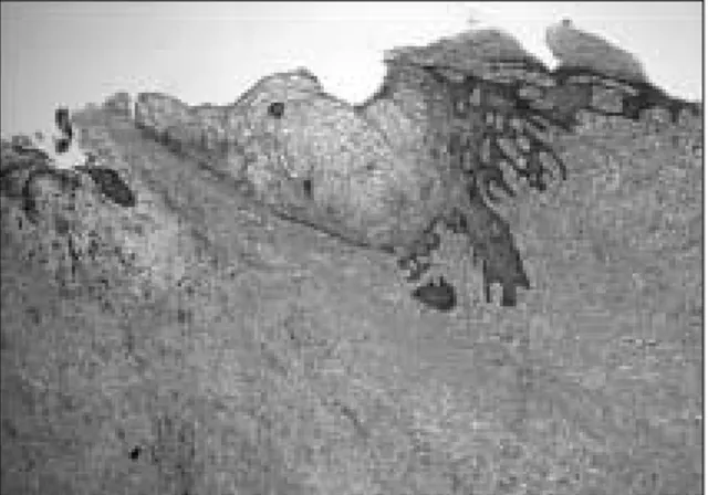
Pathologic report was ‘consistent with fibromatosis’. (Fig. 4).
Discussion
The fibromatoses are a broad group of fibrous prolifer-ations. The frequency with which fibromatoses involve
Fig. 4.Histopathological examination showing dense fibrosis in the subepithelial connective tissue (hema-toxylin-eosin, original magnification, × 40).
J. Kor. Oral Maxillofac. Surg. Vol. 33 No. 1, 2007
58
the head and neck is difficult to assess because of the pecularities of claasification used by different authors1).
They have a biologic behavior and histopathologic pat-tern that is intermediate between those of benign fibrous lesions and fibrosarcoma. A number of different forms of fibromatoses are recognized throughout the body, and they often are named on the basis of their particular clin-icopathologic features. In the soft tissue of the head and neck, these lesions are frequently called a juvenile aggressive fibromatosis or extra-abdominal desmoids. Similar lesions within the bone have been called desmo-plastic fibromas2).
The etiology of fibromatoses is still unclear. Some
explanations have been suggested like hormonal cause3),
viral theory, chromosomal alterations4), and abnormal
expression of the c-sys oncogene and of platelet-derived growth factor(PDGF)5).
Fibromatoses are uncommon soft tissue mass lesion that can occur in all anatomic sites and accountings for 6.9% of soft tissue tumors6), and among these, only 5% of
all fibromatoses are found in the head and neck region. Pathologically soft tissue fibromatoses are characterized by a cellular proliferation of spindle-shaped cells that are arranged in streaming fascicles and are associated with a variable amount of collagen. The lesion usually is poorly circumscribed and infiltrates the adjacent tissues. Hyperchromatism and pleomorphism of the cells should not be observed2).
The correct treatment is excision (radical and compart-mental, if possible) and continuous follow-up7). Difficulty
with complete excision is reflectd in the relatively high recurrence rates of fibromatoses excised from the head
and neck region. Fasching et al8) showed that
fibro-matoses recurred at a very high rate when margins were positive but also frequently when margins were consid-ered negative. Without complete excision, 12 of 14 tumors (25%) recurred. However, some literatures con-tain occassional reports of spontaneous regression and descriptions of arrested growth after incomplete resec-tion without subsequent therapy9). Siegel et al10)
suggest-ed a guideline for therapy. First, meticulous surgical excision is essential. Second, if tumor margins are known to be positive, postoperative adjunctive therapy should be considered. Finally, close follow-up is essential.
If the condition is inoperable11)or tumor margins are
known to be positive, postoperative adjunctive therapy, such as radiation therapy, should be considered. However, the possible benefits of radiation therapy need to be weighed against its potential for long- and short-term complications. Radiation therapy should be used with caution in the pediatric population because of the well-known effects of radiation on bone growth and the potential for the delayed development of a cancerous lesion10).
If radiotherapy is contraindicated, chemotherapy is an alternative. Several pharmacologic interventions such as progesterone12,13), antiestrogens, nonsteroidal
anti-inflam-matory drugs, warfarin, vitamin K, and ascorbiate14). The
use of tamoxifen or testolactone is also suggested15).
REFERENCES
1. Batsakis JG: Tumors of the head and neck (ed 2). Williams & Wilkins, 1979.
2. BW Neville et al: Oral and maxillofacial pathology (ed 1). W.B.Saunders, 1995, p 369.
3. Mackenzie DH: The fibromatoses: a clinicopathological concept. BMJ 1972; IV: 277-281.
4. Brige JA, Sreekantaiah C, Mouron B, Neff JR, Sandberg AA, Wolman SR: Clonal chromosomal abnormalities in desmoid tumors. Implications for histopathogenesis. Cancer 1992;69:430-436.
5. Palman C, Bowen-Pope DF, Broofs JJ: Platelet-derived growthfactor receptors (beta-subunit) immunoreactivity in soft tissue tumors. Lab Ivest 1992;66:108-115.
6. Krandorf MJ: Benign soft tissue tumors in a alrge referral population: distribution of specific diagnoses by age, sex, and location. AJR 1995;164:395-402.
7. Santis DD: Fibromatosis of the mandible: case report and review of previous publications. B J Oral and Maxillofac Surg 1998; 36: 384-388.
8. Fasching MC, Slaeh J, Woods JE: Desmoid tumors of the head and neck. Am J Sug 1988;156:327-31.
9. Posner MC, Shiu MH, Newsome JL, et al. The desmoid tu-mor not a benign disease. Arch Surg 1989;124:191-6. 10. Siegel NS, Braford CR: Fibromatosis of the head and neck:
A challenging lesion. Otolaryngol Head Neck Surg 2000;123:269-75.
11. Schmitt G, Mills EE, Levin V, Smit BJ, Borcker H, Pape H: Radiotherapy of aggressive fibromatosis. Eur J Cancer 1922;28A:832-835.
12. Lipschultz A, Maas M: Progesterone treatment of uterine and other abdominal fibroids induced in guinea pigs by al-pha estradiol. Cancer Res 1944;4:18-23.
13. Lanari A: Effect of progesterone on desmoid tumors. N Engl med 1983;309:123-32.
14. Waddell WR, Kirsch WM: Testolactone, sulindac, warfarin, and vitamin K1 for unresectable desmoid tumors. Am J Surg 1991;161:416-21.
15. Sciarra F: Anti-estrogens and aromatase inhibitors: tamox-ifen and testolactone. J Endocrinol Invest 1988;11:755-62.