INTRODUCTION
Mycobacterium kansasii is a slow-growing acid-fast bacillius
(AFB) and belongs to the group of environmental mycobac-teria, also known as atypical mycobacteria or nontuberculo-sis mycobacteria (NTM). Local water supplies are consid-ered as the major reservoir for the M. kansasii, and evidence of person-to-person transmission has not been reported. The most common presentation of M. kansasii infection is a chr-onic bronchopulmonary disease, which manifests typically in adult patients with chronic obstructive pulmonary disease or cystic fibrosis. In addition, M. kansasii can cause skeletal infections, skin and soft tissue infection, cervical or other lymphadenitis, and disseminated infection (1).
Disseminated infection by M. kansasii occurs almost exclu-sively in immunocompromised patients, such as solid organ transplant recipients, HIV-infected individuals, patients with hematologic malignancy, or patients receiving long-term steroid regimens (2). In the case of disseminated M. kansasii infection, involvement of multiple organs including the lungs, liver, spleen, bone marrow, lymph node (LN), bowels, cen-tral nervous system, pericardium, pleura or kidneys, has been reported (3) but disseminated M. kansasii infection associat-ed with skin involvement is not frequent (4).
Recently, we encountered a rare case of disseminated M.
kansasii infection involving multiple skin areas together with
lung and multiple LNs. To our knowledge, this is the first case of disseminated M. kansasii infection that has involved the skin in Korea. Therefore, we report this unusual case with a comprehensive review of previously reported disseminated
M. kansasii infections in non HIV-infected patients.
CASE REPORT
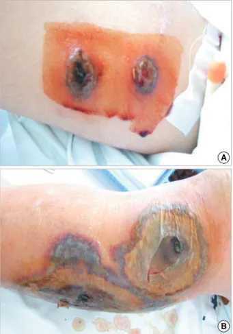
A 48-yr-old man was admitted with a 1-month history of fever and a 2-week history of dyspnea on exertion at Sever-ance Hospital in Seoul, Korea. He had a history of myelodys-plastic syndrome (MDS) diagnosed 21 months ago prior to admission and had been treated with oral glucocorticoid (pred-nisolone, 10 mg daily) with regular follow-up. A year after MDS was diagnosed, multiple erythematous tender nodules developed on both lower legs, and a skin biopsy of the calf revealed Sweet’s syndrome. He continuously had these skin lesions without complete resolution until admission. On admis-sion, several papulonodular skin lesions on his arms, chest, back, abdomen, buttocks, and legs were noted (Fig. 1). Mul-tiple LNs were palpated on the medial side of the right thigh 304
Sang Hoon Han, Kyoung Min Kim, Bum Sik Chin, Suk Hoon Choi, Han Sung Lee, Myung Soo Kim, Su Jin Jeong, Hee Kyoung Choi, Chang Oh Kim, Jun Yong Choi, Young Goo Song, and June Myung Kim Department of Internal Medicine and AIDS Research Institute, Yonsei University College of Medicine, Seoul, Korea
Address for Correspondence Jun Yong Choi, M.D.
Department of Internal Medicine, Yonsei University College of Medicine, 250 Seongsan-ro, Seodaemun-gu, Seoul 120-752, Korea Tel : +82.2-2228-1975, Fax : +82.2-393-6884 E-mail : [email protected]
Disseminated
Mycobacterium kansasii
Infection Associated with Skin
Lesions: A Case Report and Comprehensive Review of the Literature
Mycobacteruim kansasii occasionally causes disseminated infection with poorout-come in immunocompromised patients. We report the first case of disseminated M. kansasii infection associated with multiple skin lesions in a 48-yr-old male with myelodysplastic syndrome. The patient continuously had taken glucocorticoid dur-ing 21 months and had multiple skin lesions developed before 9 months without complete resolution until admission. Skin and mediastinoscopic paratracheal lymph node (LN) biopsies showed necrotizing granuloma with many acid-fast bacilli. M. kansasii was cultured from skin, sputum, and paratracheal LNs. The patient had been treated successfully with isoniazid, rifampin, ethmabutol, and clarithromycin, but died due to small bowel obstruction. Our case emphasizes that chronic skin lesions can lead to severe, disseminated M. kansasii infection in an immunocom-promised patient. All available cases of disseminated M. kansasii infection in non HIV-infected patients reported since 1953 are comprehensively reviewed. Key Words : Mycobacterium kansasii; Myelodysplastic Syndromes; Skin; Disseminated Infection
Received : 26 March 2008 Accepted : 19 September 2008
ⓒ 2010 The Korean Academy of Medical Sciences.
This is an Open Access article distributed under the terms of the Creative Commons Attribution Non-Commercial License (http://creativecommons.org/licenses/by-nc/3.0) which permits unrestricted non-commercial use, distribution, and reproduction in any medium, provided the original work is properly cited.
and left cervical area. Initial laboratory tests showed leukope-nia with a white blood cell count of 1,950/mL; severe anemia with a Hb level of 6.8 g/dL; mild thrombocytopenia with a platelet count of 113,000/mL; an elevated ESR (73 mm/hr) and C-reactive protein level (10.8 mg/dL). Chest computer tomography (CT) confirmed multiple LNs enlargement at the mediastium, paratracheal area, subcarina and right peri-hilar bronchovascular interstitial and interlobular septal thick-ening. Initially, sputum AFB smears revealed a negative find-ing. Meanwhile, both excisional LN biopsies, which were performed at the palpable LNs of the thigh and neck, and skin and mediastinoscopic paratracheal LN biopsies revealed necrotizing granuloma with many AFB. Also, an AFB smear of a pus-like discharge obtained from the paratracheal LN revealed a positive finding.
With a presumptive diagnosis of disseminated tuberculo-sis, anti-tuberculosis therapy was started with HERZ (iso-niazid[INH], rifampin [RFP], ethambutol [EMB], and pyra-zinamide[PZA]) regimens on hospital day (HD) 16. How-ever, as the skin lesions progressed rapidly and high spiking fever persisted despite HERZ treatment, we assumed he had a rapidly growing NTM such as M. Abscessus or M. fortuitum, and started him on amikacin, clarithromycin, levofloxacin and cefoxitin instead of EMB and PZA. However,
improve-ment of the skin lesions was not evident.
Three weeks after anti-tuberculosis therapy was started, a mycobacterium culture of skin and pus-like discharge obtain-ed from the paratracheal LN revealobtain-ed NTM, and repeatobtain-ed sputum mycobacterium culture also revealed NTM. At HD 43, all NTMs cultured in sputum, paratracheal LNs, and skin were identified as M. kansasii by polymerase chain reac-tion-restriction fragment length polymorphism (PCR-RFLP) of the polymorphic region of the rpoB gene. In vitro drug
Age (N=58), mean±SD 45.0±22.3 yr Sex (N=59), M:F 47 (79.7%):12 (20.3%) Underlying diseases/associated conditions (N=63)
No (previously healthy) 15 patients (23.8%) Yes (multiple, N=83) 48 patients (76.2%) Hematologic malignancy 30 (36.1%) Steroid use 19 (22.9%) Immunosuppressive agents use 8 (9.6%) Post-splenectomy 7 (8.4%)
Diabetes 4 (4.8%)
Primary immunodeficiency 3 (3.6%) Solid cancer 3 (3.6%) Autoimmiune disease 2 (2.4%) Solid organ transplantation 2 (2.4%)
Pregnancy 2 (2.4%)
Hemodialysis 1 (1.2%) Neutropenia 1 (1.2%) Renal failure 1 (1.2%) Infection site (multiple, N=208)
Lung 37 (17.8%) Lymph nodes 32 (15.4 %) Spleen 27 (13.0%) Liver 25 (12.0%) Bone marrow 20 (9.6%) Skin 12 (5.8%)
Bone and joint 9 (4.3%)
G-I tract 8 (3.8%)
Urinary tract 7 (3.4%) Cerebrospinal fluid 6 (2.9%) Male reproductive organ 6 (2.9%) Pleural fluid 5 (2.4%) Peritoneal fluid 4 (1.9%)
Abscess 2 (1.0%)
Blood, cellulitis, blood vessels, nose, tongue, 1 (Respectively, pericardium, adrenal gland, thymus 0.5%) Reported year (N=63) 1950-1959 4 (6.3%) 1960-1969 9 (14.3%) 1970-1979 17 (27.0%) 1980-1989 15 (23.8%) 1990-1999 11 (17.5%) 2000-2007 7 (11.1%) Outcome (N=63) Recovered 19 (30.2%) Not recovered 38 (60.3%) Unknown 6 (9.5%)
Table 1. Clinical characteristics of disseminated M. kansasii in-fection in non-HIV infected patients
Fig. 1. Papulonodular skin lesions on lower legs.
A
susceptibility testing of M. kansasii showed that the isolate was susceptible to RFP, EMB, PZA, streptomycin, moxiflo-xacin, and cycloserine but resistant to INH and para-aminos-alicylic acid. At HD 43, we altered the anti-mycobacterial treatment regimens to INH, RFP, EMB, and clarithromycin. Gradual improvement of the general condition and symp-toms with regression of skin lesions was noted. Sputum AFB, which was examined at HD 51, was converted into negative and mycobacterial culture of sputum did not identify any mycobacteria. However, during treatment for M. kansasii, the patient developed small bowel obstruction and ischemic colitis. Although he underwent a small bowel resection and intensive conservative management, the patient died on HD 121.
DISCUSSION
M. kansasii is the second most frequently recognized NTM
pathogen and second most frequent cause of disseminated NTM disease, after M. avium complex (MAC), in the Unites States and Japan (2, 5, 6). Furthermore, in southeast Eng-land, M. kansasii is more common than MAC (7). In South Korea, M. kansasii is the fourth most commonly isolated NTM pathogen, after MAC, M. abscessus-chelonae complex, and M. fortuitum, but its incidence has increased, especially in highly industrialized areas (8).
In agreement with previously established risk factors (2), our patient’s risk factors for disseminated M. kansasii infec-tion included a history of hematological malignancy and long-term steroid use. The patient had a disseminated M.
Patient No. Sex/ age Underlying disease/ associated conditions
Infection or isolated sites/ clinical disease
In vitro drug sus-ceptibility test
Anti-NTM Tx
regimens Outcome
Reported year [Reference] 1 F/30 Renal transplantation, Joint NA NA* Unknown 1990[9]
post-splenectomy, steroid use, cyclophosphamide
2 M/47 Metastatic clear cell carcinoma, Lung, LN NA NA* Unknown 1990[9] silicosis, DM, steroid use
3 M/48 Lymphoma, neutropenia BM NA NA* Died 1990[9] 4 M/54 Hairy cell leukemia, Lung, pleura, LN NA NA* Recovered 1990[9]
cyclophosphamide, vincristine, steroid use
5 M/40 AML, cytarabine, thioguanine BM NA NA* Unknown 1990[9] 6 M/64 CML, DM, steroid Lung, skin, bone NA NA* Died 1990[9] 7 F/72 Breast cancer Spleen NA NA* Unknown 1990[9] 8 M/65 Vasculitis, renal failure, BM, spleen, skin NA NA* Died 1990[9]
steroid use
9 F/25 Pregnancy BM, urine NA INH, RFP, STM Recovered 1991[15] 10 M/63 Hemodialysis Multiple lumbar, pre-aortic, NA INH, RFP, EMB Died 1993[16]
mediastinal LNs, liver granuloma, gastric juice, skin
11 M/38 MDS Mediastinal LN, Susceptible to INH, INH, RFP, EMB Recovered 1995[5] gastric lavage, RFP, EMB,
stool, BM granuloma, ethionamide, liver cycloserine
12 F/79 None Disseminated skin lesion, NA INH, RFP, EMB Recovered 2001[18] mediastinal LN
13 F/64 CML, PAP Lung/liver granuloma NA NA* Recovered 2003[19]
14 F/80 None Blood NA NA* Died 2006[20]
15 M/47 Malignancy of unknown origin BM NA No treatment Died 2006[20] 16 M/71 None Pleural effusion, abscess NA No treatment Died 2006[20] 17 F/26 None Vertebral osteomyelitis, Susceptible to INH, INH, RFP, EMB Recovered 2006[21]
sacroiliitis, psoas abscess, RFP, EMB, STM, KM, BM/liver graunloma, Ethionamide spleen abscess
18 M/48 MDS, Long-term steroid use Skin, mediastinal LNs, lung All susceptible except INH, RFP, EMB, Died 2007[Pre-INH, PAS Clarithromycin sent case]
Table 2. Clinical characteristics of 18 patients with disseminated M. kansasii infection reported since 1990 yr
*NA, The anti-mycobacterial treatment was performed, but their regimens were not available.
NA, not available; BM, bone marrow; LN, lymph node; PAP, pulmonary alveolar proteinosis; INH, isoniazid; RFP, rifampin; EMB, ethambutol; STM, strep-tomycin; KM, kanamycin; PAS, para-aminosalicylic acid.
kansasii infection with multiple skin lesions, as well as lung
and multiple LNs. In addition, because an abdominal CT scan revealed a splenic abscess, we speculated that splenic infection with M. kansasii was also probable. An autopsy, however, was not performed.
We comprehensively reviewed the literature written in English and available in abstract or full text form that report-ed disseminatreport-ed M. kansasii infection in non HIV-infectreport-ed patients (3, 4, 6, 9-21). Among a total of 67 cases including the present case, 4 cases were excluded from analysis because of insufficient information. Table 1 shows the characteristics of the 63 remaining cases of disseminated M. kansasii infec-tion in non-HIV infected patients. The mean age was 45 yr old and 79.7% of all patients were male. The most common underlying disease was a hematological malignancy. How-ever, the frequency of previously healthy persons with no un-derlying diseases was relatively high as 23.8%. Also, the table shows that M. kansasii caused infection in diverse visceral organs; commonly involved sites included the lungs, LNs, spleen, liver, and bone marrow. The prognosis of disseminat-ed M. kansasii infection was poor as the percentage of patients that died was 60.3%. As previously known that the presence of underlying disease and/or immunosuppression seemed to be the best predictor of outcome of disseminated M. kansasii infection (4), the mortality of patients with underlying dis-ease was higher than those without underlying disdis-ease (75% and 53.3% respectively). We summarized the clinical char-acteristics of 18 patients with disseminated M. kansasii in-fection in non HIV-infected patients reported since 1990 yr at Table 2.
The M. kansasii isolates cultured from our patient were resistant to an INH in vitro susceptibility test. The concen-trations of INH used in susceptibility testing are those cho-sen for their usefulness with M. tuberculosis. Some M. kansasii isolates may be reported resistant to INH at 0.2 or 1.0 mg/mL. However, these isolates of M. kansasii are susceptible to slight-ly higher INH concentrations, and are still susceptible to achievable blood levels. Thus, INH should be used regard-less of the in vitro susceptibility test results (8). We also used anti-mycobacterial regimens containing INH and noted a gradual improvement of skin lesions and negative sputum mycobacterial culture after this treatment.
Cutaneous NTM disease is most often caused by rapidly growing mycobacteria such as M. abscessus-chelonae complex and M. fortuitum rather than M. kansasii. It is known that M.
kansasii can rarely cause a primary cutaneous infection, which
usually results from penetrating injuries or disseminated di-sease (22). The patient described in this case had skin lesions for a long time before disseminated infection at the multi-ple LNs and lung developed. We surmised that minor local trauma by initial skin lesions of Sweet’s syndrome resulted in the inoculation of M. kansasii and caused disseminated infection in the immunocompromised condition brought about by long-term steroid use. The natural course of
untre-ated, non-disseminated skin infection by M. kansasii is one of non-serious, indolent progression. However, as seen in this case, skin infection associated with systemic dissemination in a patient with underlying disease in immunosuppressive conditions is associated with poor outcome (22).
In conclusion, our case emphasizes that chronic skin lesions can lead to severe, disseminated M. kansasii infection in an immunocompromised patient. Particular attention to the aggressive diagnostic work-up, such as biopsy, should be gi-ven in immunocompromised patients with chronic skin le-sions to diagnose infection by an unusual pathogen, such as NTM, before the infection disseminates.
REFERENCES
1. Brown-Elliott BA, Wallacc RJ. Infections Caused by
Nontuberculo-sis Mycobacteria. In: Mandell GL, Bennett JE, Dolin R, eds. Princi-ple and Practice of Infectious Disease. 6th ed. Philadelphia: Elsevi-er 2004: 2909-14.
2. Wallace RJ Jr, Cook JL, Glassroth J, Olivier KN. Diagnosis and
treat-ment of disease caused by nontuberculous mycobacteria. This offi-cial statement of the American Thoracic Society was approved by the Board of Directors, March 1997. Medical Section of the Ameri-can Lung Association.Am J Respir Crit Care Med 1997; 156: S1-25.
3. McGeady SJ, Murphey SA. Disseminated Mycobacterium kansasii
infection. Clin Immunol Immunopathol 1981; 20: 87-98.
4. Breathnach A, Levell N, Munro C, Natarajan S, Pedler S. Cutaneous
Mycobacterium kansasii infection: case report and review. Clin Infect Dis 1995; 20: 812-7.
5. Tsukamura M, Kita N, Shimoide H, Arakawa H, Kuze A. Studies
on the epidemiology of nontuberculous mycobacteriosis in Japan. Am Rev Respir Dis 1988; 137: 1280-4.
6. Lillo M, Orengo S, Cernoch P, Harris RL. Pulmonary and
dissemi-nated infection due to Mycobacterium kansasii: a decade of experi-ence. Rev Infect Dis 1990; 12: 760-7.
7. Yates MD, Pozniak A, Uttley AH, Clarke R, Grange JM. Isolation
of environmental mycobacteria from clinical specimens in south-east England: 1973-1993. Int J Tuberc Lung Dis 1997; 1: 75-80.
8. Yim JJ, Park YK, Lew WJ, Bai GH, Han SK, Shim YS.
Mycobacte-rium kansasii pulmonary diseases in Korea. J Korean Med Sci 2005; 20: 957-60.
9. Komeno T, Itoh T, Ohtani K, Kamoshita M, Hasegawa Y, Hori M, Kobayashi T, Nagasawa T, Abe T. Disseminated nontuberculous
mycobacteriosis caused by mycobacterium kansasii in a patient with myelodysplastic syndrome. Intern Med 1996; 35: 323-6.
10. Hepper NG, Karlson AG, Leary FJ, Soule EH. Genitourinary
infec-tion due to Mycobacterium kansasii. Mayo Clin Proc 1971; 46: 387-90.
11. Stewart DJ, Bodey GP. Infections in hairy cell leukemia (leukemic
reticuloendotheliosis). Cancer 1981; 47: 801-5.
12. Mead GM, Dance DA, Smith AG. Lymphadenopathy complicating
hairy cell leukaemia. A case of disseminated Mycobacterium kansasii infection. Acta Haematol 1983; 70: 335-6.
13. Bennett C, Vardiman J, Golomb H. Disseminated atypical
mycobac-terial infection in patients with hairy cell leukemia. Am J Med 1986; 80: 891-6.
14. Martin-Scapa C, Gomez-Criado C, Ortega-Nunez A, Bouza E.
Dis-seminated infection caused by Mycobacterium kansasii presenting as fever of unknown origin. Eur J Clin Microbiol 1987; 6: 501-2.
15. Ahmed MA. Promyelocytic leukaemoid reaction: an atypical
presen-tation of mycobacterial infection. Acta Haematol 1991; 85: 143-5.
16. Delclaux C, Laederich J, Adotti F, Kleinknecht D. Fatal
disseminat-ed Mycobacterium kansasii infection in a hemodialysis patient. Neph-ron 1993; 64: 155-6.
17. Flor A, Capdevila JA, Martin N, Gavalda J, Pahissa A.
Nontubercu-lous mycobacterial meningitis: report of two cases and review. Clin Infect Dis 1996; 23: 1266-73.
18. Kotb R, Dhote R, Garcia-Ricart F, Permal S, Carlotti A, Arfi C, Chri-stoforov B. Cutaneous and mediastinal lymphadenitis due to
Myco-bacterium kansasii. J Infect 2001; 42: 277-8.
19. Goldschmidt N, Nusair S, Gural A, Amir G, Izhar U, Laxer U.
Dis-seminated Mycobacterium kansasii infection with pulmonary alveo-lar proteinosis in a patient with chronic myelogenous leukemia. Am J Hematol 2003; 74: 221-3.
20. Lai CC, Lee LN, Ding LW, Yu CJ, Hsueh PR, Yang PC. Emergence
of disseminated infections due to nontuberculous mycobacteria in non-HIV-infected patients, including immunocompetent and immunocom-promised patients in a university hospital in Taiwan. J Infect 2006; 53: 77-84.
21. Tsai CW, Wang JT, Tsai CC, Yeh KH. Disseminated Mycobacterium
kansasii infection in an HIV-negative patient presenting with mim-icking multiple bone metastases. Diagn Microbiol Infect Dis 2006; 54: 211-6.
22. Razavi B, Cleveland MG. Cutaneous infection due to