1983년에 Nora 등에 의하여 처음 기술된 기괴 방골성 골연골성 증 식증(Bizarre Parosteal Osteochondromatous Proliferation, BPOP or Nora’s lesion)은 주로 수부와 족부 등의 단관골에 발생하는 양성 골성 종양이다.1) 드문 발생 빈도를 보이며, 비교적 재발 빈도가 높 고 다른 종양들과 방사선학적, 임상적, 조직학적으로 구분되는 특 징을 나타낸다. 국내에서 수부, 족부, 장골에 발생한 증례 보고가 있었으나, 중족골에 발생한 증례 보고는 극히 드물다. 저자들은 제 1 중족골에 발생하고 조직학적으로 기괴 방골성 골연골성 증 식증으로 확진 받았던 1예를 경험하여 그 임상양상 및 특성을 분 석하여 보고하고자 한다.
증례 보고
36세 남자환자가 우측 족부 제 1 중족골 두부 배측에 동통성 종괴 를 발견한 후 3개월간 종괴 크기가 증가하고 통증이 발생하여 본 원 외래에 방문하였다. 통증은 휴식 시에는 심하지 않았지만 신 발 신을 때와 보행 시에 악화되었다. 과거력상 특별한 외상력이나 감염력, 가족력은 없었고 혈액검사상 특이 소견은 없었다. 이학적 검사상 우측 제 1 중족골 두부 배측에 15×15 mm의 압통성의 고제 1 중족골에 발생한 기괴 방골성 골연골성 증식증: 증례
보고
Bizarre Parosteal Osteochondromatous Proliferation in the First Metatarsal Bone: A
Case Report
김우성 • 정유훈 • 오상훈 • 한은미*
대진의료재단 분당제생병원 정형외과, *병리과 기괴 방골성 골연골성 증식증(Nora씨 병변)은 드문 발생률을 가진 양성 종양으로 주로 수부 및 족부의 단관골에서 발생하는 것으로 알려져 있 으나 중족골에서 발생한 증례가 국내에서 보고된 경우는 매우 드물다. 저자들은 중족골에 발생한 기괴 방골성 골연골성 증식증을 경험하였기 에 문헌고찰과 함께 보고한다. 색인단어: 중족골 골종양, 기괴 방골성 골연골성 증식증, Nora씨 병변 정된 단단한 종괴가 촉지되었다. 제 1 중족골-족지골간 관절의 신 전이 30o로 제한되어 있었고, 족부의 감각이상이나 혈행 이상은 없었다. 좌측 족부 제 1중족골 두부 배측에도 3×3 mm의 고정된 단단한 종괴가 촉진되었으나 환자는 통증 및 불편감을 호소하지 않았다. 단순 방사선 촬영상 양측 제 1 중족골 두부 배측에 우측 약 12× 10 mm, 좌측 약 3×3 mm 크기의 비교적 분명한 경계를 가지는 골 성 병변이 관찰되었고 주변 피질골의 파괴는 없었다(Fig. 1). 증상 이 있는 우측 족부에만 자기공명영상촬영을 시행하였다. 자기공 명영상촬영상 제 1 중족골 두부 배측에 17×18×10 mm 크기의 골 성 돌출성 종괴가 위치해 있었고, T2 강조영상에서 고신호 강도, T1 강조영상에서 저신호 강도를 나타내었다. 종괴와 중족골 골수 와의 연결성은 없었고 피질골의 파괴를 보이지 않았다. 종괴와 장 무지신건 사이의 연부조직에 조영 증강 및 활액낭이 형성된 염증 소견이 관찰되었다(Fig. 2). 이에 대한 치료로 전신 마취하에 종괴 적출술을 시행하였다. 우 측 제 1 중족골 두부 배측에 3 cm 가량의 피부절개를 가한 후 연 부조직 박리를 하여 연골로 덮힌 골성 종괴를 확인하였고 절골도 (osteotome)를 이용하여 정상 골조직 일부를 포함하여 종양을 제 거하고 종양 주위 연부조직에 대해 변연 절제술을 시행하였다. 적 출된 종괴는 18×20×14 mm 크기의 타원형 모양의 연골 및 골성 종괴로 보였다(Fig. 3). 종괴는 피질골 및 연부조직과의 연결성이 없었으며 종괴를 절제한 중족골의 거친면은 줄을 이용하여 매끄 럽게 만들어 주었다. 환자는 특별한 문제 없이 회복되었고 종괴 접수일 2014년 10월 30일 심사수정일 2014년 11월 27일 게재확정일 2014년 11월 29일 교신저자 정유훈 경기도 성남시 분당구 서현동 255-2, 대진의료재단 분당제생병원 정형외과 TEL 031-779-0175, FAX 031-779-0176 E-mail osjungyh@naver.comCopyrights © 2014 by The Korean Bone and Joint Tumor Society
“This is an Open Access article distributed under the terms of the Creative Commons Attribution Non-Commercial License (http://creativecommons.org/licenses/by-nc/3.0/) which permits unrestricted non-commercial use, distribution, and reproduction in any medium, provided the original work is properly cited.”
105
제 1 중족골에 발생한 기괴 방골성 골연골성 증식증 에 의한 통증은 수술 후 사라졌으며 관절운동의 신전제한도 회복 되었다. 제거된 조직에 대한 병리학적 검사상에서 세포핵 이형성 이 없는 연골성 말단을 가지는 외장성 골성 병변으로 골, 연골, 섬 유성 조직이 혼재된 기괴 방골성 골연골성 증식증이 확진되었다 (Fig. 4). 술 후 18개월간 추시한 단순 방사선 촬영상 종괴의 재발 은 보이지 않았다. 좌측 족부 제 1 중족골 두부 배면에서 단순 방 사선 촬영상 발견된 종괴는 크기가 작고 호소하는 증상이 없어 추 후 크기 증가 및 증상 발생시 자기공명영상 검사 및 절제술을 시 행하기로 하였다.고 찰
기괴 방골성 골연골성 증식증(Bizarre parosteal osteochondroma-tous proliferation, Nora’s lesion)은 수부 또는 족부의 단관골에서 주로 발생하는 양성 종양으로 발생빈도가 드물다.1) Meneses 등에 의하면 약 27%는 사지의 장관골에 발생하였다고 보고하였다.2) 단순 방사선 촬영에서는 경계가 좋은 외장성의 골성 돌출 음영 을 보이며 피질골의 붕괴 및 기저부에 있는 골구조의 변화가 관찰 되지 않는다. 자기공명영상이 가장 유용한 영상 검사로 T1 강조영 상에서 저신호 강도, T2 강조영상 및 조영 증강 영상에서 고신호 강도를 나타내며 주변 연부조직의 침범이 없고 인접골의 피질골 손상 및 수질과의 연계성이 없는 것이 특징이다.3) 본 증례에서 자 기공명영상 검사상 종괴 말단부 주변조직으로 조영증강을 보인 것은 종괴에 의한 주변 연부조직 및 장무지 신전건의 자극으로 인 한 염증으로 보인다. 조직학적 소견상 병변은 연골, 골, 방추상 세포로 구성되어 있 으며 연골은 방추상 세포나 섬유성 세포 배경 안에 정렬되어 있 다. 하지만 연골 세포의 비정형은 드물다. 또한 불규칙한 연골의 골화 성숙을 보이며 골소주는 불규칙하게 배열되고 양성의 형태
Figure 1. Arrow at plain radiograph shows a bony mass in the both 1st metatarsal
head dorsal area.
Figure 2. The MRI shows that the tumor is homogenous appearance and there is no bony deconstruction, medulla abnormality, continuity to bone marrow, that suggests bizarre parosteal osteochondromatous proliferation. (A) T1 enhanced image (B) T2 enhanced image (C) Enhancement of soft tissue and bursa around the tumor in the MRI with intravenous gadolinium contrast suggests inflammation of soft tissue due to irritation from tumor.
A B C
Figure 3. Gross specimen photo shows the 6.8×4.8×2.5 cm sized bony mass with cartilaginous cap.
를 보여 가골과 유사한 모습을 보일 수 있다. 방추상 세포는 섬유 조직을 형성하여 골주 사이에 존재하며 비전형 세포의 출현은 없 다.4-6) 본 증례에서도 이와 같은 조직학적 양상을 보였다. 기괴 방골성 골연골성 증식증은 양성종양이지만 특징적으로 재발 빈도가 높아 다른 악성 및 양성 병변과의 감별진단이 중요하 다. 그 중 특히 가장 발생빈도가 높은 양성 골 종양인 골연골종과 특징이 유사하므로 감별진단이 필요하며 그 외에도 방골성 골육 종, 피질골 인접 골막 연골종, 이소성 연골 골화, 화골성 근염, 반 응성 골막염, 조갑화 이골종 등과 감별해야 한다.7) 골연골종은 연골세포가 좀더 평행한 골소강 공간 내에 배열되 어 있으며 종괴의 골수강이 숙주골의 골수강과 연결되어 있다. 피 질골 인접 골막 연골종은 인접 피질골의 침식과 경화가 특징적으 로 일어난다. 방골성 골육종은 주로 장골에서 발생하며 수부에서 도 발생할 수 있으나 조직학적으로 기괴 방골성 골연골성 증식증 에 비해 골소주의 배열이 좀더 규칙적이고 비전형 세포분열상을 보이는 섬유세포와 유골이 풍부한 섬유성 기질과 혼재되어 있다. 화골성 근염은 연골모가 없고 주변부에서 성숙한 골화 소견을 보 인다. 반응성 골막염의 경우 기괴 방골성 골연골성 증식증보다 좀 더 많은 섬유성 비전형을 보이며 층판배열의 골막반응과 하부 피 질골의 손상이 없는 인접 피질골 연부조직 석회화를 보이는 것이 특징이다.5-7) 기괴 방골성 골연골성 증식증은 악성화 되지 않기 때문에 종괴 의 단순 국소 절제술로 치료가 가능하다. Choi 등은 조직 검사상 기괴 방골성 골연골성 증식증 주위에 섬유육종이 동시에 발생한 1예를 보고하였지만 기괴 방골성 골연골성 증식증이 악성화된 증 례 보고는 없었다.8) Meneses 등에 의하면 40명의 기괴 방골성 골 연골성 증식증 환자 중 14명(55%)이 한번 재발하였고, 5명(12.5%) 이 두 번 재발, 3명(7.5%)이 세 번 재발한 것으로 보고 되었다.2) Nora 등에 의하면 대부분의 재발이 2년 이내였으나 최장 추시 기 간 13년만에 재발한 경우도 보고 되었다.1) 따라서 재발에 대하여 장기간 추시 관찰이 필요하다. 요약하면, 기괴 방골성 골연골성 증식증은 불규칙적이고 기괴 한 모양의 연골을 포함한 양성 병변으로, 영상학적 및 조직병리학 적으로 다른 유사 종양과 감별이 되며, 높은 재발률을 가지지만 악성 종양화 하지 않기 때문에 국소절제술로 치료가 가능한 병변 이다.
참고문헌
1. Nora FE, Dahlin DC, Beabout JW. Bizarre parosteal osteo-chondromatous proliferations of the hands and feet. Am J Surg Pathol. 1983;7:245-50.
2. Meneses MF, Unni KK, Swee RG. Bizarre parosteal osteochon-dromatous proliferation of bone (Nora's lesion). Am J Surg Pathol. 1993;17:691-7.
3. Torreggiani WC, Munk PL, Al-Ismail K, et al. MR imaging features of bizarre parosteal osteochondromatous proliferation of bone (Nora's lesion). Eur J Radiol. 2001;40:224-31.
4. Kim BH, Park YK, Kim YW, Yang MH. Bizarre Parosteal Os-teochondromatous Proliferation: A report of five cases. Korean J Pathol. 1996;30:733-8.
5. van der Walt JD, Ryan JF. Parosteal osteogenic sarcoma of the hand. Histopathology. 1990;16:75-8.
6. Chung YK, Shin SI, Kim SW, Kim KW. Bizarre parosteal os-teochondromatous proliferation of hand -a case report-. J Ko-rean Bone and Joint Tumor Soc. 1998;4:166-9.
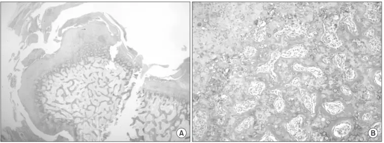
Figure 4. The specimen was examined histologically, the lesion is composed of a cartilaginous cap, calcified cartilage matrix, cancellous bone, spindle cell that compose fibrous tissue. (A) H&E, ×12.5, (B) H&E, ×100.
107
제 1 중족골에 발생한 기괴 방골성 골연골성 증식증
7. Gruber G, Giessauf C, Leithner A, et al. Bizarre parosteal os-teochondromatous proliferation (Nora lesion): a report of 3 cases and a review of the literature. Can J Surg. 2008;51:486-9.
8. Choi JH, Gu MJ, Kim MJ, Choi WH, Shin DS, Cho KH. Fibro-sarcoma in bizarre parosteal osteochondromatous prolifera-tion. Skeletal Radiol. 2001;30:44-7.
Bizarre Parosteal Osteochondromatous Proliferation
in the First Metatarsal Bone: A Case Report
Woo-Sung Kim, Yu-Hun Jung, Sang-Hun Oh, and Eun-Mee Han*
Departments of Orthopedic Surgery, *Pathology, Bundang Jesaeng General Hospital, Daejin Medical Center, Seongnam, Korea
Bizarre parosteal osteochondromatous proliferation (Nora’s lesion) is a rare benign tumor and known to be primarily occur in the small tubular bone of the hands and feet. However, it is very unusual to be reported that it occurs in metatarsal bone in Korea. Thus, we report this tumor of metatarsal bone including the literature review because we have experienced this example.
Key words: tumor of metatarsal bone, bizarre parosteal osteochondromatous proliferation, Nora’s lesion
Received October 30, 2014 Revised November 27, 2014 Accepted November 29, 2014 Correspondence to: Yu-Hun Jung
Department of Orthopaedic Surgery, Bundang Jesaeng General Hospital, Daejin Medical Center, 255-2, Seohyun-dong, Bundang-gu, Seongnam 463-774, Korea TEL: +82-31-779-0175 FAX: +82-31-779-0176 E-mail: osjungyh@naver.com
Copyrights © 2014 by The Korean Bone and Joint Tumor Society
“This is an Open Access article distributed under the terms of the Creative Commons Attribution Non-Commercial License (http://creativecommons.org/licenses/by-nc/3.0/) which permits unrestricted non-commercial use, distribution, and reproduction in any medium, provided the original work is properly cited.”