Lack of Effect of Dexamethasone on Growth of
Orientia Tsutsugamushi Gilliam in Mouse L929 Cells
Chang Oh Kim,
1* Ae Jung Huh,
2* Joon Sup Yeom,
3Kkot Sil Lee,
4Bum Sik Chin,
1Sang Hoon Han,
1Su Jin Jeong,
1Jun Yong Choi,
1Young Goo Song,
1and June Myung Kim
11Department of Internal Medicine and AIDS Research Institute, Yonsei University College of Medicine, Seoul; 2Department of Internal Medicine, National Health Insurance Corporation Ilsan Hospital, Goyang; 3Department of Internal Medicine, Kangbuk Samsung Hospital, Sungkyunkwan University School of Medicine, Seoul; 4Department of Internal Medicine, Kwandong University Myongji Hospital, Kwandong University College of Medicine, Goyang, Korea.
Received: June 17, 2010 Revised: August 10, 2010 Accepted: September 28, 2010
Corresponding author: Dr. June Myung Kim, Department of Internal Medicine, Yonsei University College of Medicine, 250 Seongsan-ro, Seodaemun-gu, Seoul 120-752, Korea.
Tel: 82-2-2228-1946, Fax: 82-2-393-6884 E-mail: jmkim@yuhs.ac
*Chang Oh Kim and Ae Jung Huh contributed equally to this work.
∙ The authors have no financial conflicts of interest.
© Copyright:
Yonsei University College of Medicine 2011
This is an Open Access article distributed under the terms of the Creative Commons Attribution Non-Commercial License (http://creativecommons.org/ licenses/by-nc/3.0) which permits unrestricted non-commercial use, distribution, and reproduction in any medium, provided the original work is properly cited.
Purpose: Previous studies and our own clinical experience suggest that concurrent corticosteroid treatment for severe rickettsial disease with multiorgan failure may improve the clinical course or reduce mortality. However, the use of corticoste-roids as adjunctive treatment for rickettsial diseases is controversial. We attempted to determine the influences of corticosteroid on the growth of Orientia tsutsuga-mushi in vitro to justify and evaluate the clinical applicability of corticosteroid in rickettsial disease. Materials and Methods: L929 cells were infected with Orien-tia tsutsugamushi Gilliam. Dexamethasone was added to the cells at final concen-trations of 101 and 107 pg/mL. Cultures were incubated at 35°C and processed for flow cytometry on the 6th day after addition of dexamethasone. Results: Observa-tion on the 6th day after treatment with dexamethasone in infected cultures re-vealed that there was no difference in fluorescence intensity among the treatment wells. Treatment of the cells with dexamethasone at concentrations of 101 and 107 pg/mL showed no influence on the growth of Orientia tsutsugamushi. Conclu-sion: Our results to show that isolated corticosteroid does not enhance the replica-tion of Orientia tsutsugamushi in vitro. Concurrent use of anti-inflammatory or immunosuppressive doses of corticosteroids in conjunction with antibiotics may not have detrimental effects on the course of scrub typhus.
Key Words: Orientia tsutsugamushi, L929 cells, corticosteroids, interferon
INTRODUCTION
The causative agent of scrub typhus, Orientia tsutsugamushi, is obligate intracellu-lar bacteria proliferating within eukaryotic cells.1 Several studies on the effect of interferon on the growth of rickettsia have been conducted, and interferon-γ has been reported to inhibit the growth of rickettsia in vitro; however, its direct effect on recovery from acute infection and prevention of recurrence has not yet been clearly clarified.2,3
tor maintained at 5% CO2, and the cell proliferation level was examined.
After more than 90% of cells were infected, the cultured L929 cells were collected using a scraper and the collected cell suspension was disrupted by glass dounce (tight piston, Wheaton Inc., Millville, NJ, USA). The disrupted cell solu-tion was centrifuged at 500×g for 5 min, cell debris was re-moved, and the supernatant was collected. The supernatant was centrifuged at 1,000×g for 10 min. Precipitates were collected and resuspended in DMEM medium, aliquoted as 1 mL, stored at -80°C in liquid nitrogen, and used in the ex-periments.
Antibody and indirect immunofluorescence staining
Anti-Orientia tsutsugamushi Gilliam mouse serum was ob-tained by injecting C3H mice 2-3 times with rickettsia at 2-week intervals and sampling after 4 weeks. Using an anti-Orientia tsutsugamushi Gilliam mouse serum with the titer higher than 1 : 640 and FITC-conjuated goat anti-mouse IgG (Cappel Laboratories, Cochranville, PA, USA) diluted 1 : 100 in PBS, rickettsia-infected cells were stained by in-direct immunofluorescent antibody method and examined under a fluorescent microscope (microscope trinocular, Olympus, Tokyo, Japan). Infection levels of the L929 cells were assessed and flow cytometry (FACSCalibur, Becton Dickinson, San Jose, CA, USA) was used to evaluate in-fected cell proliferation.
Addition of corticosteroids
To evaluate the effect of corticosteroids on the proliferation of cells themselves, 1 mL DMEM medium containing dexa-methasone (Sigma Chemical Co., St. Louis, MO, USA) at fi-nal concentrations of 101, 103, 105, and 107 pg/mL was add-ed to the L929 cells culturadd-ed as a monolayer in six wells and by varying the culture period, cell viability level was measured by MTT assay and hemocytometer.7
To examine the effects of rickettsia infection, rickettsia suspension was added to L929 cells, cultured at 35°C for 1 hr, and under the infected conditions, 1 mL DMEM medi-um with dexamethasone (101 to 107 pg/mL) was added and cultured for six days.
Interferon-γ administration
Monolayers of L929 cells in 6-well plates (Corning Glass Works, Corning, NY, USA) received maintenance DMEM medium containing an appropriate amount of interferon-γ (Serotec, Oxford, UK) and were incubated for 24 hrs. After Supplemental administration of corticosteroids for
rick-ettsial diseases is still controversial, and definitive data to substantiate its beneficial or detrimental effects are not suf-ficient. Physicians can be confronted with the dilemma of initiating immunosuppressive corticosteroid treatment in a critically ill patient, for which the tentative diagnoses in-clude immunologically mediated anemia and/or immunity-mediated thrombocytopenia or severe rickettsial disease.4
Indeed, in some studies, the simultaneous administration of corticosteroid to severe rickettsia-infected patients with concomitant multiorgan impairment has been reported to be of help to the improvement of the clinical course or reduc-tion of mortality.4-6 But in a clinical course, the direct effects of drugs and many diverse factors may be involved and it is difficult to evaluate the pure effect of drugs in clinical stud-ies. The possibility that corticosteroids could stimulate the proliferation of pathogens in some infectious diseases could be a limiting factor to the additional administration of corti-costeroids.4
Therefore, we objectively evaluated the effects of the ad-ministration of corticosteroids on the growth rate of Orien-tia tsutsugamushi in an L929 cell line infected in vitro.
MATERIALS AND METHODS
Cell culture
Murine L929 cells provided by the American Type Culture Collection (CCL1, Rockville, MD, USA) and kept at the De-partment of Microbiology and Immunology at Seoul Nation-al University were obtained and used. L929 cells were cul-tured in Dulbecco’s modified Eagle’s medium (DMEM, Gibco BRL, Grand Island, NY, USA) containing 10% (vol/ vol) fetal bovine serum (FBS), penicillin (100 unit/mL), streptomycin (100 μg/mL), and 2 mM L-Glutamine in a 37°C incubator maintained 5% CO2. When the cells grew and covered 80-90% of the bottom of culture flasks, they were either infected with rickettsia or a subculture was per-formed.
Rickettsia
In 6-well plates (Corning Glass Works, Corning, NY, USA), 106 cells per well were added, cultured for 24-36 hr, and the L929 cells formed a monolayer that was infected with Orientia tsutsugamushi Gilliam at a density of 0.5 particle per cell. Subsequently, DMEM medium was added, con-taining 5% FBS, the cells were incubated in a 35°C
incuba-RESULTS
Cellular morphology of L929 cells after Orientia tsutsugamushi infection and the addition of corticosteroids
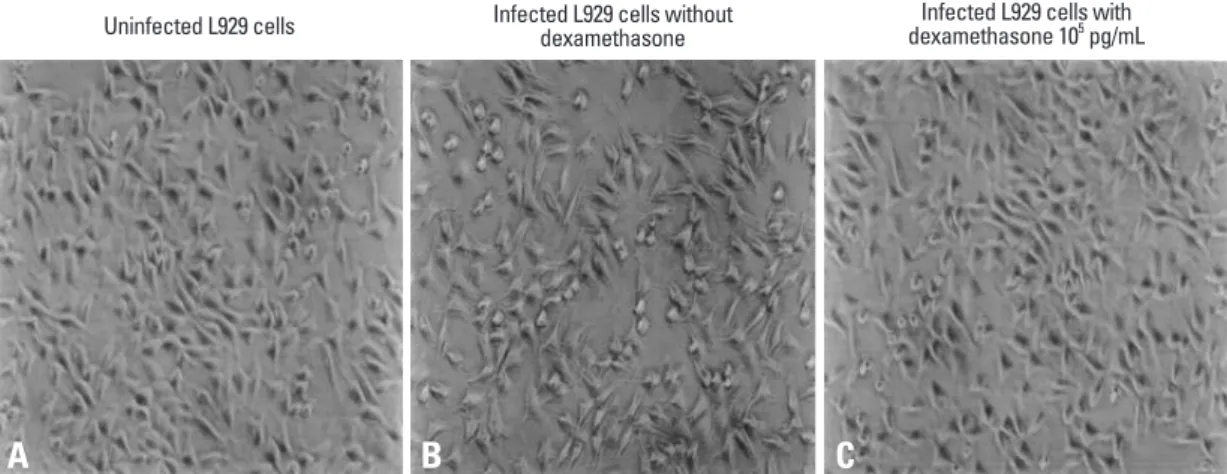
Considering the results of previous studies regarding mor-phological changes of mouse fibroblasts prior to and after Orientia tsutsugamushi infection, uninfected L929 cells, L929 cells infected with Orientia tsutsugamushi, and L929 cells treated with corticosteroids after infection were cul-tured.10 At 24-hr intervals between 24 and 96 hrs, the gross morphology of the L929 cells was examined by phase con-trast microscopy. No differences were detected in the num-ber of cells or morphology according to the status of Orien-tia tsutsugamushi infection or corticosteroid doses (Fig. 1). Alteration of growth of L929 cells according to corticosteroid concentration
To examine the effect of corticosteroids on the proliferation of L929 cells to be infected Orientia tsutsugamushi, cells were cultured for 10 days with various concentrations of cor-ticosteroids and the number of cells was checked. The results showed that during 1-7 days of culture, cell proliferation was not affected by the presence or absence of corticosteroid, nor was it influenced by the dosage of corticosteroid (Fig. 2). Af-ter day 3, it was observed that cell proliferation decreased markedly because cells were in a fully saturated state.
Effect of corticosteroid on Orientia tsutsugamushi growth?
Corticosteroid concentration was raised from 101 pg/mL to 107 pg/mL by 10-fold. The growth level of Orientia tsutsuga-aspiration of the medium, the cells were inoculated with the
rickettsia at multiplicity of infection and absorbed for 1 hr at 35°C. Unabsorbed rickettsiae were aspirated off, and each culture was fed with 1 mL of maintenance medium contain-ing interferon-γ. Untreated control cultures were inoculated with rickettsia and treated with maintenance medium alone. Cultures were incubated at 35°C and processed for flow cy-tometry at various intervals.8
Preparation of samples used for flow cytometry
The fixation and staining of cells infected with rickettsia was performed as suggested in previous studies.8,9 Summarizing briefly, after rickettsial infection and addition of corticoste-roid, cells cultured for a certain time were washed with PBS, PBS was added to cell precipitates obtained by the dissocia-tion of cells using 0.25% trypsin-0.1% etylene-diamine tet-raacetate-PBS, centrifuged, and resuspended to 106 cells/ mL. Cell precipitates obtained by centrifugation at 5,000 rpm for 15 sec were resuspended with 4°C PBS 100 μL and -30°C methanol 900 μL was added and kept at -30°C for more than 15 min. These cells were stored at -30°C until staining. As described above, the fixed cells were stained by the indirect immunofluorescent antibody method, washed with PBS, and used for flow cytometry analysis.
Flow cytometry measurement
Rickettsia particles within L929 cells appearing as green fluorescence were measured by EPICS Elite flow cytome-ter (Coulcytome-ter Corp., Hialeah, FL, USA) and 20,000 cells were analyzed to find the fluorescence level of each sam-ple. The excitation wave length was 488 nm, and data were presented as fluorescence intensity, the cellular frequency of its log scale.
Fig. 1. Morphological changes of L929 cells after steroid treatment (phase contrast microscopy, ×100). L929 cells uninfected with Orientia tsutsugamushi (A), infected L929 cells (B), and L929 cells treated with dexamethason 105 pg/mL (C) after infection were cultured. At 24-hr
intervals from 24 hr to 96 hr, gross morphology of L929 cells was examined by phase contrast microscopy.
Uninfected L929 cells Infected L929 cells without dexamethasone dexamethasone 10Infected L929 cells with 5 pg/mL
er, diverse serotypes are present. Among these, the standard serotyes are Gilliam, Karp, and Kato, with various serotypes having been reported in different countries. In Korea, Gil-liam, Karp, and Boryung are the major serotypes, and the Boryung serotype has been isolated most abundantly.11 As shown by recent clinical studies, the clinical manifestation of patients and laboratory results are differ greatly and opinions suggesting that this is due to serological differenc-es have been reported. In the literature, all previous report-ed deaths were causreport-ed by the Gilliam serotype. This sug-gests the possibility that the severity of clinical manifestation is influenced by serotype.12,13 The Gilliam serotype associ-ated with severe rickettsial disease was used in our experi-ments because the use of corticosteroids was considered in cases suspected to be severe rickettsial disease. Although it was not shown in our results, similar results have been ob-mushi was evaluated by flow cytometry and the results
showed that differences in the growth of Orientia tsutsuga-mushi with or without the addition of corticosteroids as well as added dosages were not detected (Fig. 3). To evaluate the reliability of our results, experiments that assessed the effects of interferon on the growth of Orientia tsutsugamushi by flow cytometry reported in other studies were used as the control group in our study and, similar to the previously re-ported results, reduction of the growth of Orientia tsutsuga-mushi in response to interferon-γ administration was con-firmed (Fig. 4).8
DISCUSSION
Orientia tsutsugamushi is classified as one species;
howev-Fig. 2. Effect of steroid concentrations on the growth of L929 cells. L929 cells were cultured for 10 days with various concentrations of
corticosteroids and the number of cells was enumerated. The results showed that during 1-7 days of culture.
0 200 400 600 800 1,000 1,200 Ce ll v ia bi lit y ( 10 3/m L) 1 day
Culture days after steroid treatment
2 day 3 day 4 day 5 day 6 day 7 day
Without dexamethasone Dexamethasone 101 pg/mL
Dexamethasone 103 pg/mL
Dexamethasone 105 pg/mL
Dexamethasone 107 pg/mL
Fig. 3. Effect of steroid on the growth of Orientia tsutsugamushi. Corticosteroid concentration was raised from 101 pg/mL to 107 pg/mL by
10-fold. The growth level of Orientia tsutsugamushi was evaluated by flow cytometry.
0 20 40 60 80 100 120 Co un ts 100 101 102 FL1-H 103 104 Dexamethasone 101 pg/mL Dexamethasone 102 pg/mL Dexamethasone 103 pg/mL Dexamethasone 104 pg/mL Dexamethasone 105 pg/mL Dexamethasone 106 pg/mL 120 0 Co un ts 101 102 103 104 105 FL1-H dexamethasone 101 pg/mL 120 0 Co un ts 101 102 103 104 105 FL1-H dexamethasone 103 pg/mL 120 0 Co un ts 101 102 103 104 105 FL1-H dexamethasone 105 pg/mL 120 0 Co un ts 101 102 103 104 105 FL1-H dexamethasone 107 pg/mL Without steroid With steroid
A
B
peripheral steroid resistance, etc. Recently, effects on the im-provement of symptoms, disease duration, and mortality have been reported.19 However, in severe sepsis, short-term administration of high-dose corticosteroids is not recom-mended except in cases such as typhoid fever, pneumocysti-tis carinii pneumonia in acquired immune deficiency syn-drome, or bacterial encephalomeningitis in children.20 The outcome of treatment with corticosteroids in bacterial infec-tions is difficult to discuss completely. Time of administra-tion, dosage, maintenance time of corticosteroids as well as antibiotics, causative pathogens, and characteristics of the infectious disease should be considered.
In rickettsial diseases, administration of corticosteroids is frequently applied to Rocky Mountain Spotted Fever (RMSF) that shows ocular symptoms including uveitis, ocular hem-orrhage, exudate retinosis, etc.21 In addition, it has been re-ported that in severe RMSF infection accompanying initial endotoxemia, hyperazotemia, or hypoproteinemia, cases in which cortisone and antibiotics were administered together had better treatment outcomes than those cases treated with antibiotics alone.5 There have been animal-modeled experi-ments to show the theoretical basis for this. Because of the similarity of diseases between humans and dogs, Rickettsia rickettsii infected animal models with the using dogs is a proper methodology for human infection research.22 Based on recent animal-modeled research on corticosteroid adju-vant therapy, it has been reported that concurrent use of corticosteroids and doxycycline confers no clinically rele-vant detrimental effects.4
However, when we tried to apply positive treatment re-tained in experiments using the Karp serotype.
For the treatment of scrub typhus, administration of 100 mg of doxycycline twice a day for 3-7 days is recommend-ed.14 This treatment has been reported to have excellent re-sults, and the mortality rate has decreased noticeably from 50% before use of antibiotics was introduced. Recently, in the northern area of Thailand, Orientia tsutsugamushi patho-gen showing resistance to doxycyclin was isolated and showed a tendency of delayed response to antibiotics, but this did not raise mortality.15 Although not frequently encoun-tered, other substitutions could not be suggested for severe rickettsial diseases besides antibiotic and conservative treat-ments. In some patients, progression to severe infection with involvement of major organs (such as resulting in encephali-tis, myocardiencephali-tis, kidney injury, and interstitial pneumonitis) occurred before the antibiotics took effect. Administration of corticosteroid to such patients showed inconsistent results.16 In severe rickettsial disease with immune mediated thrombo-cytopenia and hemolytic anemia, it is difficult for physicians to decide to use corticosteroids at the immunosuppressive prednisolone dose (2.0 mg/kg).4
In terms of the use of corticosteroids for bacterial diseas-es, the most investigated area is septic shock. Prior to the 1940s, corticosteroid treatment had been administered em-pirically to severe septic shock patients. Upon summarizing the results of clinical studies conducted for the objective evaluation of effects, its effects remian unclear.17,18 Despite these results, in catecholamine-dependent septic shock, low-dose corticosteroid treatments have been continuously in-vestigated because of relative adrenal function deterioration,
Fig. 4. Effect of IFN-γ on the growth of Orientia tsutsugamushi. To evaluate the stability of our results, experiments that assessed the ef-fect of interferon-γ on the growth of Orientia tsutsugamushi by flow cytometry were used as the control group.
0 40 80 120 160 200 Co un ts 100 101 102 103 104 FL1-H IFN 1 ng/mL 0 40 80 120 160 200 Co un ts 100 101 102 103 104 FL1-H IFN 100 ng/mL 0 40 80 120 160 200 Co un ts 100 101 102 103 104 FL1-H IFN 10 ng/mL 0 40 80 120 160 200 Co un ts 100 101 102 103 104 FL1-H IFN 1,000 ng/mL
Rocky Mountain spotted fever. N Engl J Med 1952;246:962-6. 6. Choi HC, Wie SH, Lee SY, Kim SI, Park SK, Jung YJ, et al. Use of
high-dose steroid in a case of scrub typhus with acutely progressive local neurologic symptoms. Korean J Infect Dis 2002;34:391-5. 7. Hansen MB, Nielsen SE, Berg K. Re-examination and further
de-velopment of a precise and rapid dye method for measuring cell growth/cell kill. J Immunol Methods 1989;119:203-10.
8. Tasneen R, Yoshiie K, Ohkawa T, Oda H. High concentration of recombinant murine interferon-beta enhances the growth of Ori-entia (formerly Rickettsia) tsutsugamushi Gilliam in mouse L929 cells. Acta Virol 1997;41:77-82.
9. Laffin J, Lehman JM. Detection of intracellular virus and viral products. Methods Cell Biol 1990;33:271-84.
10. Tsuruhara T, Urakami H, Tamura A. Surface morphology of Rick-ettsia tsutsugamushi-infected mouse fibroblasts. Acta Virol 1982; 26:506-11.
11. Chang WH. Occurence of tsutsugamushi disease and prototypes of Rickettsia tsutsugamushi in Korea. J Korean Med Assoc 1988; 31:601-7.
12. Peck KR, Shin HS, Pai HJ, Chung MH, Oh MD, Song YW, et al. A comparative clinical study of scrub typhus seen in rural area and at Seoul National University Hospital. Korean J Infect Dis 1991; 23:155-62.
13. Choe K. Clinical manifestataions of scub typhus. J Korean Med Assoc 1988;31:608-11.
14. Song JH, Lee C, Chang WH, Choi SW, Choi JE, Kim YS, et al. Short-course doxycycline treatment versus conventional tetracy-cline therapy for scrub typhus: a multicenter randomized trial. Clin Infect Dis 1995;21:506-10.
15. Watt G, Chouriyagune C, Ruangweerayud R, Watcharapichat P, Phulsuksombati D, Jongsakul K, et al. Scrub typhus infections poorly responsive to antibiotics in northern Thailand. Lancet 1996; 348:86-9.
16. Brown GW. Scrub typhus; pathogenesis and clinical syndrome. In: Walker DH, editor. Biology of rickettsial disease. Vol 1. Boca Raton, FL: CRC Press; 1988. p.93-100.
17. Lefering R, Neugebauer EA. Steroid controversy in sepsis and septic shock: a meta-analysis. Crit Care Med 1995;23:1294-303. 18. Cronin L, Cook DJ, Carlet J, Heyland DK, King D, Lansang MA,
et al. Corticosteroid treatment for sepsis: a critical appraisal and meta-analysis of the literature. Crit Care Med 1995;23:1430-9. 19. Briegel J, Forst H, Haller M, Schelling G, Kilger E, Kuprat G, et
al. Stress doses of hydrocortisone reverse hyperdynamic septic shock: a prospective, randomized, double-blind, single-center study. Crit Care Med 1999;27:723-32.
20. Annane D. Resurrection of steroids for sepsis resuscitation. Mi-nerva Anestesiol 2002;68:127-31.
21. Davidson MG, Breitschwerdt EB, Nasisse MP, Roberts SM. Ocu-lar manifestations of Rocky Mountain spotted fever in dogs. J Am Vet Med Assoc 1989;194:777-81.
22. Walker DH. The role of host factors in the severity of spotted fe-ver and typhus rickettsioses. Ann N Y Acad Sci 1990;590:10-9. 23. Takami A, Yamauchi H, Asakura H, Ishiyama K, Nakao S.
Tsutsu-gamushi disease (scrub typhus)-associated hemophagocytic syn-drome. Int J Hematol 2002;75:337-8.
sults from RMSF to scrub typhus, there was one serious limitation. We were not able to control the direct risk of corticosteroid for the growth of Orientia tsutsugamush, be-cause it could be-cause adverse effects. Although one impor-tant limitation of our study is that in vitro experiments on the effects of dexamethasone on cells in culture cannot be extrapolated to the complex in vivo situation, our results do have meaning in showing that corticosteroid itself does not increase the growth of Orientia tsutsugamush, and also in reporting that the concurrent use of anti-inflammatory or immunosuppressive doses of corticosteroid in conjunction with antimicrobials did not lead to adverse influences on the course of scrub typhus, because it could not induce bac-terial growth. Recently, there have been some research into the positive effects of corticosteroid in rickettsial disease.6,23 After this study, more clinical research is needed, such as animal model experiments or various experiments to find the proper dosages, times and medication duration for corti-costeroids as treatment for serious rickettsial disease.
ACKNOWLEDGEMENTS
We thank Ik-Sang Kim and Myung-Suk Huh of Seoul Na-tional University for their assistance in performing this re-search.
REFERENCES
1. Tamura A, Takahashi K, Tsuruhara T, Urakami H, Miyamura S, Sekikawa H, et al. Isolation of Rickettsia tsutsugamushi antigeni-cally different from Kato, Karp and Gilliam strains from patients. Microbiol Immunol 1984;28:873-82.
2. Jerrells TR, Turco J, Winkler HH, Spitalny GL. Neutralization of lymphokine-mediated antirickettsial activity of fibroblasts and macrophages with monoclonal antibody specific for murine inter-feron-gamma. Infect Immun 1986;51:355-9.
3. Hanson B. Susceptibility of Rickettsia tsutsugamushi Gilliam to gamma interferon in cultured mouse cells. Infect Immun 1991;59: 4125-33.
4. Breitschwerdt EB, Davidson MG, Hegarty BC, Papich MG, Grin-dem CB. Prednisolone at anti-inflammatory or immunosuppres-sive dosages in conjunction with doxycycline does not potentiate the severity of Rickettsia rickettsii infection in dogs. Antimicrob Agents Chemother 1997;41:141-7.
5. Workman FB, Hightower JA, Borges FJ, Furman JE, Parker RT. Cortisone as an adjunct to chloramphenicol in the treatment of