INTRODUCTION
Hepatocellular carcinoma (HCC) is the fifth leading cause of
cancer mortality worldwide.
1,2The main cause of HCC is
cir-rhosis, which originates from infections caused by chronic
hepatitis B virus, hepatitis C virus, or alcohol consumption.
3,4Received: August 27, 2018
Corresponding author: Mijin Yun, MD, PhD, Department of Nuclear Medicine,
Yon-sei University College of Medicine, 50-1 YonYon-sei-ro, Seodaemun-gu, Seoul, 03722, Korea. Tel: 82-2-2123 5851, Fax: 82-2-365-5882, E-mail: yunmijin@yuhs.ac
•The authors have no financial conflicts of interest. © Copyright: Yonsei University College of Medicine 2018
This is an Open Access article distributed under the terms of the Creative Com-mons Attribution Non-Commercial License (https://creativecomCom-mons.org/licenses/ by-nc/4.0) which permits unrestricted non-commercial use, distribution, and repro-duction in any medium, provided the original work is properly cited.
Cancer Metabolism as a Mechanism of Treatment
Resistance and Potential Therapeutic Target
in Hepatocellular Carcinoma
Misu Lee
1,2, Haeyong Ko
1, and Mijin Yun
11Department of Nuclear Medicine, Severance Hospital, Yonsei University College of Medicine, Seoul;
2Division of Life Science, College of Life Science and Bioengineering, Incheon National University, Incheon, Korea.
Various molecular targeted therapies and diagnostic modalities have been developed for the treatment of hepatocellular
carcino-ma (HCC); however, HCC still recarcino-mains a difficult carcino-malignancy to cure. Recently, the focus has shifted to cancer metabolism for the
diagnosis and treatment of various cancers, including HCC. In addition to conventional diagnostics, the measurement of
en-hanced tumor cell metabolism using F-18 fluorodeoxyglucose (18F-FDG) for increased glycolysis or C-11 acetate for fatty acid
synthesis by positron emission tomography/computed tomography (PET/CT) is well established for clinical management of
HCC. Unlike tumors displaying the Warburg effect, HCCs vary substantially in terms of 18F-FDG uptake, which considerably
re-duces the sensitivity for tumor detection. Accordingly, C-11 acetate has been proposed as a complementary radiotracer for
de-tecting tumors that are not identified by 18F-FDG. In addition to HCC diagnosis, since the degree of 18F-FDG uptake converted
to standardized uptake value (SUV) correlates well with tumor aggressiveness, 18F-FDG PET/CT scans can predict patient
out-comes such as treatment response and survival with an inverse relationship between SUV and survival. The loss of tumor
sup-pressor genes or activation of oncogenes plays an important role in promoting HCC development, and might be involved in the
“metabolic reprogramming” of cancer cells. Mutations in various genes such as TERT, CTNNB1, TP53, and Axin1 are responsible
for the development of HCC. Some microRNAs (miRNAs) involved in cancer metabolism are deregulated in HCC, indicating that
the modulation of genes/miRNAs might affect HCC growth or metastasis. In this review, we will discuss cancer metabolism as a
mechanism for treatment resistance, as well as an attractive potential therapeutic target in HCC.
Key Words:
Hepatocellular carcinoma, cancer metabolism, positron emission tomography/computed tomography (PET/CT),
drug resistance
pISSN: 0513-5796 · eISSN: 1976-2437 Yonsei Med J 2018 Dec;59(10):1143-1149
https://doi.org/10.3349/ymj.2018.59.10.1143
Patients with early-stage HCC are often asymptomatic, and it
is usually detected at intermediate or advanced stages in
which patients cannot be treated by curative hepatic resection
or liver transplantation.
5In addition, although surgical
treat-ment for early HCC has improved patient outcome, the risk of
recurrence remains substantial and there is still no curative
therapy for advanced HCC. Therefore, there is an increasing
need for effective early diagnosis and development of novel
therapeutics for HCC patients. Owing to its important role in
metabolic reprogramming during carcinogenesis, cancer
me-tabolism has gained popularity in the fields of cancer
diagno-sis and therapy. This review summarizes the current state of
research related to cancer metabolism, to help identify
poten-tial new therapeutic targets for HCC.
CLINICAL APPLICATIONS OF
METABOLIC IMAGING IN HCC
Alpha-fetoprotein (AFP) was the first glycoprotein identified as
a marker of HCC, and this protein is used to screen for this
par-ticular disease.
6However, approximately 30% of HCC patients
maintain normal AFP levels, and some HCC patients also have
relatively low levels of AFP.
7To overcome these problems,
sev-eral imaging techniques are used to diagnose patients with
sus-pected HCC, such as ultrasound, computed tomography (CT),
magnetic resonance imaging (MRI), positron emission
tomog-raphy/computed tomography (PET/CT), and angiography.
Of the imaging modalities, clinical efficacy of functional
imag-ing based on cancer metabolism for the assessment of HCC has
been actively investigated. F-18 fluorodeoxyglucose (18F-FDG),
a surrogate for enhanced glucose metabolism, has been used
widely for HCC. The uptake mechanism and biochemical
path-way of 18F-FDG metabolism has been extensively studied in
vi-tro and in vivo; transport through cell membrane via glucose
transporter isoform 1 (GLUT1) and intracellular phosphorylation
by hexokinase (HK) have been identified as key steps for
subse-quent accumulation in HCC.
8-13Many studies have shown
up-regulation of GLUT1 in HCC, but not in non-tumor liver
tis-sue.
14,15Amann, et al.
15demonstrated a positive correlation
between GLUT1 expression and Ki-67 labeling index in patients
with HCC, suggesting that its expression is associated with
ad-vanced tumor stage and poor differentiation. In addition, poor
survival has been reported in patients with high tumor GLUT1
expression, based on The Cancer Genome Atlas (TCGA) data
set.
16A relationship between enhanced FDG uptake and
dysreg-ulation of epithelial-mesenchymal transition-related proteins
was demonstrated in HCC through in vitro and patient tissue
ex-periments.
16Representative of the underlying biological
charac-teristics of tumor, 18F-FDG PET/CT images are predictive of
tu-mor recurrence or survival after various treatments.
17Despite displaying increased glycolysis even with the presence
of oxygen, the so-called Warburg effect, HCCs are notorious for
exhibiting a wide spectrum of 18F-FDG uptake capabilities,
con-siderably reducing the sensitivity of tumor detection.
Alternative-ly, C-11 acetate has been proposed as a radiotracer for detecting
tumors that are not identified based on 18F-FDG uptake (Figs. 1
and 2). Acetate is a source of acetyl-CoA, and it plays an essential
role in regulating the activity and expression of proteins involved
in regulation of intracellular biomass, lipogenesis, and
acetyla-tion.
18Acetate was shown to be utilized by tumors as an
alterna-tive nutrient under low cellular glucose uptake conditions, and
C-11 acetate accumulation in tumors has been found to be
asso-ciated with tumor progression.
19HCC has been reported to use
acetate as a substrate for fatty acid biosynthesis through
up-regu-lation of acetyl-CoA synthase and monocarboxylate transporter
(MCT).
20Recent studies have indicated that MCT1 is a novel
im-port system of acetate in non-glycolytic HCC tumors. Indeed, Fig.
3 shows various expressions of GLUT1 and MCT1 in HCC
pa-tients with different levels of 18F-FDG and 11C-acetate uptake. It
was demonstrated that absorption of acetate by MCT1 promotes
oxidative phosphorylation and lipid metabolism in
non-glycolyt-ic HCC tumors.
21Accordingly, combining 18F-FDG PET/CT with
C-11 acetate PET/CT could be useful to provide relevant
infor-mation on prognostic and molecular heterogeneity.
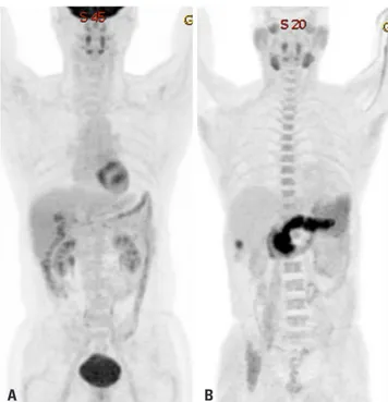
A
B
Fig. 1. Hepatocellular carcinoma positive for F-18 fluorodeoxyglucose (A), but negative for C-11 acetate (B).
A
B
Fig. 2. Hepatocellular carcinoma negative for F-18 fluorodeoxyglucose (A), but positive C-11 acetate (B).
DIFFERENTIAL GENE EXPRESSION THAT
ALTERS METABOLISM IN HCC CELLS
HCC is a heterogeneous disease, both clinically and from a
molecular standpoint. Different risk factors such as hepatitis
virus infection, aflatoxin exposure, or alcohol abuse are linked
to specific pathways, and these can be strongly associated with
certain types of HCC. Based on the results of HCC tumor
se-quencing, different driver genes and associated oncogenic
pathways have been identified, based on the composition of
tumor source.
22-25Therefore, heterogeneity should be
investi-gated to determine the etiological cause and affected pathways
of HCC. High levels of heterogeneity are clinically relevant, as
they lead to inconsistent treatment outcomes. Recently, deep
sequencing/next generation sequencing has provided new
sights into the complex molecular pathogenesis of HCC,
in-cluding the identification of novel oncogenic pathways and
driver genes.
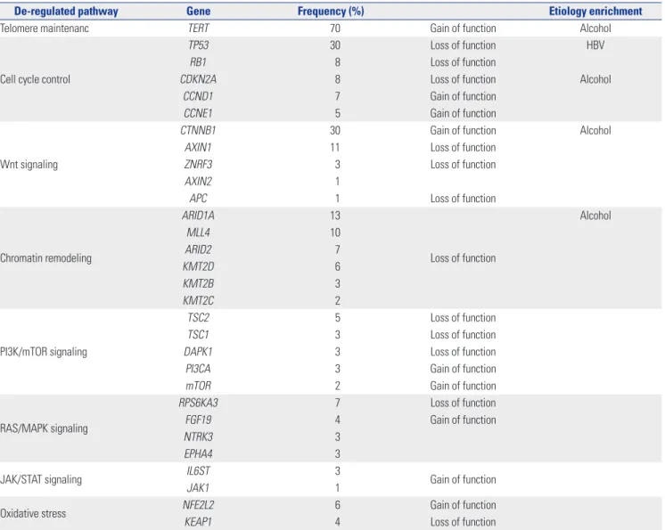
25-33Aberrant telomerase reverse transcriptase
(TERT) activation is the most common somatic alteration
ob-served in HCC (~70%). In addition to TERT, CTNNB1, TP53,
and Axin1 are mutated at high frequency in HCC. Table 1
sum-marizes the most relevant mutations in HCC.
Warburg effect occurs downstream of survival signaling
path-ways, which are altered by loss of tumor suppressor genes or
acti-vation of oncogenes such as c-Myc, Ras, Akt, TP53, and
HIF-1α.
34-38c-Myc was reported to result in mouse liver tumors with
elevated glycolysis
39. HIF-1
α, a major transcription factor involved
in hypoxic response of cancer cells,
40has been shown to play an
important role in several cancers by promoting tumorigenesis,
and might also be involved in the “metabolic reprogramming” of
cancer cells.
41,42This activates the transcription of genes encoding
angiogenic cytokines and growth factors, such as VEGF and
gly-colytic enzymes including hexokinase1 (HK1), hexokinase2
(HK2), glyceraldehyde-3-phosphate dehydrogenase (GAPDH),
and pyruvate kinase (PKM).
43-45Moreover, HIF-1α enhances
che-moresistance and radioresistance, whereas it suppresses
differ-entiation and apoptosis in HCC.
46,47As a result, elevated HIF-1α
levels are associated with increased patient mortality and
metas-tasis in various tumors, including HCC.
48-50Certain gene mutations or loss-of-heterozygosity events alter
metabolism in a HIF-1α-dependent manner. Although
muta-tions in PTEN gene rarely occur in HCC, frequent loss of
hetero-zygosity of PTEN allele has been identified (in 20−30% of HCC
patients).
51,52The loss of PTEN plays a critical role in HCC
pro-C
A
B
D
Fig. 3. Differences in the expression of glucose transport 1 (A and C) and monocarboxylate transporter 1 (B and D) in hepatocellular carcinoma (HCC) samples, based on 18F-fluorodeoxyglucose and 11C-acetate uptake. Human HCC samples were used. Immunohistochemistry (IHC) was performed as described previously.16 After antigen retrieval, IHC was performed using indicated antibodies. Scale bars: 40 μm.
gression and patient outcome by increasing HIF-1α synthesis
and stability.
53,54The modulation of HIF-1α expression by
epider-mal growth factor/phosphatidylinositol 3-kinase (PI3K)/PTEN/
AKT/FRAP pathway has implications in tumor angiogenesis.
55A
few studies have shown that HIF-1 can be activated by Ras and
membrane type-1 matrix metalloproteinases under normal
oxy-gen conditions in cancer cells, providing new insight into
regula-tion of cancer glycolysis beyond hypoxic condiregula-tion.
56,57Bufalin, a
cardiotonic steroid, was shown to suppress tumor invasion and
metastasis by targeting HIF-1α via PI3K/AKT/mTOR pathway,
and thus has potential for HCC targeted therapy.
58Numerous microRNAs (miRNAs) have been shown to be
as-sociated with HCC. Six miRNAs have been consistently reported
to be dysregulated in HCC, when compared to their expressions
in non-tumorous tissue.
59-61For example, miR-122 and miR-199a,
which act as tumor suppressors by regulating the expression of
cyclin G and components of PAK4/Raf/MEK/ERK pathway, are
downregulated in this disease. Conversely, miR-21, miR-221,
miR-222, and miR-224, which target various molecules including
PTEN, SMAD4, CDKN1B, and CDKN1C, are upregulated in
HCC. Some miRNAs that regulate cancer metabolism are also
dysregulated in HCC. miR-34a plays a major role in regulation of
cellular metabolism by targeting SIRT1, a key NAD-dependent
deacetylating enzyme involved in a wide range of metabolic
pro-cesses including lipid metabolism, glucose metabolism, and
ex-pression of other metabolic regulators.
62,63This molecule inhibits
cellular glycolysis by targeting HK1/2 and glucose-6-phosphate
isomerase. miR-23a directly targets the key gluconeogenic
en-zyme glucose-6-phosphatase catalytic subunit (G6PC), and is
significantly upregulated in primary human HCC.
NEW TREATMENTS TARGETING CANCER
METABOLISM
Although sorafenib has limited efficacy, it is still the only
stan-dard treatment available for advanced HCC with portal vein
invasion or extrahepatic spread.
64,65However, other molecules
Table 1. List of the Most Relevant Mutations in Hepatocellular Carcinoma
De-regulated pathway Gene Frequency (%) Etiology enrichment
Telomere maintenanc TERT 70 Gain of function Alcohol
Cell cycle control
TP53 30 Loss of function HBV
RB1 8 Loss of function
CDKN2A 8 Loss of function Alcohol
CCND1 7 Gain of function
CCNE1 5 Gain of function
Wnt signaling
CTNNB1 30 Gain of function Alcohol
AXIN1 11 Loss of function
ZNRF3 3 Loss of function
AXIN2 1
APC 1 Loss of function
Chromatin remodeling ARID1A 13 Loss of function Alcohol MLL4 10 ARID2 7 KMT2D 6 KMT2B 3 KMT2C 2 PI3K/mTOR signaling TSC2 5 Loss of function TSC1 3 Loss of function
DAPK1 3 Loss of function
PI3CA 3 Gain of function
mTOR 2 Gain of function
RAS/MAPK signaling
RPS6KA3 7 Loss of function
FGF19 4 Gain of function
NTRK3 3
EPHA4 3
JAK/STAT signaling IL6ST 3 Gain of function
JAK1 1
Oxidative stress NFE2L2 6 Gain of function
are being developed for targeted therapy. Recent studies on
metabolic regulation of cancer cell growth and metastasis
have been actively performed. 2-deoxy-D-glucose (2-DG), a
glucose analogue that is able to suppress glycolysis by
com-petitively inhibiting HK2, has an effect on HCC growth.
66,67The combination of conventional therapy and 2-DG has been
reported to synergistically inhibit the proliferation of
sorafenib-sensitive and sorafenib-resistant HCC cells.
683-bromopyru-vate (3-BP) directly inhibits HK2 activity and glycolysis
path-way. In vitro and in vivo studies have demonstrated the
anticancer effects of 3-BP on HCC, and consequently, this
drug has been approved by the FDA.
69,70Facilitative glucose transporters (GLUTs) have emerged as
key factors that are required for increasing glucose uptake by
cancer cells.
14,15Therefore, small molecules targeting GLUT1
will inhibit cancer cell growth or metastasis by reducing
glu-cose uptake. To increase GLUT1 targeting specificity,
deriva-tives of GLUT1 inhibitor, such as fasentin
71,72and WZB117,
73have been investigated. Regulation of hypoxia by molecules,
including HIF-1, is an attractive potential therapeutic target
for HCC as well as other cancers. The HIF-1α mRNA
antago-nist EZN-2968, a novel inhibitor of hypoxia-induced gene
ac-tivation, is currently in Phase I trial for HCC patients.
CONCLUSIONS
Metabolic reprogramming is essential for angiogenesis,
prolif-eration, invasion, and metastasis of cancer. It is also associated
with de-differentiation, anti-apoptotic properties, and
resis-tance to conventional chemotherapy and radiotherapy. In the
future, cancer metabolism would represent an attractive
po-tential therapeutic target. With their development, PET/CT
scans combined with various metabolic radiotracers will offer
clinical importance in selecting patients who would benefit
from novel drugs targeting different pathways related to cancer
metabolism.
ACKNOWLEDGEMENTS
This research was supported by the National Research
Founda-tion of Korea (Seoul, Korea ; grant number
NRF-2016R1E1A1A01943303, NRF-2015R1D1A1A01057737), and was
supported by an Incheon National University Research Grant
(Incheon, Korea) in 2016.
ORCID
Mijin Yun https://orcid.org/0000-0002-1712-163X
REFERENCES
1. Ferlay J, Shin HR, Bray F, Forman D, Mathers C, Parkin DM. Esti-mates of worldwide burden of cancer in 2008: GLOBOCAN 2008. Int J Cancer 2010;127:2893-917.
2. Ferlay J, Soerjomataram I, Dikshit R, Eser S, Mathers C, Rebelo M, et al. Cancer incidence and mortality worldwide: sources, meth-ods and major patterns in GLOBOCAN 2012. Int J Cancer 2015; 136:E359-86.
3. Velázquez RF, Rodríguez M, Navascués CA, Linares A, Pérez R, Sotorríos NG, et al. Prospective analysis of risk factors for hepato-cellular carcinoma in patients with liver cirrhosis. Hepatology 2003;37:520-7.
4. Fattovich G, Stroffolini T, Zagni I, Donato F. Hepatocellular carci-noma in cirrhosis: incidence and risk factors. Gastroenterology 2004;127(5 Suppl 1):S35-50.
5. Bruix J, Sherman M, American Association for the Study of Liver Diseases. Management of hepatocellular carcinoma: an update. Hepatology 2011;53:1020-2.
6. Song PP, Xia JF, Inagaki Y, Hasegawa K, Sakamoto Y, Kokudo N, et al. Controversies regarding and perspectives on clinical utility of biomarkers in hepatocellular carcinoma. World J Gastroenterol 2016;22:262-74.
7. Cameron AM. Screening for viral hepatitis and hepatocellular cancer. Surg Clin North Am 2015;95:1013-21.
8. Phelps ME, Huang SC, Hoffman EJ, Selin C, Sokoloff L, Kuhl DE. Tomographic measurement of local cerebral glucose metabolic rate in humans with (F-18)2-fluoro-2-deoxy-D-glucose: valida-tion of method. Ann Neurol 1979;6:371-88.
9. Gambhir SS, Czernin J, Schwimmer J, Silverman DH, Coleman RE, Phelps ME. A tabulated summary of the FDG PET literature. J Nucl Med 2001;42(5 Suppl):1S-93S.
10. Stokkel MP, Draisma A, Pauwels EK. Positron emission tomogra-phy with 2-[18F]-fluoro-2-deoxy-D-glucose in oncology. Part IIIb: therapy response monitoring in colorectal and lung tumours, head and neck cancer, hepatocellular carcinoma and sarcoma. J Cancer Res Clin Oncol 2001;127:278-85.
11. Iwata Y, Shiomi S, Sasaki N, Jomura H, Nishiguchi S, Seki S, et al. Clinical usefulness of positron emission tomography with fluo-rine-18-fluorodeoxyglucose in the diagnosis of liver tumors. Ann Nucl Med 2000;14:121-6.
12. Khan MA, Combs CS, Brunt EM, Lowe VJ, Wolverson MK, Solo-mon H, et al. Positron emission tomography scanning in the eval-uation of hepatocellular carcinoma. J Hepatol 2000;32:792-7. 13. Böhm B, Voth M, Geoghegan J, Hellfritzsch H, Petrovich A,
Scheele J, et al. Impact of positron emission tomography on strat-egy in liver resection for primary and secondary liver tumors. J Cancer Res Clin Oncol 2004;130:266-72.
14. Medina RA, Owen GI. Glucose transporters: expression, regula-tion and cancer. Biol Res 2002;35:9-26.
15. Amann T, Maegdefrau U, Hartmann A, Agaimy A, Marienhagen J, Weiss TS, et al. GLUT1 expression is increased in hepatocellular carcinoma and promotes tumorigenesis. Am J Pathol 2009;174: 1544-52.
16. Lee M, Jeon JY, Neugent ML, Kim JW, Yun M. 18F-Fluorodeoxy-glucose uptake on positron emission tomography/computed to-mography is associated with metastasis and epithelial-mesen-chymal transition in hepatocellular carcinoma. Clin Exp Metastasis 2017;34:251-60.
17. Hwang SH, Lee JW, Cho HJ, Kim KS, Choi GH, Yun M. Prognostic value of metabolic tumor volume and total lesion glycolysis on preoperative 18F-FDG PET/CT in patients with very early and
early hepatocellular carcinoma. Clin Nucl Med 2017;42:34-9. 18. Schug ZT, Vande Voorde J, Gottlieb E. The metabolic fate of
ace-tate in cancer. Nat Rev Cancer 2016;16:708-17.
19. Ho CL, Yu SC, Yeung DW. 11C-acetate PET imaging in hepatocel-lular carcinoma and other liver masses. J Nucl Med 2003;44:213-21. 20. Bjornson E, Mukhopadhyay B, Asplund A, Pristovsek N, Cinar R,
Romeo S, et al. Stratification of hepatocellular carcinoma patients based on acetate utilization. Cell Rep 2015;13:2014-26.
21. Jeon J, Lee M, Whang S, Kim JW, Cho A, Yun M. Regulation of ac-etate utilization by monocarboxylate transporter 1 (MCT1) in he-patocellular carcinoma (HCC). Oncol Res 2018;26:71-81. 22. Breuhahn K, Gores G, Schirmacher P. Strategies for
hepatocellu-lar carcinoma therapy and diagnostics: lessons learned from high throughput and profiling approaches. Hepatology 2011;53:2112-21. 23. Thorgeirsson SS. Genomic decoding of hepatocellular carcinoma.
Gastroenterology 2006;131:1344-6.
24. Wang K, Lim HY, Shi S, Lee J, Deng S, Xie T, et al. Genomic land-scape of copy number aberrations enables the identification of oncogenic drivers in hepatocellular carcinoma. Hepatology 2013; 58:706-17.
25. Kan Z, Zheng H, Liu X, Li S, Barber TD, Gong Z, et al. Whole-ge-nome sequencing identifies recurrent mutations in hepatocellu-lar carcinoma. Genome Res 2013;23:1422-33.
26. Totoki Y, Tatsuno K, Covington KR, Ueda H, Creighton CJ, Kato M, et al. Trans-ancestry mutational landscape of hepatocellular car-cinoma genomes. Nat Genet 2014;46:1267-73.
27. Schulze K, Imbeaud S, Letouzé E, Alexandrov LB, Calderaro J, Re-bouissou S, et al. Exome sequencing of hepatocellular carcino-mas identifies new mutational signatures and potential therapeu-tic targets. Nat Genet 2015;47:505-11.
28. Fujimoto A, Totoki Y, Abe T, Boroevich KA, Hosoda F, Nguyen HH, et al. Whole-genome sequencing of liver cancers identifies etiological influences on mutation patterns and recurrent muta-tions in chromatin regulators. Nat Genet 2012;44:760-4.
29. Guichard C, Amaddeo G, Imbeaud S, Ladeiro Y, Pelletier L, Maad IB, et al. Integrated analysis of somatic mutations and focal copy-number changes identifies key genes and pathways in hepatocel-lular carcinoma. Nat Genet 2012;44:694-8.
30. Huang J, Deng Q, Wang Q, Li KY, Dai JH, Li N, et al. Exome se-quencing of hepatitis B virus-associated hepatocellular carcino-ma. Nat Genet 2012;44:1117-21.
31. Cleary SP, Jeck WR, Zhao X, Chen K, Selitsky SR, Savich GL, et al. Identification of driver genes in hepatocellular carcinoma by exome sequencing. Hepatology 2013;58:1693-702.
32. Ahn SM, Jang SJ, Shim JH, Kim D, Hong SM, Sung CO, et al. Ge-nomic portrait of resectable hepatocellular carcinomas: implica-tions of RB1 and FGF19 aberraimplica-tions for patient stratification. Hepatology 2014;60:1972-82.
33. Nault JC, Zucman-Rossi J. TERT promoter mutations in primary liver tumors. Clin Res Hepatol Gastroenterol 2016;40:9-14. 34. Jones RG, Thompson CB. Tumor suppressors and cell
metabo-lism: a recipe for cancer growth. Genes Dev 2009;23:537-48. 35. Bensaad K, Tsuruta A, Selak MA, Vidal MN, Nakano K, Bartrons
R, et al. TIGAR, a p53-inducible regulator of glycolysis and apop-tosis. Cell 2006;126:107-20.
36. Dang CV, Le A, Gao P. MYC-induced cancer cell energy metabo-lism and therapeutic opportunities. Clin Cancer Res 2009;15: 6479-83.
37. Shim H, Dolde C, Lewis BC, Wu CS, Dang G, Jungmann RA, et al. c-Myc transactivation of LDH-A: implications for tumor metabo-lism and growth. Proc Natl Acad Sci U S A 1997;94:6658-63. 38. Levine AJ, Puzio-Kuter AM. The control of the metabolic switch in
cancers by oncogenes and tumor suppressor genes. Science
2010;330:1340-4.
39. Méndez-Lucas A, Li X, Hu J, Che L, Song X, Jia J, et al. Glucose ca-tabolism in liver tumors induced by c-MYC can be sustained by various PKM1/PKM2 ratios and pyruvate kinase activities. Cancer Res 2017;77:4355-64.
40. Semenza GL, Wang GL. A nuclear factor induced by hypoxia via de novo protein synthesis binds to the human erythropoietin gene enhancer at a site required for transcriptional activation. Mol Cell Biol 1992;12:5447-54.
41. Semenza GL, Roth PH, Fang HM, Wang GL. Transcriptional regu-lation of genes encoding glycolytic enzymes by hypoxia-inducible factor 1. J Biol Chem 1994;269:23757-63.
42. Firth JD, Ebert BL, Pugh CW, Ratcliffe PJ. Oxygen-regulated con-trol elements in the phosphoglycerate kinase 1 and lactate dehy-drogenase A genes: similarities with the erythropoietin 3’ en-hancer. Proc Natl Acad Sci U S A 1994;91:6496-500.
43. Rigiracciolo DC, Scarpelli A, Lappano R, Pisano A, Santolla MF, De Marco P, et al. Copper activates HIF-1α/GPER/VEGF signal-ling in cancer cells. Oncotarget 2015;6:34158-77.
44. Xia X, Lemieux ME, Li W, Carroll JS, Brown M, Liu XS, et al. Inte-grative analysis of HIF binding and transactivation reveals its role in maintaining histone methylation homeostasis. Proc Natl Acad Sci U S A 2009;106:4260-5.
45. Hamaguchi T, Iizuka N, Tsunedomi R, Hamamoto Y, Miyamoto T, Iida M, et al. Glycolysis module activated by hypoxia-inducible factor 1alpha is related to the aggressive phenotype of hepatocel-lular carcinoma. Int J Oncol 2008;33:725-31.
46. Daskalow K, Rohwer N, Raskopf E, Dupuy E, Kühl A, Lodden-kemper C, et al. Role of hypoxia-inducible transcription factor 1alpha for progression and chemosensitivity of murine hepato-cellular carcinoma. J Mol Med (Berl) 2010;88:817-27.
47. Lau CK, Yang ZF, Ho DW, Ng MN, Yeoh GC, Poon RT, et al. An Akt/hypoxia-inducible factor-1alpha/platelet-derived growth factor-BB autocrine loop mediates hypoxia-induced chemoresis-tance in liver cancer cells and tumorigenic hepatic progenitor cells. Clin Cancer Res 2009;15:3462-71.
48. Krishnamachary B, Zagzag D, Nagasawa H, Rainey K, Okuyama H, Baek JH, et al. Hypoxia-inducible factor-1-dependent repres-sion of E-cadherin in von Hippel-Lindau tumor suppressor-null renal cell carcinoma mediated by TCF3, ZFHX1A, and ZFHX1B. Cancer Res 2006;66:2725-31.
49. Yang MH, Wu MZ, Chiou SH, Chen PM, Chang SY, Liu CJ, et al. Direct regulation of TWIST by HIF-1alpha promotes metastasis. Nat Cell Biol 2008;10:295-305.
50. Zhang L, Huang G, Li X, Zhang Y, Jiang Y, Shen J, et al. Hypoxia in-duces epithelial-mesenchymal transition via activation of SNAI1 by hypoxia-inducible factor-1α in hepatocellular carcinoma. BMC Cancer 2013;13:108.
51. Wang L, Wang WL, Zhang Y, Guo SP, Zhang J, Li QL. Epigenetic and genetic alterations of PTEN in hepatocellular carcinoma. Hepatol Res 2007;37:389-96.
52. Peyrou M, Bourgoin L, Foti M. PTEN in liver diseases and cancer. World J Gastroenterol 2010;16:4627-33.
53. Rahman MA, Kyriazanos ID, Ono T, Yamanoi A, Kohno H, Tsuchiya M, et al. Impact of PTEN expression on the outcome of hepatitis C virus-positive cirrhotic hepatocellular carcinoma pa-tients: possible relationship with COX II and inducible nitric ox-ide synthase. Int J Cancer 2002;100:152-7.
54. Zundel W, Schindler C, Haas-Kogan D, Koong A, Kaper F, Chen E, et al. Loss of PTEN facilitates HIF-1-mediated gene expression. Genes Dev 2000;14:391-6.
55. Tian T, Nan KJ, Wang SH, Liang X, Lu CX, Guo H, et al. PTEN reg-ulates angiogenesis and VEGF expression through
phosphatase-dependent and -inphosphatase-dependent mechanisms in HepG2 cells. Carci-nogenesis 2010;31:1211-9.
56. Chen C, Pore N, Behrooz A, Ismail-Beigi F, Maity A. Regulation of glut1 mRNA by hypoxia-inducible factor-1. Interaction between H-ras and hypoxia. J Biol Chem 2001;276:9519-25.
57. Sakamoto T, Niiya D, Seiki M. Targeting the Warburg effect that arises in tumor cells expressing membrane type-1 matrix metal-loproteinase. J Biol Chem 2011;286:14691-704.
58. Wang H, Zhang C, Xu L, Zang K, Ning Z, Jiang F, et al. Bufalin sup-presses hepatocellular carcinoma invasion and metastasis by tar-geting HIF-1α via the PI3K/AKT/mTOR pathway. Oncotarget 2016;7:20193-208.
59. Wang Y, Ren J, Gao Y, Ma JZ, Toh HC, Chow P, et al. MicroRNA-224 targets SMAD family member 4 to promote cell proliferation and negatively influence patient survival. PLoS One 2013;8:e68744. 60. Wang Y, Lee CG. MicroRNA and cancer--focus on apoptosis. J
Cell Mol Med 2009;13:12-23.
61. Fornari F, Gramantieri L, Ferracin M, Veronese A, Sabbioni S, Calin GA, et al. MiR-221 controls CDKN1C/p57 and CDKN1B/ p27 expression in human hepatocellular carcinoma. Oncogene 2008;27:5651-61.
62. Lee J, Kemper JK. Controlling SIRT1 expression by microRNAs in health and metabolic disease. Aging (Albany NY) 2010;2:527-34. 63. Lee J, Padhye A, Sharma A, Song G, Miao J, Mo YY, et al. A
path-way involving farnesoid X receptor and small heterodimer part-ner positively regulates hepatic sirtuin 1 levels via microRNA-34a inhibition. J Biol Chem 2010;285:12604-11.
64. Qin Y, Lu Y, Wang R, Li W, Qu X. SL1122-37, a novel derivative of sorafenib, has greater effects than sorafenib on the inhibition of human hepatocellular carcinoma (HCC) growth and prevention of angiogenesis. Biosci Trends 2013;7:237-44.
65. Llovet JM, Ricci S, Mazzaferro V, Hilgard P, Gane E, Blanc JF, et al.
Sorafenib in advanced hepatocellular carcinoma. N Engl J Med 2008;359:378-90.
66. Aft RL, Zhang FW, Gius D. Evaluation of 2-deoxy-D-glucose as a chemotherapeutic agent: mechanism of cell death. Br J Cancer 2002;87:805-12.
67. Zhang Y, Huang F, Wang J, Luo H, Wang Z. 2-DG-regulated RIP and c-FLIP effect on liver cancer cell apoptosis induced by TRAIL. Med Sci Monit 2015;21:3442-8.
68. Tesori V, Piscaglia AC, Samengo D, Barba M, Bernardini C, Scate-na R, et al. The multikiScate-nase inhibitor Sorafenib enhances glycoly-sis and synergizes with glycolyglycoly-sis blockade for cancer cell killing. Sci Rep 2015;5:9149.
69. Geschwind JF, Georgiades CS, Ko YH, Pedersen PL. Recently elu-cidated energy catabolism pathways provide opportunities for novel treatments in hepatocellular carcinoma. Expert Rev Anti-cancer Ther 2004;4:449-57.
70. Ko YH, Verhoeven HA, Lee MJ, Corbin DJ, Vogl TJ, Pedersen PL. A translational study “case report” on the small molecule “energy blocker” 3-bromopyruvate (3BP) as a potent anticancer agent: from bench side to bedside. J Bioenerg Biomembr 2012;44:163-70.
71. Schimmer AD, Thomas MP, Hurren R, Gronda M, Pellecchia M, Pond GR, et al. Identification of small molecules that sensitize re-sistant tumor cells to tumor necrosis factor-family death recep-tors. Cancer Res 2006;66:2367-75.
72. Wood TE, Dalili S, Simpson CD, Hurren R, Mao X, Saiz FS, et al. A novel inhibitor of glucose uptake sensitizes cells to FAS-induced cell death. Mol Cancer Ther 2008;7:3546-55.
73. Liu Y, Cao Y, Zhang W, Bergmeier S, Qian Y, Akbar H, et al. A small-molecule inhibitor of glucose transporter 1 downregulates glycolysis, induces cell-cycle arrest, and inhibits cancer cell growth in vitro and in vivo. Mol Cancer Ther 2012;11:1672-82.