determine the feasibility of beta-emitting radionuclides for the treatment of skin cancers in clinical trial.
MATERIALS AND METHODS
Production of Holmium-166 Skin Patch
Holmium-165 (N03) 3.5H2 0 (53.5 mg) dispensed in 0.4 ml of distilled water was reacted with 198 mg of NaBH4 in 2 ml of 0.2 M NaOH, which produced macroaggregates of holmium particles.
Uniform size holmium particles (1—6 @)were obtained by ultra somfication, centrifugation, washing with water and acetone, drying at room temperature and finally microsieving of the ‘65Ho macroaggregates. The particles were uniformly affixed on an adhesive tape (approximately 50
@ in thickness) and then coated
with a polyethylene microfilm (100
@ in thickness) to avoid
leakage of the particles. The patches were bombarded in a nuclear reactor at a neutron flux of 1 X 10¹³n/cm2.sec to convert ‘65Hoto
@Ho,which is a beta emitter (Emax = 1.84 MeV) with a half life of 26.9 hr. Holmium-l66 also emits gamma photons (5.4% of 0.081 MeV and 0.9% of 1.38 MeV).
D@
Radiation dose from a ‘@Hoskin patch to skin tissue was estimated macrodosimetrically.The Monte Carlo code EGS4 was chosen for electron-gamma transport simulations.
Calculation Model. The geometrical model for simulation is shown in Figure 1. A cylinder is sectioned into two regions: one represents the skin tissue and the other represents the air over the outermost skin layer. The coin-like patch, containing 37 MBq ‘@Ho,was applied to the affected site of the skin. The transport medium is defmed large enough to cover the electron range corresponding to the endpoint energy of the ‘66Hobeta spectrum. The holmium patch was the source organ and the target organ was designed to be a 1-mm height cylinder of the same radius as the source patch. The target is located at an 1-mm distance from the skin surface toward the deep skin layer perpendicular to the alr-skin tissue boundary. Liquid water is used to simulate the particle transport in skin tissue.
Computer Simulation. EGS4 allows tracing electrons and pho
tons down to 10 keV and 1 keV, respectively. Since the electron range corresponding to 10 keV is about 2.5 @.tmin liquid water, the cutoff energy at 10 keV is low enough to describe the local energy deposition from the 1-mm height target volume. Simulation is started by assigning the initial energy, direction and location to a beta particle emission. The energy of the beta particle emitted is chosen at random in the ‘@Hobeta energy spectrum. The initial direction of the emission is also chosen at random, over the 4T space. In controlling the transport simulation, the following pars metric values were involved: IRAYL 1, ESTEPE = 0.02, ECUT = 10 keV and PCUT 1 keV. The number ofhistories was chosen at 100,000for the outer four depth values (target at 0.5—3.5 mm depth). For a greater target depth, the number ofhistones were increased up to 4,000,000 so as to obtain the energy deposition Skin cancer is the most common malignancy in humans.Therapeu
tic modalities for skin cancer are local destruction, radiotherapy and surgery. External radiation therapy leads to good results, however, generally5-6 wk of treatment is needed to deliver optimal radiation dose to tumors. In this study, a beta-emitting radiOnUClide,lasHo, impregnated in a specially designed patch, was used on superficial skin cancersand Bowen'sdiseasefor localirradiation.Methods Ten mice with chemically induced skin tumors were studied. Five millimeter size patches containing 22.2—72.15MBq (0.6-1.95 mCi)
lesHowereappliedtothetumorsurfacefor1-2hr.Inahumantrial,
patientswith squamous-call carcinoma(n = 3), basalcell carcinoma (n = 1) and Bowen'sdisease(n = 1) were treatedv@thpatches containing 273.8—999MBq (74-27 mCi) of lasHo
@ 3@mm to 1 hr.
Pathologicexaminationwas performed 4-7 wk after treatment in an animalmodel.SFdnbiopsywas performed8 wk post-treatmentin four patients. Results Tumor destruct@n was seen 1 wk post treatment, however, radiationdermatitis or ulcerationdeveloped at
thesiteofradionuclide
application.
Thosereactions
healed
gradually
withfibrosisorepftheliahzation,
whichwasconfirmed
pathoIog@ally.
Nosignificant
adversereactionto radiation
exceptsubcutaneous
fibrosis was found. Conclusion: Superficial skin tumors could be successfullytreated by topical application of beta-emitting radionu clides.
Key Words skin cancer@radlotherapy radionuclide; holmium-166
J NuciMed1997;38,@697-7O2
‘i;@
eclassical
approaches
tothe
treatment
ofskin
cancer,
which
is the most common malignancy in humans, are local destruc tion, surgery and radiotherapy (1—2).Each method has its advocates; among them, radiotherapy is indicated when plastic repair is not easy due to its wide involvement. Radiotherapy with low-energy orthovoltage radiographs or megavoltage dcc trons has long been used in the treatment of malignant skin cancers (3). However, several factors such as total dose, fractionation regimens, field size and beam quality affect the treatment outcomes. In general, a total dose ranging 35—70Gy with daily fractionations lying in the 2.0—3.5Gy is accounted for the optimal therapeutic regimen (4—6).Instead of external irradiation, radon or radioactive cobalt applied as a surface mould, and interstitial radiation with radium needles, gold grain or iridium wire have been used (2—3).However, those methods are not widely used at present, since proper preparation of the moulds is not easy and interstitial implantation needs an invasive technique. In this study, a specially desiped skin patch uniformly impregnated with a beta-emitter, @Ho,was developed for topical application. The aim of this study was to evaluate the tissue response to beta rays in an animal model andReceivedJul. 9, 1996; revisionaccepted Sep. 11, 1996.
For osrrespondence or reprints contact Jong Doo Lee, MD, DMsion of Nudear Mediane, Dept of Disgnos@cRadiOlOgy,Yonsd University Medical College, 134, Shincheon-dong,Seodaemun-gu,Seoul,120-752,Korea.
RADIONUCLIDE THERAPY OF SKIN CANCERS •Lee et al. 697
Radionuclide Therapy of Skin Cancers and Bowen's
Disease Using a Specially Designed Skin Patch
Jong Doo Lee, Kwang Kyun Park, Min-Geol Lee, Eun-Hee Kim, Kyung un Rhim, Jong Tae Lee, Hyung Sik Yoo, Young Mi Kim, Kyung Bae Park and Jae Rok Kim
Departments ofDiagnostic Radiology, Nuclear Medicine, Oral Biology and Dermatology, Yonsei University Medical College, Seoul; Department ofDermatology, Cyclotron Application Laboratory, Korea Cancer Center Hospital, Seoul; and
Air source patch @ I > Radius (@) 0. 5. L Target @ @ 1 nun Skin Depth (mm)
tape
Holmium
particles
Tumor
SIch depth (mm)Absorbed dose (Gy)Dose fractionCumulated dose fractionRelative percent dose0.50.3501e + 02(±0.68%)0.6231e + 000.6218e + 00100.01.50.1253e + 02(±1.2%)0.2230e + 000.8461e + 0035.782.50.5425e + 01(±0.82%)0.9648e —010.9425e + 0015.483.50.2214e + 01(±2.3%)0.3937e —010.9819e + 006.3194.50.7751e + 00(±2.8%)0.1378e —010.9957e + 0022125.50.2037e + 00(±2.9%)0.3623e —020.9993e + 000.58156.50.3526e —01(±4.0%)0.6271e —030.9999e + 000.10067.50.3171e —02(±11.0%)0.5640e —040.l000e + 010.0091 FiGURE2. Schematicdrawing of a ‘@Hoskin patch.with 22.2 MBq and 29.6 MBq, and 1 hr (n = 5) with 44.4 MBq and 48. 1 MBq (radiationdose ranged42—45Gy at the tumor surface). In one mouse, the tumorwas treatedfor 19 hr with 72.15 MBq, which is about 30 times higher dose, to evaluate tissue response to excessive radiation. The mice were killed at 2 (n = 3), 4 (n = 5) and 7 wk (n = 2) after treatment for pathologic examinations.
In the human trials, informedconsents were obtained. The tumor tissue as well as surrounding normal tissue 0.5 cm beyond the tumor margin were included in the irradiation field. A patch uniformly impregnatedwith 273.8—999MBq of 166Howas applied to the tumor surface (Fig. 2). The total treatment time ranged from 30 miii to 1 hr to have approximately 50 Gy in radiation absorbed dose. Two months after the treatment, skin biopsy on multiple sites were performed in four patients.
RESULTS An@umaIModels
Ten mice with squamous-cell carcinoma (n = 3) or kerato acanthoma (n = 7) were successfully treated with ‘66Hoskin patches without adverse injury on the underlying soft tissue or internal organs. Between 1 and 2 wk after the completion of of ‘66Hotherapy, destruction of the tumor tissue was noted, however, acute radiation dermatitis or skin ulceration were developed in all cases. Pathologically, there was no viable tumor tissue but infiltration of inflammatory cells with edema and exudate (Fig. 3C). The radiation dermatitis or ulceration gradually healed with regeneration of epithelium until 7 wk post-therapy (Fig. 4). In one case, the tumor (8 mm) was larger than the patch size (5 mm) (Fig. 5A). At 4 wk post-therapy, tumor necrosis was found only at the patch application site. Discrete demarcation between irradiated and nonirradiated areas was seen microscopically (Fig. 5B,C). Another mouse, in which the tumor size was about 4 mm in diameter and thickness, was treated for 19 hr with 72.15 MBq. The estimated radiation dose was about 30 times higher than usual therapeutic dose. The tumor was completely destroyed with muscle necro sis beneath the skin layer up to 3 mm in depth (Fig. 6). No adverse injury was seen in underlying deep muscle or internal organs.
FIGURE1.GeometiicalmodelforEGS4simulations.
records of over 2000. The approximate formulas provided by Prestwitch Ctal. (7) were used to describe the beta energy spectrum and calculated the electron range in liquid water.
The dose estimates for 37 MBq hours of ‘66Howere 35.01 Gy at 0.5 mm, 12.56Gy at 1.5mm and 5.4 Gy at 2.5 mm in depth (Table 1).Although ‘@Hoemits gamma photons, they are ofa low enough photon yield to resultin significantradiationdoses. Therefore,photon emissions from ‘@Howere excluded from the simulation.
AnimalModel of SkinTumors
Eight ICR and two hairless mice were used in this study; 15 mM of 12-O-tetradecanoyl-l3-acetate (TDA) in 0.1 ml of acetone was topically applied to the skin of the mice. Then, 2 @molof 2'-(4-nitrophenoxy) oxirane (NPO) in 0.2 ml of acetone was applied once a week for two consecutive weeks and followed by TDA twice a week for 35 wk. Squamous-cellcarcinomas (n = 3) and keratoacanthomas (n = 7) were developed. The tumor size was 4—8mm in diameter and 3—4mm in thickness.
HumanSubjects
Five patients were used in this study (four women, one man; age range 41—95yr). Three patients with superficial squamous-cell carcinoma on the scalp, face and neck, respectively, one patient with basal cell carcinoma at the nasolabial fold and one with Bowen's disease at toe were included. These patients had no distant metastasis and tumor infiltration was confined to epidermal or dermal layers.
Treatment of SIdn Tumors
In the animal models, 5-mm size patches were applied over the skin tumors and firmly affixed with adhesive tape for 2 hr (n = 4)
TABLE I
Dose Estimates for 1 mCi Hours of Holmium-166 Uniformly Distributed in the Patch
personal use only.
by YONSEI UNIV. WONJU COLLEGE OF MEDICINE on February 14, 2019. For
jnm.snmjournals.org
.,.@
@-. -
\
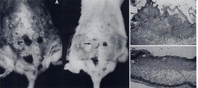
...@ .-,... :,, \\ • . ... @-. .?@ @ - ‘ @ ‘ , @:• @ @ -@eFiGURE3. Chemicallyinducedsquamous-cellcarcinomainan ICR mousewastreatedwithleaHopatch(arrow,1@.Pretreatmentbiopsyshowedirregular
proliferationand invasionof the tumor cells into dermis (B: HE,40x), but complete destructionof tumor cells was demonstratedon post-treatment biopsy (C:HE,40X).
on her neck was treated with 273.8 MBq (7.4 mCi) of ‘66Hofor 40 mm. Complete destruction of tumor was seen with almost normal skin color and texture over the irradiated field (Fig.
8A-D). Re-epithelializationwas observedat 3 mo post-treat
ment (Fig. 8E-F).
A 41-yr-old man suffered from recurrent Bowen's disease on the fifth toe despite cryosurgery or skin graft excision. Re cently, a 2-cm lesion recurred and was treated with 740 MBq (20 mCi) of 166Hofor 30 mm. After treatment, no residual lesion or recurrence was observed during the 13-mo follow-up period (Fig. 9).
DISCUSSION
Radionuclide therapy is a unique cancer treatment modality, which is alternative for or adjuvant to external radiation and chemotherapy. Recently, expanding availability of suitable Clinical Cases
Successful tumor destruction was seen in all subjects. Des quamation, erythema or ulceration developed between 1—2wk post-therapy. These acute radiation reactions were gradually healed with minimal fibrosis within 1 mo. There was no adverse reaction or recurrence during 8 —20mo follow-up periods.
A 89-yr-old woman had a 2-cm squamous-cell carcinoma on her scalp (Fig. 7A). Skin biopsy demonstrated irregular down ward proliferation of epidermis and invasion of atypical cells with hyperchromatic and large nuclei. The tumor was treated with 555 MBq (15 mCi) of ‘°6Hofor 40 mm. One month after the treatment, tumor size decreased considerably (Fig. 7B). Complete destruction of the tumor with re-epithelialization was demonstrated on follow-up biopsy (Fig. 7C). Allopecia was the only postirradiation complication.
A 75-yr-old woman with a 1.3-cm squamous-cell carcinoma
FIGURE4.(A)Holmium-166patchwasappliedtoa skintumorofanICRmouse.(B)Erythemaattheirradiatedregionandulcerationattumorsitewere
developedwithin 1 wk, but postradiationskin reactionswere graduallyimproved (C: 1 mo, D: 7 wk post-treatment).Pathologicexaminationdemonstrated regenerationof epitheliumwithfibrosisandlossof subcutaneousfat tissue(E:HE,40X).
1@ r@ @.-. I @ , @ @ ‘ j.
FIGURE5.(@Au8-mmkeratoacanthomawastreatedwitha 5-mmleaHopatch.(B)Tumordestructionisseenonlyattheirradiatedarea.(C)Pathology
depicted sharp demarcationbetween irradiatedand nonirradiatedareas(arrows). radiopharmaceuticals in oncology and endocrinology enable the use of radionuclide therapy. In nuclear oncology, specific tumor-seeking radiopharmaceuticals are being used since they can deliver radiation doses selectively into the target tissues.
In terms of the methodology of radionuclide therapy, intra venous or intra-arterial injection and intracavitary instillation of radiotracers are the preferable methods (8). However, beta emitters such as 90Sr, 32P and gamma emitters such as 182Ta, 1251and 60Co have been used for topical application to
treat ophthalmologic disease (9, 10). Specially designed appli cators have been used for ocular melanoma at the Royal Marsden Hospital (9). The applicators are still being used for the treatment of corneal vascularization. In this article, a beta-emitting radionuclide, @Ho,impregnated in a skin patch, was used for local radiation therapy of superficial skin cancer and Bowen's disease.
Holmium-166 emits beta-rays (Emax = 1.84 MeV) with a maximum soft-tissue range of 8.7 mm (average 2.1 mm) (1 1).
‘ ,-@— ___@___j
I
_@i I
A
I
FiGURE6. A keratoacanthomawas treated withan extremelyhighradiationdose
@ beforetreatment).Compiatetumor necrosis was found, but there was
a hardmassbeneaththes1@nlayer(arrow)dueto musdenecrosisandfibrosis(B,C).
FiGURE7. An89-yr-oldwomanwitha
2-cmsquamouscarcinomaon thescalp @A)wastreatedwith15mCi1@Ho.Tumor was completely destroyed, but allopecia developed (B; 3 mo after treatment).Bi opsy showed compiate regenerationof epitheliumwithoutviabletumorcells(C: HE,bOX). .... p ‘‘ . t@ . :‘ -@
personal use only.
by YONSEI UNIV. WONJU COLLEGE OF MEDICINE on February 14, 2019. For
jnm.snmjournals.org
B
( .,/@
O
D
FIGURE8@A 75-yr-oldwomanwitha b.3-cmsquamouscarcinomaontheleftneck
@ Radiationdermatitiswasseenwithin1 mopost-therapy(B),but
gradualhealingof the radiationreactionwasdemonstratedwithoutcom@ation(C:2 mo post-Tx,D:3 mo post-Tx).Irregularproliferationof tumortissue (E; HE, 40X) seen on pretreatmentbkpsy was completely destroyed vtñthskin regeneration(F; HE,40X).
Due to its favorable physical characteristics, ‘66Hohas been used for internal radiation therapy ofprimary hepatoma or liver metastasis and for radiation synovectomy (12—14).
In our study, all skin tumors of both humans and animals were successfully treated using the ‘66Hoskin-patch in a relatively short period without injury to the surrounding normal tissue. On the basis of our study, radionuclide treatment with beta-emitters has some advantages over external radiation therapy, since the former does not need expensive therapeutic units and the procedure is simple and noninvasive. Production of ‘66Hois simple and less expensive compared with @°Yor 89Sr, since 165Ho is a naturally abundant element. In addition, radionuclide treatment may play an important role in patients with multiple lesions when excision or external radiotherapy of the lesions is not possible. Another advantage is that there is no adverse effect on underlying bone and soft tissue due to the physical characteristics of beta-rays, high linear energy transfer and rapid fall off. Our study in animals had minimal injury to the adjacent tissues, except fibrosis confined to the beta-ray penetration length, despite an extremely high radiation dose (Fig. 6B,C). Although acute forms of skin reactions to radiation such as erythema, desquamation and ulceration may occur, it usually heals gradually with epithelial regeneration or fibrosis. Hyperpigmentation of irradiated skin may develop as a chronic reaction within a couple of months after completion of the irradiation, but this reaction will fade. It may also progress to a vitiligo appearance in some patients (3). Skin necrosis is a serious late complication of radiation, but 90% of the subjects are known to have healed spontaneously (15). Therefore, long-term follow-up is required to evaluate chronic complica tions and secondary malignancy.
In the treatment of large protruding tumors, beta emitters have a limitation due to their limited penetration range (maxi mum 8.6 mm). Ninety percent ofthe energy deposits within 2.1 mm and 10% within 2.1—8.6mm (11). For these subjects, external radiotherapy is preferable, but a higher radiation dose or fractionated treatment using beta emitters may be effective. Therapeutic response to the fractionated treatment in large
FiGURE9. A 2-cm recurrentBowen's diseaseon the fifth toe (arrowheads) in a 4b-yr-oldman was completelycured by the leaHopatch (A@before therapy; B, 3 mo post-therapy).M area of skin discoloration can be seen antenorto the lesson(arrow)due to pnor excision and sidn graft.
RADIONUCLIDE THERAPY OF SKIN CANCERS •Lee et al. 701
‘l'.'
tumors is being evaluated in our hospital. Therefore, beta emitters such as ‘66Ho,32P, @°Yor 89Sr can be used in the treatment of superficial malignant tumors of the skin.
ACKNOWLEDGMENTS
We thank J.W. Chang and C.H. Kim for excellent technical assistance and E.W. Park and K.H. Han for prepairing the patches. This study was supported by Korea Atomic Energy Research Institute grant 92C-l2l.
REFERENCES
I. Wilder RB, Kittelson JM, Shimm DS. Basal cell carcinoma treated with radiation therapy.Cancer l991;68:2134—2137.
2. Mackie RM. Squamous-cell carcinoma of the skin. In: Champion RH, Burton JL, Ebling FiG, eds. Texthook of dermatology, 5th ed. London: Blackwell Scientific Publications l992;1497—l504.
3. Shimm DS, Cassady JR. The Skin. In: Cox JD, ed. Moss ‘radiation oncology:
rationale, technique and results, 7th ed. St. Louis: Mosby-Year Book Inc. l994;99—
118.
4. Shimm DS, Wilder RB. Radiation therapy for squamous cell carcinoma of the skin.
Am J ClinOncol l991;14:383—386.
5. Petrovich Z, Parker RG, Luxton 0, Kuisk H, Jepson J. Carcinoma of the lip and
selected sites of head and neck skin: a clinical study of 896 patients. Radiother Oncol
1987;8:l 1—17.
6. Nevrkla E, Newton KA,. A survey ofthe treatment of200 casesofbasal cell carcinoma
(1959—1966inclusive).Br J Dermatol1974:91:429—433.
7. PrestwitchWV, NunesJ, KwokCS.Betadosepointkernelsfor radionuclidesof
potential use in radioimmunotherapy. J Nucl Med 1989;30:1036—l046. 8. Hoefnagel CA. Radionuclide therapy revisited. Eur I Nuci Med l991;18:408—431.
9. Lederman M. Some application of radioactive isotopes in ophthalmology. Br J
Radiology 1956;29:1—23.
10. Packer5, Rotman M. Radiotherapyofchoroidal melanomawith iodine.125. Ophthal.
mology 1980;87:582—590.
11. Johnson LS, Yanch JC, Shortkroff 5, Barnes CL, Sitzer Al, Sledge CB. Beta-particle dosimetry in radiation synovectomy. Eur J Nucl Med l995;22:977—988. 12. Mumper Ri, Ryo UY, Jay M. Neutron.activatedholmium-166-poly(L.lacticacid)
microspheres. A potential agent for the internal radiation therapy of hepatic tumors.
J NuclMed 199l;32:2139—2143.
13. Turner iN, Claringbold PG. Klemp PEG, Ct al. Holmium-166 microsphereliver radiotherapy: a preclinical SPECT dosimetry in the pig. NucI Med Commun 1994;15: 545—553.
14. Johnson LS, Yanch JC. Absorbed dose profiles for radionuclides of frequent use in
radiation synovectomy. Arthritis Rheum 1991;34:l521—1530.
15. Traenkle HL. A studyoflate radiationnecrosisfollowing therapyofskin cancer.Arch Dermatol 1995;72:446—453.
oncologists to help select patients for continued therapy in spite of a reduced ejection fraction. Our results argue that use of fixed criteria may be too restrictive.
KeyWordscardiotoxicity;leftventricleejectionfraction;equilibrium
gated radionuclideangiography
J NucIMed1997;3&702-.705
Cardiac
toxicity
due
to
chemotherapy
with
anthracyclines
and
anthraquinones is a serious adverse effect associated withsubstantial morbidity in some patients who are fortunate enough to survive their cancer (1—10).Recommendations for the “safe― use of anthracyclines have included either not exceeding a defined cumulative dose (e.g., for doxorubicin 400 mg/rn2) or ensuring adequate cardiac function by limiting treatment to patients with ejection fractions >50% (usually defined by quantitative radionuclide angiography). The threshold dose is an unreliable predictor of cardiotoxic risk and, furthermore, varies with the mode ofanthracycline administration [i.e., bolus versus continuous infusion (11)], as well as the exact drug administered. Pharmacologic advances in cancer chemotherapy have resulted in new ways of safely administering higher cumulative doses of anthracyclines. Dextrazoxane ameliorates anthracycline-induced cardiotoxicity and has recently been approved for use in women with metastatic breast cancer who have received a cumulative dose of doxorubicin of >300 mg/m2 (12). Liposomal doxorubicin preparations have also received recent approval and result in significantly less cardio toxicity (13). A study of endomyocardial biopsies of patients receiving liposomal doxorubicin administered to cumulative doses of up to 860 mg/m2 revealed no evidence of significant anthracycline specific cardiotoxicity (14). Thus, the cumulative dose criteria for establishing safe limits of anthracycline expo sure will likely decline in clinical value while objective studies Decreased left ventricular ejection fractkn (LVEF)is a ralativecon
traindication for the use of potentially cardiotoxic chemotherapy.A resting LVEF of 50% is usually used as the lower limit of normal values.The decision to change chemotherapy,however,is complex and is affected by many factors, including ejection fraction. Methods: To determine how LVEF data were used by clinical oncologists in clinical decision making, we performed a retrospec tive analysisof patients referredfor ejection fraction measurements from the hematology/oncologydMsions of Stanford Unrversftyfrom March 1992 through March 1995. The records of 565 patients treated with potentially cardiotoxic chemotherapy were evaluated. Results: LVEFs <50% were found in 153 patients. The charts of patientswith reduced ejectionfractions were reviewedto determine if the radionuclidemeasurementresulted in either discontinuationof the cardiotoxic agent or substitution of a less cardiotoxic drug or mode of administration.Thesespecific changes in therapy occurred in only 43 of the 153 (28%) patients with ejection fractions balow 50%; 24 of the 43 (57%) had ejectionfractions 40%. Patlentswith lower ejection fraction values were more likely to have their therapy changed than those with LVEFs close to normal. Patients with ejection fractions s30 generally had cardiotoxic agents discontin ued. Of patients who had a resting LVEF<50% and whose therapy was not changed,8b% had a normal increasein LVEFwith exercise. Conclusion: In clink@alpractice at our institution, ejection fraction <50% is not used as an absolute contraindication to cardiotoxic chemotherapy.When the LVEFis lessthan 40%, potentiallycardio toxic therapy is most often discontinued or omitted. Radionuclide evidence of cardiac reserve may account for decisions to continue cardiotoxic agents despite ejection fractions <50% in the majority of patients. Further study will be needed to establish standard criteria Reserve function, as measured by the change in ejection fraction from rest to stress may be an important parameterused by
Recdved Jul. 9, 1996; revision accepted Sep. 11, 1996.
For correspondence or reprints contact: H. Wdliam Strauss, MD, DMsion of Nuclear
Medkine, Dept of Radk@ogy, Stanford Unrversity Med@ Center, Am. HOlOl, Stan ford, CA 94035-5281.
Clinical Decision Making Based on Radionuclide
Determined Ejection Fraction in Oncology Patients
Nan-Jing Peng, Ranjana Advani, Susan Kopiwoda, George Fisher and H. William StraussDepartment ofNuclear Medicine, Kaohsiung Veterans General Hospital, Kaohsiung, Taiwan; Division ofNuclear Medicine, Departments of Oncology and Radiology, Stanford University Medical Center, Stanford, Ca4fornia
personal use only.
by YONSEI UNIV. WONJU COLLEGE OF MEDICINE on February 14, 2019. For
jnm.snmjournals.org
1997;38:697-702.
J Nucl Med.
Kim, Kyung Bae Park and Jae Rok Kim
Jong Doo Lee, Kwang Kyun Park, Min-Geol Lee, Eun-Hee Kim, Kyung Jin Rhim, Jong Tae Lee, Hyung Sik Yoo, Young Mi
Designed Skin Patch
Radionuclide Therapy of Skin Cancers and Bowen's Disease Using a Specially
http://jnm.snmjournals.org/content/38/5/697
This article and updated information are available at:
http://jnm.snmjournals.org/site/subscriptions/online.xhtml
Information about subscriptions to JNM can be found at:
http://jnm.snmjournals.org/site/misc/permission.xhtml
Information about reproducing figures, tables, or other portions of this article can be found online at: